Submitted Successfully!
To reward your contribution, here is a gift for you: A free trial for our video production service.
Thank you for your contribution! You can also upload a video entry or images related to this topic.
Version Summary Created by Modification Content Size Created at Operation
1 -- 2140 2022-04-30 04:51:02 |
2 format change Meta information modification 2140 2022-05-05 11:21:44 |

Video Upload Options

Do you have a full video?

Confirm

Are you sure to Delete?
Cite
If you have any further questions, please contact Encyclopedia Editorial Office.
Nakano, R.; Shiomi, H.; , .; Aizawa, N.; Enomoto, H. Immune Checkpoint Inhibitor-Related Pancreatitis. Encyclopedia. Available online: https://encyclopedia.pub/entry/22530 (accessed on 28 July 2024).
Nakano R, Shiomi H,  , Aizawa N, Enomoto H. Immune Checkpoint Inhibitor-Related Pancreatitis. Encyclopedia. Available at: https://encyclopedia.pub/entry/22530. Accessed July 28, 2024.
Nakano, Ryota, Hideyuki Shiomi,  , Nobuhiro Aizawa, Hirayuki Enomoto. "Immune Checkpoint Inhibitor-Related Pancreatitis" Encyclopedia, https://encyclopedia.pub/entry/22530 (accessed July 28, 2024).
Nakano, R., Shiomi, H., , ., Aizawa, N., & Enomoto, H. (2022, April 30). Immune Checkpoint Inhibitor-Related Pancreatitis. In Encyclopedia. https://encyclopedia.pub/entry/22530
Nakano, Ryota, et al. "Immune Checkpoint Inhibitor-Related Pancreatitis." Encyclopedia. Web. 30 April, 2022.
Immune Checkpoint Inhibitor-Related Pancreatitis
Edit

The indications for immune checkpoint inhibitors (ICIs) have expanded to include carcinomas of various organs. ICIs include drugs that target programmed cell death-1 (PD-1), programmed cell death ligand 1 (PDL-1), and cytotoxic T-lymphocyte-associated antigen 4 (CTLA-4). The indications for these drugs have been expanded to include many types of cancer, as efficacies have been reported for malignant melanoma and lung, kidney, head and neck, stomach, liver, ovarian, and pancreatic cancers .

ICI-related pancreatitis irAEs EUS ERCP

1. Introduction

During ICI therapy, blood amylase and lipase levels are often elevated [1]. The significance of these elevated pancreatic enzyme levels in the absence of symptoms is unclear, and aggressive treatment is often not warranted. Therefore, it is not recommended to discontinue ICI therapy based on elevated pancreatic enzymes alone [2][3][4]. Some cases of acute pancreatitis with a symptomatic elevation of pancreatic enzymes during ICI therapy have been reported [2][5][6], and the NCCN recommends evaluation for pancreatitis, including imaging studies, if the pancreatic enzyme elevations persist [7]. If the onset of acute pancreatitis is confirmed, the NCCN recommends treatment, including rapid infusion and pain management, and consultation with a specialist for moderate or more severe pancreatitis [7]. Nevertheless, because ICI-related pancreatitis is quite rare and there have been few reported cases, its imaging characteristics and appropriate treatment are largely unknown. Although cases of severe pancreatitis are rare, deaths have been reported [8].

2. Incidence

ICI-related pancreatitis has been reported as a rare irAE [5][9][10][11]. According to previous reports, the incidence of ICI-related pancreatitis has been reported to be approximately 0.3–3.9% [1][5][9][10][11]. Tirumani et al. reported that in 147 melanoma patients treated with ipilimumab, 46 (31%) developed irAEs, and the incidence of irAE pancreatitis was less than 1% [9]. Additionally, Friedman et al. reported that in a retrospective study of 119 patients with melanoma treated with nivolumab and ipilimumab, the incidence of pancreatitis was 1.7%, occurring in 20% of patients with highly elevated amylase and lipase [10]. Moreover, George et al. found that CTLA-4 inhibitors had a significantly higher incidence of pancreatitis than PD-1 inhibitors (3.98% vs. 0.94%, p < 0.05) and that combination therapy with CTLA-4 inhibitors and PD-1 inhibitors resulted in a relatively higher incidence of pancreatitis than monotherapy [12]. All reported cases in which radiographic or endoscopic images of ICI-related pancreatitis were available are shown in Table 1. The onset of pancreatitis varied from 20 days to more than one year after the start of ICI therapy, depending on the case.
Table 1. Radiographic and endoscopic images of ICI-related pancreatitis.

No.

Ref.

Sex

Age

ICI

CT Findings

MRI Findings

EUS Findings

ERCP Findings

Imaging Type

1

Ofuji et al. [13]

M

82

pembrolizumab

diffuse enlargement

diffuse restricted diffusion diffuse enlargement narrowing of the MPD

hypoechoic enlargement hyperechoic spots

NA

autoimmune

pancreatitis

2

Dehghani et al. [14]

M

63

nivolumab

focal enlargement fat stranding

focal restricted diffusion late enhancement

NA

NA

autoimmune

pancreatitis

3

Das et al. [15]

M

47

nivolumab

diffuse enlargement diffuse fat stranding

NA

NA

NA

acute interstitial

pancreatitis

4

Das et al. [15]

F

70

nivolumab

focal enlargement subtle fat stranding

NA

NA

NA

acute interstitial

pancreatitis

5

Das et al. [15]

F

50

pembrolizumab

NA

focal enlargement abrupt cut-off of the CBD

NA

NA

autoimmune

pancreatitis

6

Das et al. [15]

F

64

nivolumab

diffuse enlargement heterogenous enhancement fat stranding

NA

NA

NA

acute interstitial

pancreatitis

7

Das et al. [15]

F

56

ipilimumab nivolumab

NA

NA

NA

NA

autoimmune

pancreatitis

8

Capurso et al. [16]

F

76

pembrolizumab

MPD dilation

MPD dilation focal restricted diffusion

hypoechoic solid lesion stiff at elastography stenosis of the MPD

NA

autoimmune

pancreatitis

9

Saito et al. [17]

M

72

nivolumab

diffuse enlargement

NA

NA

NA

acute interstitial

pancreatitis

10

Kakuwa et al. [18]

M

70

pembrolizumab

mild diffuse enlargement MPD dilation

NA

NA

NA

autoimmune

pancreatitis

11

Tanaka et al. [19]

F

70

nivolumab

NA

diffuse enlargement focal restricted diffusion

diffuse hypoechoic enlargement

skipped narrowing of the MPD

autoimmune

pancreatitis

Clinical characteristics and imaging findings of ICI-related pancreatitis cases in which radiographic and endoscopic images are available. Abbreviations: Ref., reference; ICI, immune checkpoint inhibitor; CT, computed tomography; MRI, magnetic resonance imaging; EUS, endoscopic ultrasonography; ERCP, endoscopic retrograde cholangiopancreatography; PET-CT, positron emission tomography combined with computed tomography; MPD, main pancreatic duct; CBD, common bile duct; NA, not available.

3. Diagnosis

3.1. Clinical Symptoms

Similar to the clinical manifestations of acute pancreatitis in general [20], symptoms such as abdominal pain, back pain, nausea and vomiting, diarrhea, and fever have been reported in ICI-related pancreatitis [21]. Epigastric pain was the most frequent symptom in 39% of patients with ICI-related pancreatitis, and many patients had multiple symptoms [22]. However, a certain number of patients were also observed to have pancreatitis noted on imaging but were completely asymptomatic [22].

3.2. Blood Examination

Elevated blood pancreatic enzymes are most frequently observed in ICI-related pancreatitis [22]. In patients undergoing treatment with anti-CTLA-4 antibodies and anti- PD-1 antibodies, grades 3–4 of the Common Terminology Criteria for Adverse Events (CTCAE) and elevated levels of serum amylase and lipase were reported in 1–8% [10][23]. Blood lipase levels are considered useful for diagnosing acute pancreatitis, as they have better sensitivity and specificity than amylase levels [24]. Friedman et al. reported that ICI-related pancreatitis occurred in 20% of patients with elevated amylase and 6.7% of patients with elevated lipase levels with a CTCAE grade ≥3 [10]. Abu-Sbeih et al. also reported that among patients with ICI-related pancreatitis, those with clinical symptoms had significantly higher blood lipase levels than asymptomatic patients (p = 0.032) [22]. However, many cases of clinically undiagnosed pancreatitis have been observed, even in patients with lipase levels above grade 3. These data indicate that in most cases, lipase increase related to anti-PD1 or anti-PD-L1 is not associated with a significant clinical event. [1][22]. These results suggest that elevated pancreatic enzymes alone are not the basis for the development of ICI-related pancreatitis and that imaging evaluation is important for diagnosis. Autoimmune antibodies, including immunoglobulin G4 (IgG4), have been reported to be within normal ranges [13][14].

3.3. Radiology Images

The imaging features of ICI-related pancreatitis have been reported to include two patterns: one resembling acute interstitial pancreatitis and the other resembling autoimmune pancreatitis (Table 1). The imaging features of ICI-related pancreatitis, which resemble those of acute interstitial pancreatitis, include an enlarged pancreas on contrast-enhanced CT, poor contrast, and increased lipid concentrations [22]. Hofmann et al. reported that abdominal CT consistently revealed reduced lobulation, tissue swelling, and reduced tissue contrast enhancement in the pancreatic body and tail [5]. In contrast, Das et al. reported that diffuse (n = 14) or localized (n = 11) pancreatic enlargement was noted as an imaging finding in ICI-related pancreatitis and that a pattern consistent with autoimmune pancreatitis was present in 4/25 (16%) patients, while no patients developed necrotizing pancreatitis or pancreatic pseudocysts [15]. Regarding MRI, a few characteristic findings of ICI-related pancreatitis have been reported. ICI-associated pancreatitis with imaging findings similar to those of focal type 2 autoimmune pancreatitis has been noted on MRI, including signal restriction on diffusion-weighted images and contrast enhancement in the late phase of gadolinium contrast [14][16] (Table 1). Capurso et al. reported that the pancreatic head showed signal limitation on diffusion-weighted images and stenosis of the main pancreatic duct at the pancreatic head [16]. It has also been reported in several studies that ICI-related pancreatitis has a strong uptake of 18F-fluorodeoxyglucose positron emission tomography combined with CT (18F-FDG-PET/CT) [14][17][18] (Table 1). During ICI therapy, FDG-PET is often performed to evaluate the effect of treatment on the primary tumor, and asymptomatic cases of ICI-related pancreatitis incidentally diagnosed using FDG-PET have also been reported [14][17]. Radiology imaging reports of ICI-related pancreatitis are very limited. However, based on the reported cases of these imaging features, ICI-related pancreatitis can be classified into two major patterns: cases with imaging findings similar to acute interstitial pancreatitis and cases with imaging findings similar to type 2 autoimmune pancreatitis. During ICI treatment, when characteristic imaging findings of acute interstitial pancreatitis or type 2 autoimmune pancreatitis are observed, ICI-related pancreatitis should be considered a differential diagnosis (Table 1).

4. Endoscopic Findings

4.1. EUS

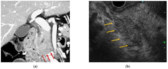
EUS has been established as a standard diagnostic imaging modality for pancreatic tumors and inflammatory pancreatic diseases and has become an indispensable modality for the detailed observation of pancreatic diseases. Some EUS images of ICI-related pancreatitis have been reported as similar to those characteristic of autoimmune pancreatitis [13][16][19][25][26] (Table 1 and Figure 1). Tanaka et al. reported nivolumab-related pancreatitis with imaging findings similar to diffuse-type autoimmune pancreatitis, and EUS showed diffuse hypoechoic enlargement of the pancreas with a hypoechoic, patchy, and heterogeneous parenchyma [19]. Ofuji et al. reported that in pembrolizumab-related pancreatitis, EUS showed an enlarged pancreas with hypoechogenicity and scattered hyperechoic spots [13]. Capurso et al. reported EUS findings mimicking those of focal-type autoimmune pancreatitis. Their report showed a 2 cm hypoechoic solid lesion of the pancreatic neck, stiffness on elastography, and low vascularity after the administration of a contrast agent in EUS images [16]. Some cases of ICI-related pancreatitis have been reported to be similar to the CT and EUS images of autoimmune pancreatitis [15]. In the diagnosis of autoimmune pancreatitis, numerous characteristic EUS findings have been reported for both diffuse and focal types. The diffuse type is characterized by pancreatic enlargement with diffuse hypoechoic images having a sausage-like appearance. Conversely, a capsule-like hypoechoic zone at the edge of the mass or spot-like hyperechoic findings in the pancreas may be observed in the focal type. These EUS findings are assumed to reflect a high degree of inflammatory cell infiltration. Distinguishing focal type pancreatitis from pancreatic cancer is particularly difficult because both appear as hypoechoic masses. Compared to pancreatic cancer, autoimmune pancreatitis may appear as multiple masses, and the duct penetration sign, which shows the pancreatic duct within the mass, is considered useful for diagnosing autoimmune pancreatitis.
Healthcare 10 00763 g001 550
Figure 1. Endoscopic ultrasonography and contrast-enhanced computed tomography (CT) images of immune checkpoint inhibitor (ICI)-related pancreatitis. (a) Focal pancreatic enlargement at the pancreatic head with a mass-like lesion and poor contrast enhancement (arrowhead). (b) Focal hypoechoic mass-like findings with internal hyperechoic spots from the pancreatic head to the pancreatic uncinate process (arrowhead).

4.2. ERCP

Generally, ERCP is not performed to diagnose acute pancreatitis, except in cases of gallstone pancreatitis. There are few reports of ERCP performed for ICI-related pancreatitis, and imaging findings of such ERCP procedures are almost unknown. Tanaka et al. reported the endoscopic diagnosis of nivolumab-related pancreatitis using ERCP. According to these, pancreatic duct findings by ERCP showed a relatively long narrowing with a skip in the main pancreatic duct, which was very similar to the findings in autoimmune pancreatitis [19]. Irregular narrowing of the main pancreatic duct is considered a characteristic of autoimmune pancreatitis on pancreatic ductal angiography with ERCP [27]. Irregular narrowing of the main pancreatic duct is defined as a diffuse or localized finding of a relatively long series of narrowed and irregular pancreatic ducts rather than the localized obstruction or stenosis characteristic of pancreatic ductal carcinoma. In addition, mild dilatation of the pancreatic duct of the pancreatic tail is also characteristic of autoimmune pancreatitis compared to pancreatic ductal carcinoma [28].

4.3. Histopathological Findings

EUS-guided fine-needle aspiration (EUS-FNA) has been shown to have a high diagnostic capability for pancreatic masses and is currently performed as an essential test for their differential diagnosis. In the absence of surgical resection, EUS-FNA is the only modality used for histological evaluation of pancreatic disease. In recent years, with the improvement in puncture needle technology, the number of specimens that can be collected by EUS-FNA has been increasing, making it possible to perform a thorough pathological evaluation. Pathological studies using EUS-FNA have reported several cases of ICI-related pancreatitis [16][25][26][29]. According to the pathological findings of autopsy cases of ICI-related pancreatitis, along with the pathological findings of pembrolizumab necrotizing acute pancreatitis, significantly more CD8+ T cells than CD4+ T cells were detected in the remaining pancreatic parenchyma [8]. A report of EUS-FNA for pembrolizumab-related pancreatitis also showed infiltration of T-lymphocytes, with a predominance of CD8+ cells over CD4+ cells [29]. Ofuji et al. reported that EUS-FNA was performed for ICI-related pancreatitis with a 25-G needle puncture, and cytological examination showed infiltration of inflammatory cells composed mainly of neutrophils, which is a presentation similar to that seen in type 2 autoimmune pancreatitis [13]. Song et al. reported that when EUS-FNA was performed for nivolumab-related pancreatitis, EUS images showed mass-forming pancreatitis, with lymphocytes infiltrating the surrounding pancreatic duct and fibrosis in the stroma and no plasma cells. These authors also highlighted the similarities between nivolumab-related and type 2 autoimmune pancreatitis [26].

5. Management

Steroid therapy is generally used to treat irAEs that develop during ICI therapy. The NCCN guidelines recommend that ICI therapy continuation should be considered when there is no evidence of pancreatitis and only elevated amylase and lipase levels are observed [7]. For moderate pancreatitis, the NCCN recommends interruption of ICI therapy and initiation of methylprednisolone or prednisolone at 0.5–1.0 mg/kg/day, and for severe pancreatitis, complete discontinuation of ICI therapy and steroids at 1–2 mg/kg/day. However, Abu-Sbeih et al. reported that steroid therapy for ICI-associated pancreatitis did not affect short-term improvement in lipase levels or shorten hospital stay, making the efficacy of steroid therapy controversial [22]. In nearly all reported cases, ICI-related pancreatitis showed an eventual improvement by interruption of ICI therapy or steroid therapy. A severe acute case of ICI-related pancreatitis was diagnosed after the administration of nivolumab followed by pazopanib. According to the report, methylprednisolone was administered, and conservative treatment for acute pancreatitis was initiated, but the patient’s general condition rapidly worsened, resulting in a fatality [8].

References

  1. Michot, J.-M.; Ragou, P.; Carbonnel, F.; Champiat, S.; Voisin, A.-L.; Mateus, C.; Lambotte, O.; Annereau, M. Significance of Immune-related Lipase Increase Induced by Antiprogrammed Death-1 or Death Ligand-1 Antibodies: A Brief Communication. J. Immunother. 2018, 41, 84–85.
  2. Naidoo, J.; Page, D.B.; Li, B.T.; Connell, L.C.; Schindler, K.; Lacouture, M.E.; Postow, M.A.; Wolchok, J.D. Toxicities of the anti-PD-1 and anti-PD-L1 immune checkpoint antibodies. Ann. Oncol. 2015, 26, 2375–2391.
  3. Postow, M.A. Managing Immune Checkpoint-Blocking Antibody Side Effects. Am. Soc. Clin. Oncol. Educ. Book 2015, 35, 76–83.
  4. Kumar, V.; Chaudhary, N.; Garg, M.; Floudas, C.S.; Soni, P.; Chandra, A.B. Current Diagnosis and Management of Immune Related Adverse Events (irAEs) Induced by Immune Checkpoint Inhibitor Therapy. Front. Pharmacol. 2017, 8, 49.
  5. Hofmann, L.; Forschner, A.; Loquai, C.; Goldinger, S.M.; Zimmer, L.; Ugurel, S.; Schmidgen, M.I.; Gutzmer, R.; Utikal, J.; Göppner, D.; et al. Cutaneous, gastrointestinal, hepatic, endocrine, and renal side-effects of anti-PD-1 therapy. Eur. J. Cancer 2016, 60, 190–209.
  6. Cramer, P.; Bresalier, R.S. Gastrointestinal and Hepatic Complications of Immune Checkpoint Inhibitors. Curr. Gastroenterol. Rep. 2017, 19, 3.
  7. Thompson, J.A.; Schneider, B.J.; Brahmer, J.; Andrews, S.; Armand, P.; Bhatia, S.; Budde, L.E.; Costa, L.; Davies, M.; Dunnington, D.; et al. NCCN Guidelines Insights: Management of Immunotherapy-Related Toxicities, Version 1.2020. J. Natl. Compr. Cancer Netw. 2020, 18, 230–241.
  8. Tanaka, T.; Sakai, A.; Shiomi, H.; Masuda, A.; Kobayashi, T.; Tanaka, S.; Nakano, R.; Shigeoka, M.; Koma, Y.-I.; Kodama, Y. An autopsy case of severe acute pancreatitis induced by administration of pazopanib following nivolumab. Pancreatology 2021, 21, 21–24.
  9. Tirumani, S.H.; Ramaiya, N.H.; Keraliya, A.R.; Bailey, N.D.; Ott, P.A.; Hodi, F.S.; Nishino, M. Radiographic Profiling of Immune-Related Adverse Events in Advanced Melanoma Patients Treated with Ipilimumab. Cancer Immunol. Res. 2015, 3, 1185–1192.
  10. Friedman, C.F.; Clark, V.; Raikhel, A.V.; Barz, T.; Shoushtari, A.N.; Momtaz, P.; Callahan, M.K.; Wolchok, J.D.; Chapman, P.B.; Hellmann, M.D.; et al. Thinking Critically About Classifying Adverse Events: Incidence of Pancreatitis in Patients Treated With Nivolumab + Ipilimumab. JNCI J. Natl. Cancer Inst. 2016, 109, djw260.
  11. Clamon, G.; Patel, R.; Mott, S. Pancreatitis associated with newer classes of antineoplastic therapies. J. Community Support. Oncol. 2017, 15, e135–e141.
  12. George, J.; Bajaj, D.; Sankaramangalam, K.; Yoo, J.W.; Joshi, N.; Gettinger, S.; Price, C.; Farrell, J.J. Incidence of pancreatitis with the use of immune checkpoint inhibitors (ICI) in advanced cancers: A systematic review and meta-analysis. Pancreatol. 2019, 19, 587–594.
  13. Ofuji, K.; Hiramatsu, K.; Nosaka, T.; Naito, T.; Takahashi, K.; Matsuda, H.; Ohtani, M.; Imamura, Y.; Ishizuka, T.; Nakamoto, Y. Pembrolizumab-induced autoimmune side effects of colon and pancreas in a patient with lung cancer. Clin. J. Gastroenterol. 2021, 14, 1692–1699.
  14. Dehghani, L.; Mikail, N.; Kramkimel, N.; Soyer, P.; Lebtahi, R.; Mallone, R.; Larger, E. Autoimmune pancreatitis after nivolumab anti–programmed death receptor-1 treatment. Eur. J. Cancer 2018, 104, 243–246.
  15. Das, J.P.; Postow, M.A.; Friedman, C.F.; Do, R.K.; Halpenny, D.F. Imaging findings of immune checkpoint inhibitor associated pancreatitis. Eur. J. Radiol. 2020, 131, 109250.
  16. Capurso, G.; Archibugi, L.; Tessieri, L.; Petrone, M.C.; Laghi, A.; Arcidiacono, P.G. Focal immune-related pancreatitis occurring after treatment with programmed cell death 1 inhibitors: A distinct form of autoimmune pancreatitis? Eur. J. Cancer 2018, 95, 123–126.
  17. Saito, H.; Ono, K. Nivolumab-induced Pancreatitis: An Immune-related Adverse Event. Radiol. 2019, 293, 521.
  18. Kakuwa, T.; Hashimoto, M.; Izumi, A.; Naka, G.; Takeda, Y.; Sugiyama, H. Pembrolizumab-related pancreatitis with elevation of pancreatic tumour markers. Respirol. Case Rep. 2020, 8, e00525.
  19. Tanaka, T.; Sakai, A.; Kobayashi, T.; Masuda, A.; Shiomi, H.; Kodama, Y. Nivolumab-related pancreatitis with autoimmune pancreatitis-like imaging features. J. Gastroenterol. Hepatol. 2019, 34, 1274.
  20. Banks, P.A.; Bollen, T.L.; Dervenis, C.; Gooszen, H.G.; Johnson, C.D.; Sarr, M.G.; Tsiotos, G.G.; Vege, S.S.; Acute Pancreatitis Classification Working Group. Classification of acute pancreatitis—2012: Revision of the Atlanta classification and definitions by international consensus. Gut 2013, 62, 102–111.
  21. Di Giacomo, A.M.; Danielli, R.; Guidoboni, M.; Calabrò, L.; Carlucci, D.; Miracco, C.; Volterrani, L.; Mazzei, M.A.; Biagioli, M.; Altomonte, M.; et al. Therapeutic efficacy of ipilimumab, an anti-CTLA-4 monoclonal antibody, in patients with metastatic melanoma unresponsive to prior systemic treatments: Clinical and immunological evidence from three patient cases. Cancer Immunol. Immunother. 2009, 58, 1297–1306.
  22. Abu-Sbeih, H.; Tang, T.; Lu, Y.; Thirumurthi, S.; Altan, M.; Jazaeri, A.A.; Dadu, R.; Coronel, E.; Wang, Y. Clinical characteristics and outcomes of immune checkpoint inhibitor-induced pancreatic injury. J. Immunother. Cancer 2019, 7, 31.
  23. Weber, J.S.; D’Angelo, S.P.; Minor, D.; Hodi, F.S.; Gutzmer, R.; Neyns, B.; Hoeller, C.; Khushalani, N.I.; Miller, W.H., Jr.; Lao, C.D.; et al. Nivolumab versus chemotherapy in patients with advanced melanoma who progressed after anti-CTLA-4 treatment (CheckMate 037): A randomised, controlled, open-label, phase 3 trial. Lancet Oncol. 2015, 16, 375–384.
  24. Vissers, R.J.; Abu-Laban, R.B.; McHugh, D.F. Amylase and lipase in the emergency department evaluation of acute pancreatitis. J. Emerg. Med. 1999, 17, 1027–1037.
  25. Janssens, L.; Takahashi, N.; Majumder, S. Pancreatic Atrophy in Nivolumab-Associated Pancreatitis Mimics Autoimmune Pancreatitis. Pancreas 2021, 50, e28–e29.
  26. Song, Z.; Shih, J.; Seid, D.S. Rare Case of Nivolumab-Induced Chronic Pancreatitis. Clin. Gastroenterol. Hepatol. 2021.
  27. Sugumar, A.; Levy, M.J.; Kamisawa, T.; Webster, G.J.; Kim, M.; Enders, F.; Amin, Z.; Baron, T.H.; Chapman, M.H.; I Church, N.; et al. Endoscopic retrograde pancreatography criteria to diagnose autoimmune pancreatitis: An international multicentre study. Gut 2011, 60, 666–670.
  28. Wakabayashi, T.; Kawaura, Y.; Satomura, Y.; Watanabe, H.; Motoo, Y.; Okai, T.; Sawabu, N. Clinical and imaging features of autoimmune pancreatitis with focal pancreatic swelling or mass formation: Comparison with so-called tumor-forming pancreatitis and pancreatic carcinoma. Am. J. Gastroenterol. 2003, 98, 2679–2687.
  29. Suda, T.; Kobayashi, M.; Kurokawa, K.; Matsushita, E. Simultaneous occurrence of autoimmune pancreatitis and sclerosing cholangitis as immune-related adverse events of pembrolizumab. BMJ Case Rep. 2021, 14, e243360.
More
Information
Contributors MDPI registered users' name will be linked to their SciProfiles pages. To register with us, please refer to https://encyclopedia.pub/register : , , , ,
View Times: 967
Revisions: 2 times (View History)
Update Date: 05 May 2022
1000/1000
Video Production Service