Submitted Successfully!
To reward your contribution, here is a gift for you: A free trial for our video production service.
Thank you for your contribution! You can also upload a video entry or images related to this topic.
Version Summary Created by Modification Content Size Created at Operation
1 -- 5827 2022-04-20 08:33:44 |
2 layout Meta information modification 5827 2022-04-21 03:34:41 | |
3 layout Meta information modification 5827 2022-04-22 09:06:34 |

Video Upload Options

Do you have a full video?

Confirm

Are you sure to Delete?
Cite
If you have any further questions, please contact Encyclopedia Editorial Office.
Ma, X.; , .; Wang, J.; Liu, Z.; Xi, J.; Ye, L. Polymeric Hydrogel Carrier for Nerve Repair. Encyclopedia. Available online: https://encyclopedia.pub/entry/21977 (accessed on 22 July 2024).
Ma X,  , Wang J, Liu Z, Xi J, Ye L. Polymeric Hydrogel Carrier for Nerve Repair. Encyclopedia. Available at: https://encyclopedia.pub/entry/21977. Accessed July 22, 2024.
Ma, Xiaoyu, , Jin Wang, Zongjian Liu, Jianing Xi, Lin Ye. "Polymeric Hydrogel Carrier for Nerve Repair" Encyclopedia, https://encyclopedia.pub/entry/21977 (accessed July 22, 2024).
Ma, X., , ., Wang, J., Liu, Z., Xi, J., & Ye, L. (2022, April 20). Polymeric Hydrogel Carrier for Nerve Repair. In Encyclopedia. https://encyclopedia.pub/entry/21977
Ma, Xiaoyu, et al. "Polymeric Hydrogel Carrier for Nerve Repair." Encyclopedia. Web. 20 April, 2022.
Polymeric Hydrogel Carrier for Nerve Repair
Edit

Nerve regeneration and repair still remain a huge challenge for both central nervous and peripheral nervous system. Although some therapeutic substances, including neuroprotective agents,clinical drugs and stem cells, as well as various growth factors, are found to be effective to promote nerve repair, a carrier system that possesses a sustainable release behavior, in order to ensure high on-site concentration during the whole repair and regeneration process, and high bioavailability is still highly desirable. Hydrogel, as an ideal delivery system, has an excellent loading capacity and sustainable release behavior, as well as tunable physical and chemical properties to adapt to various biomedical scenarios; thus, it is thought to be a suitable carrier system for nerve repair. 

hydrogel nerve repair carrier

1. Introduction

Nerve repair still remains a huge challenge in the clinic. Recently, more and more researchers have paid attention to the significant effects of biomaterials such as hydrogels on nerve repair. The nervous system is divided into the central nervous system (CNS), which is composed of the brain nerve and spinal cord, and peripheral nervous systems (PNS) [1][2]. As shown in Figure 1, hydrogels are widely used in various nervous systems, including the CNS and PNS, but are particularly useful in repairing brain nerves after stroke.
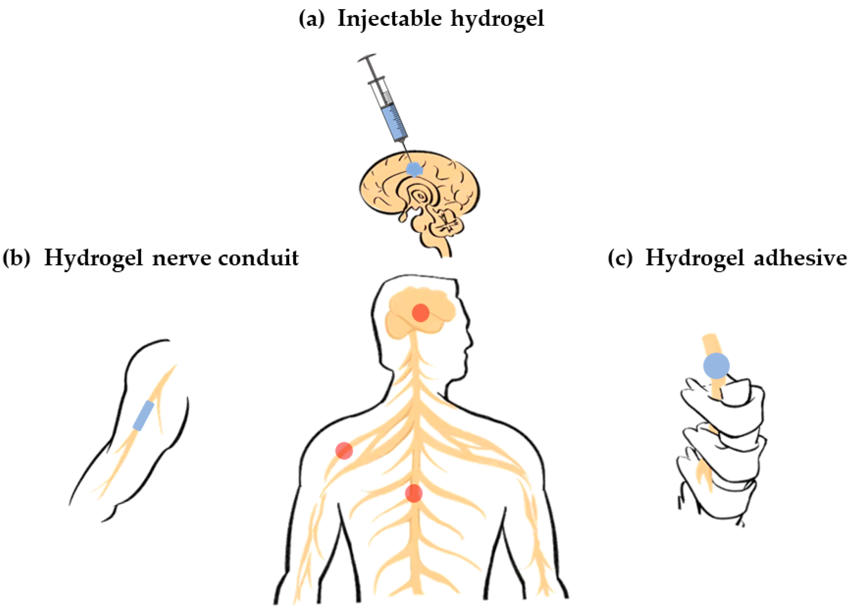
Figure 1. Various hydrogel carriers for nerve repair. (a) Injectable hydrogel carriers for brain nerve repair (central nervous system); (b) Hydrogel carriers as conduits for peripheral nerve repair (peripheral nervous system); (c) Hydrogel carriers as adhesive for spinal cord repair (central nervous system).
Recently, the incidence of stroke worldwide has sharply increased. It has a very poor prognosis, including a high mortality and long-term disability. Stroke patients often show difficulties with speaking, cognitive impairment, memory loss, dementia, and depression, which indicate the damage and loss of brain nerves [3][4][5]. Thus, it is believed that brain nerve repair after stroke plays a vital role in patient rehabilitation and body function reconstruction. Currently, stroke could be treated to some extent in the acute phase in the clinic [6]. However, the therapeutic time window is so narrow that the most patients miss it. Thus, rehabilitative therapies in the sub-acute phase and chronic phase that promote nerve repair and functional recovery are very important in stroke treatment. Although some approaches, including neuroprotective agents, stem cells and immune T-cells, as well as various growth factors, are shown to be effective in animal models, some concerns still remain [7][8][9]. As these therapeutic substances are usually intravenously injected, it is difficult to reach the brain site and maintain enough local concentration due to the presence of the blood –brain barrier in sub-acute phase and chronic phase and the lack of targeting ability [10]. Although therapeutic substances could also be delivered by cephalotomy or craniotomy to endow a high initial concentration, they would be absorbed quickly by the cerebrospinal fluid, leading to a sharp decrease in local concentration. Consequently, a biocompatible carrier system, which could effectively deliver therapeutic substances and maintain their long-term retention in the brain site, is highly desirable for stroke treatment in the sub-acute and chronic phases.
Hydrogel is a water-swollen and cross-linked polymeric network produced by the simple reaction of one or more monomers [11]. They possess a degree of flexibility that ios very similar to natural tissues due to their large water content and exhibit a 3D structure that emulates the properties of the native extracellular matrix (ECM) within the CNS [12][13]. A hydrogel is considered to be an ideal carrier system for various biomedical applications including nerve repair. Extensive studies have revealed that hydrogel carriers exhibit an excellent restorative function against stroke-induced brain damage [14][15]. Thus, it is necessary to make a comprehensive summary to provide useful insights and technical enlightenment for researchers to optimize the design and fabrication of hydrogel carriers in order to achieve better therapeutic effects in nerve repair, which might be translated into the clinic in the future. Furthermore, hydrogels that can be injected in form of liquid and gelate in situ by some external stimuli, including heat, light, magnetism, etc., have great potential to be very suitable carriers for brain nerve repair after stroke. The liquid formed during the injection greatly simplifies both the loading and the delivering of various therapeutic substances. Then, the in situ gelation causes sustainable release behavior to maintain long-term retention in brain, resulting in a high bioavailability, as well as good therapeutic effect. On the other hand, the field of brain nerve repair after stroke urgently needs a breakthrough in both science and technology to drive it from the lab to the clinic due to the sharp increase in the number of stroke patients worldwide.

2. Classification of Hydrogel Carriers for Nerve Repair

A hydrogel exhibits several advantages in its application as a nerve repair material. First of all, it is an ideal carrier that can load cells, drugs, nutrients, growth factors and other bioactive components, as well as maintain its activity for a long time to promote nerve repair. Second, it has good biodegradability so that it can provide support for the growth of new tissues at the initial stage of repair and provide space for nerve regeneration through gradual degradation. Third, it has a higher moisture content and better biocompatibility, making it a suitable substrate for endogenous or transplanted cell growth. Forth, it has an adjustable mechanical strength, which can flexibly fit the modulus of the damaged nerve tissue. Finally, it has abundant pore space that is conducive to cell growth and material exchange. Therefore, the application of hydrogels in the field of nerve repair has been widely studied.
From different angles, hydrogels have various classification standards. For instance, based on their physical shape, hydrogels can generally be classified as blocks, microspheres, fibrous, and films. Based on their physical structure, hydrogels can be divided into three categories: amorphous, crystalline and semicrystalline phases, which is a mixture of amorphous and crystalline. Based on the presence or absence of electrical charge located on the cross-linked chains, hydrogels can be divided into four categories: nonionic, ionic, amphoteric electrolyte and zwitterionic. Based on the cross-linking mechanism, hydrogels can be divided into two categories. The chemical cross-linked hydrogel network is a permanent connection. The physical cross-linked hydrogel network is a transient connection that arises from molecular/polymer chain entanglement or under the action of weak interactions, such as hydrogen bonding and electrostatic interactions. Based on their polymeric composition, hydrogels can be divided into three categories. Homopolymeric hydrogels are referred to as polymer networks derived from a single species of monomer. Copolymeric hydrogels are referred to as polymer networks that comprise two or more different monomer species with at least one hydrophilic component. Multipolymer interpenetrating polymeric hydrogels are referred to as polymer networks made of two independent cross-linked components where one component is a cross-linked polymer and other component is a non-cross-linked polymer [11].
Based on their source, hydrogels can be divided into natural-based biomaterials and synthetic biomaterials. The source of the polymer has a greater impact on the biocompatibility and safety of the hydrogel when considered as a functional material for biological tissues. Therefore, this research introduces the classification of hydrogels based on their sources as shown in Figure 2.
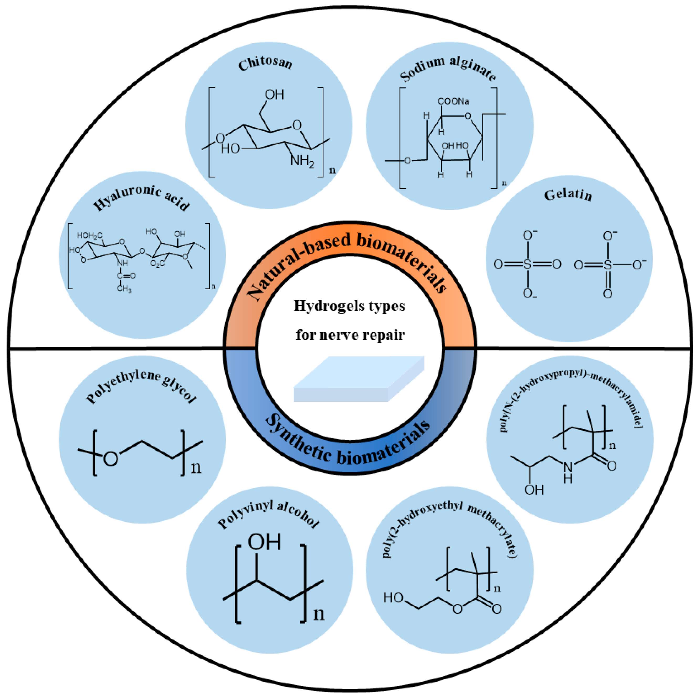
Figure 2. Classification of hydrogel carriers for nerve repair based on their sources.

2.1. Natural-Based Biomaterials

Natural-based biomaterials refer to biomaterials generated under natural conditions, usually in the form of naturally occurring macromolecular substances derived from animals, plants or humans. Natural-based biomaterials can be gradually degraded into small molecular substances existing in the body through hydrolysis, enzymatic hydrolysis and other methods under the physiological environment of the body, and then completely absorbed or excreted through metabolism without causing toxic side effects on the body itself. Because of their large availability, large quantities of natural-based biomaterials are accessible at reasonable prices. Thus, they have been widely used as hydrogels for nerve repair. However, their mechanical properties are usually poor, and some of them are insoluble in water or common organic solvents. It is difficult to purify them for large-scale production [16]. The following will briefly introduce and discuss some natural-based biomaterials used in nerve repair.
Hyaluronic acid (HA) is a macromolecular linear polysaccharide and the main component of an extracellular matrix, especially abundant in the central system. It is involved in the regulation of a variety of cell activities. Its unique molecular structure and physicochemical properties endows it with a variety of important physiological functions in the body [17]. Thus, it has been widely used for nerve repair. In vitro experiments have shown that HA hydrogel can promote strong neurite outgrowth [18]. Neurite is an elongated part extending from the cell body of a nerve cell (neuron), and can be divided into dendrites and axons. The dendrite is the entrance that receives information, and the axon is the exit that sends out information. Both are used together for information transfer between cells. HA hydrogels also support the viability of neural precursor cells [19][20], neural stem cells [21] and Schwann cells [22], maintain their activities, and enable them to proliferate and differentiate. HA hydrogels have a high water-absorbency and enzyme degradability. In vivo, they usually exhibit rapid erosion and degradation behavior. They mainly form a hydration open mesh at the site of injury to inhibit the formation of a glial scar. Glial scarring is a compensatory reaction product after injury. In the early post-injury period, glial scarring can limit the spread of inflammation. However, with the progression of the disease, a glial scar can form a physical and chemical barrier, secrete a variety of axon regeneration inhibitory factors, and hinder the recovery of neural function. By reducing its formation, glial scarring can promote neuroprotection and functional recovery. In addition, HA hydrogels support the cell infiltration of cells into the hydrogel and angiogenesis [23]. Broguiere cross-linked high-molecular-weight hyaluronic acid and transglutaminase to prepare a new type of hydrogel, which could be covalently bound to the protein in the body when it was injected in vivo, so that the hydrogel could covalently bind to brain or spinal cord defects. Thus, it showed a fast neurite outgrowth, axonal and dendritic speciation, strong synaptic connectivity in 3D networks, and rapidly occurring and long-lasting coordinated electrical activity [24]. The healing potential of HA hydrogels may be partly attributed to hyaluronic acid promoting interactions between hydrogels and the central nervous system [25].
Collagen is the most important extracellular water-insoluble fibrin. It is the skeletal component of the extracellular matrix and the most abundant protein in the human body. Collagen hydrogel can be used as the inner filling of nerve conduits to repair broken nerve tissue [26] or independent tissue engineering material to fill tissue defect [27]. It is an excellent carrier of neural stem cells, mesenchymal stem cells and other cells. It can be used for cell transplantation to repair brain or spinal cord injury [28][29]. It can also be used as a sustained-release drug carrier [30].
Gelatin is obtained by the thermal denaturation of collagen. Gelatin has a relatively low antigenicity compared with collagen. The body’s immune system response can be avoided or reduced during its use. Animal-derived gelatin has been widely studied in medical applications due to its biocompatibility, plasticity and adhesion. Gelatin hydrogel is beneficial for cell adhesion and activity, and has significant effects on nerve healing and reconstruction [31]. Experimental results have shown that the nerve conduit containing gelatin hydrogel and a basic fibroblast growth factor promoted axonal regeneration after a 15 mm sciatic nerve injury in rats. The nerve regeneration rate was significantly increased, and a large number of regenerated nerve axons were induced [32]. However, this effect was not as good as autologous transplantation [33]. Due to its weak mechanical strength and rapid degradation, most gelatins will be prepared in a combination with other biomaterials to improve the comprehensive properties of hydrogels [34][35][36].
Fibrin is a protein formed during blood coagulation and is also a component of the extracellular matrix. Fibrin has an obvious advantage over other types of protein hydrogels in the repair of the nervous system: when autologous fibrin is used, it has a complete biocompatibility [37]. Fibrin hydrogel can improve the survival rate of neural stem cells [38][39], support the neurogenic differentiation of mesenchymal stem cells and the regeneration of axons [40]. It also has a satisfactory effect in loading drugs [41]. In order to overcome the application limitation of fibrin, Robinson crosslinked it with genipin to reduce its degradation rate, so that the hydrogel scaffold could support the neural aggregates derived from neural progenitor cells and induce neurite outgrowth [42].
Keratin is a kind of fibrin with abundant sources and biological activities [43]. Keratin hydrogel not only has a good cell adhesion [44] but can also maintain the high activity of cells [45][46]. Keratin hydrogel also has a high nerve induction effect [47][48]. In a mouse tibial nerve model, keratin hydrogel significantly improved the electrophysiological recovery in the early stage. At 6 months, the keratin hydrogel group even produced electrical and histological results superior to the empty conduits and sensory nerve autografts [49]. In 1 cm rat sciatic nerve injury, keratin hydrogel could promote debris clearance in the distal stump by Schwann cell activation [50].
Chitosan is formed by the deacetylation of chitin in crustacean shells. It is a widely used tissue engineering material with a low production cost. It has the potential to interact with regeneration-related cells and neural microenvironments, so as to improve axon regeneration and reduce the formation of neuroma. Chitosan can effectively maintain the biological activity of loaded cells and promote their proliferation and differentiation [51], such as promoting the growth of Schwann cells [52], supporting the differentiation of neural progenitor cells [53], and inducing mesenchymal stromal cells to differentiate into Schwann-like cells [54]. It allows a functional and morphological nerve regeneration similar to autologous nerve transplantation [55]. Jafar prepared chitosan scaffolds containing human endometrial stem cells and platelet plasma. It promoted nerve fiber regeneration and angiogenesis and prevented the formation of scar tissue in a rat spinal cord injury model [56]. In addition, chitosan oligosaccharide, the degradation product of chitosan, showed a neuroprotective effect in the process of peripheral nerve regeneration. It could promote cell proliferation, prevent cell apoptosis and accelerate peripheral nerve regeneration [57][58][59].
Alginate is a polysaccharide found in brown algae. It has an antioxidant capacity and can protect nerve cells from H2O2 damage [60][61]. Alginate hydrogel can not only promote the adhesion and proliferation of nerve cells [62], but also enhance the differentiation of pluripotent cells (mouse embryonic stem cells) into neural lineages [63]. It can also trigger the regeneration of up and down axon fibers and effectively form synapses, which play a supporting role in axon regeneration [64]. However, alginate hydrogel still has some limitations. Since mammals do not produce endogenous seaweed enzymes, the alginate hydrogel implanted in vivo may take several months to complete degradation. This poor degradation will reduce the penetration ability of cells [65]. In addition, pure alginate hydrogel is bioinert and cannot promote the activity of encapsulated cells [66]. For its applications, it can be improved by compounding with other materials [67], or adding substances such as nanofibers [68].
Silk fibroin and sericin are two main proteins in silk. Sericin has neurotrophic and neuroprotective effects, can promote axon extension and branching, and prevent the cell death of primary neurons in a hypoxia environment [69][70]. Sericin hydrogel can promote the adhesion and differentiation of neurons and facilitate the repair of nerves [71]. In addition, the degradation products of sericin protein are rich in glycine and serine, which play an important role as neurotransmitters in neurotransmission [72]. Silk fibroin has excellent nerve biocompatibility and can support nerve regeneration. It has a high potential in nerve tissue engineering. At present, it is mainly used as a nerve conduit to bridge damaged nerves by electrospinning [73][74][75]. Silk fibroin hydrogel has good biocompatibility with ganglion, and is beneficial for maintaining the activity of Schwann cells [76], promoting axonal growth and guiding axonal sprouting [77].

2.2. Synthetic Biomaterials

Polyethylene glycol (PEG) is a stable, nontoxic and biocompatible polymer synthetic material with a wide range of clinical applications. When PEG is used for spinal cord repair, it has many advantages, such as no accumulation in the body, inhibiting the formation of vacuoles and scars, reducing inflammation and so on [78]. PEG can be used to synthesize hydrogels and modify other hydrogels [79]. When repairing sciatic nerve transection injury in rats, PEG hydrogels can effectively reduce scar tissue formation [80], support axon regeneration [81], and promote limb function by promoting more and more myelin regeneration, enabling a faster recovery [82]. Myelin refers to the protective shell formed by oligodendrocytes on the outside of nerve fibers. Its main function is to protect nerves and participate in the conduction of nerve impulses. Changing the end groups of PEG and introducing different functional groups, such as toluene sulfonate, amino group, carboxyl group and aldehyde group, can further expand the application field of PEG. By introducing biological ingredients, such as natural biomaterials and nutritional factors, into synthetic hydrogels, PEG can effectively improve the function of nerve cells in inert PEG hydrogels, and promote the survival and growth of nerve cells [83][84][85]. Zhang prepared hydrogels made from graphene oxide and four-arm polyethylene glycol terminated with two acetyl glycerol and loaded with the anti-inflammatory agent, diacerein. The hydrogel was delivered to the spinal cord in rats, causing injury by a minimally invasive injection. Free diacerein can minimize inflammatory responses and prevent the formation of an inhibitory microenvironment. Graphene oxide gives proper conductivity to hydrogels, which promotes the neuronal growth and remyelination of axons [86].
Methacrylate-based hydrogels are widely used in the field of nerve repair. Hydrogels based on poly(2-hydroxyethyl methacrylate) (PHEMA) and poly[N-(2-hydroxypropyl)-methacrylamide] (PHPMA) belong to a group of synthetic, highly biocompatible polymers [87]. The high water content of the PHEMA hydrogel and its mechanical compatibility with host tissues allows it to restore the anatomical continuity of damaged nerve structures. Porous PHEMA hydrogels inhibit scarring [88] and provide scaffolds for the growth of connective tissue elements, blood vessels, neurofilaments, and Schwann cells [89]. In addition, it can also be used as a platform for continuous drug delivery. The PHEMA also has some disadvantages. First, as a non-degradable polymer, hydrogel calcification and prolonged inflammatory responses might limit long-term axonal regeneration [90]. Biodegradable hydrogels were prepared by compounding PHEMA with other polymers. Pertici combined PHEMA with PLA to obtain block copolymer. The PLA-b-PHEMA block copolymer can indeed display desired properties, such as biodegradability and an improvement in rat motor function, which were observed after implantation following a thoracic spinal cord hemisection in rats [91]. In addition, PHEMA itself is not adhesive or attractive to neurons, so it can be modified using substances with biorecognition sites, thereby improving the biocompatibility of the hydrogels. Šárka used laminin-derived peptide sequences to modify PHEMA to promote cell adhesion and neural differentiation [92]. The modification of PHEMA hydrogels with lysine can prolong the release time of its loading factor, increase the survival rate of neurons, and induce favorable biological reactions [93].
PHPMA is more biocompatible than PHEMA [94]. The PHPMA hydrogel implantation into the hemisected T10 rat spinal cord induced locomotor and neurophysiological improvements [95]. Six months after the PHPMA hydrogel was implanted into the spinal cord of transected adult cats, the interface formed between the hydrogel and the spinal stumps prevented scar formation. The presence of the hydrogel implant led to a considerable reduction in damage to distal caudal portions of the spine and provided an adequate environment for growth of myelinated fibers [96]. Peptides- and aminosugar-modified PHPMA hydrogels could increase adhesion properties with host neural tissue, promote the migration and reorganization of local wound repair cells, and be filtrated by host nonneuronal cells [97]. Woerly prepared a PHPMA hydrogel with a bulk-modified saccharidic portion of ganglioside GM3 (The 3′-sialyllactose is a bioactive epitope recognized by many cell surface receptors on cells). The modified hydrogel was well-tolerated by the neural tissue and had the capacity to store endogenous self-migrating stem cells and, for a proportion of them, to support their neural differentiation in a non-neurogenic region [98]. Similar to PHEMA, PHPMA is also a non-degradable polymer, which can conjugate with degradable polymers to expand its application range [99].
Polyvinyl alcohol (PVA) is a crystalline polymer prepared from polyvinyl acetate. Although PVA can be hydrolyzed, it cannot be completely degraded, which is the main disadvantage limiting its application in biomedical fields. PVA conduit does not support the adhesion and proliferation of human neuroblastoma cells in vitro [100]. However, in vivo experiments showed that PVA hydrogel prevented the migration of inflammatory cells after the laminectomy of cats. At the same time, it reduced the formation of scar tissue [101]. Heang synthesized PVA/HA hydrogels, which can promote the differentiation of human-bone-marrow mesenchymal stem cells into specific cell types by adjusting the hardness of hydrogels [102].
In short, natural-based biomaterials possess an excellent biocompatibility and some may have a bioactive sequence in the moiety, which can benefit nerve repair to some extent. However, there are slight differences in the performances of natural-based biomaterials between different batches as they are extracted from animals and plants, limiting their further translation into the clinic. Thus, it is important for natural-based biomaterials to pay attention to maintaining performance uniformity.

3. Hydrogels as Carriers for Nerve Repair

3.1. Cell Carrier

As an ideal carrier system for nerve repair, hydrogels have a good biocompatibility and mechanical strength, which can transport multiple cells and release regulatory factors over a long time period in damaged parts [37]. Currently, cell transplantation is considered to be one of the most promising methods in nerve repair. Neural progenitor/stem cells, mesenchymal stem cells and Schwann cells, etc., are widely used in nerve repair [103].
Stem cells are a kind of cell that can self-renew and differentiate into a variety of cells. The main goals of stem cell therapy are to regenerate axons, prevent apoptosis and replace lost cells. Neural progenitor/stem cells were the first stem cells to show immunomodulatory properties and one of the most studied and promising stem cell subtypes [104]. Nowadays, the results of in vivo studies indicate that, in nerve injury models of zebrafish embryos, hydrogels can significantly promote nerve growth by introducing neural progenitor cells/stem cells [105]. Moreover, the use of hydrogels to transport neural stem cells can also effectively improve the survival rate of these cells. At the same time, neural stem cells are more evenly distributed in the damaged nerve area. As shown in Figure 3, Zou transplanted collagen scaffolds loaded with human federal spinal-cord-derived neural stem cells into rat spinal cord transection injury. It was found that neural progenitor cells/stem cells also have a stronger neural differentiation tendency and can effectively reduce the formation of a glial scar and inflammation in Figure 3b [106]. Zhang expanded the application of nonionic and thermoresponsive diblock copolypeptide hydrogels as vehicles for transplanting cell suspensions into the central nervous system. Hydrogels support the long-term viability of suspended cells in vitro, and their viscosity can prevent cell sedimentation. The thermoresponsiveness of hydrogels allows for neural stem cell suspensions to be easily injected as liquids at room temperature. At body temperature, liquids self-assembled into hydrogels with a stiffness tuned to that of central nervous system tissue. Compared with the direct injection of neural stem cells into the central nervous system through small-bore cannulae, hydrogels significantly increased the survival of neural stem cells grafted into a healthy central nervous system by threefold. Neural stem cells, injected into hydrogels of the injured central nervous system of mice, gave rise to new neural cells that distributed throughout large tissue lesions and supported the regrowth of host nerve fibers into and through lesion core tissues, which were devoid of host neural cells [107]. Neural progenitor cells have been shown to promote cell-contact-mediated axon regeneration [108].
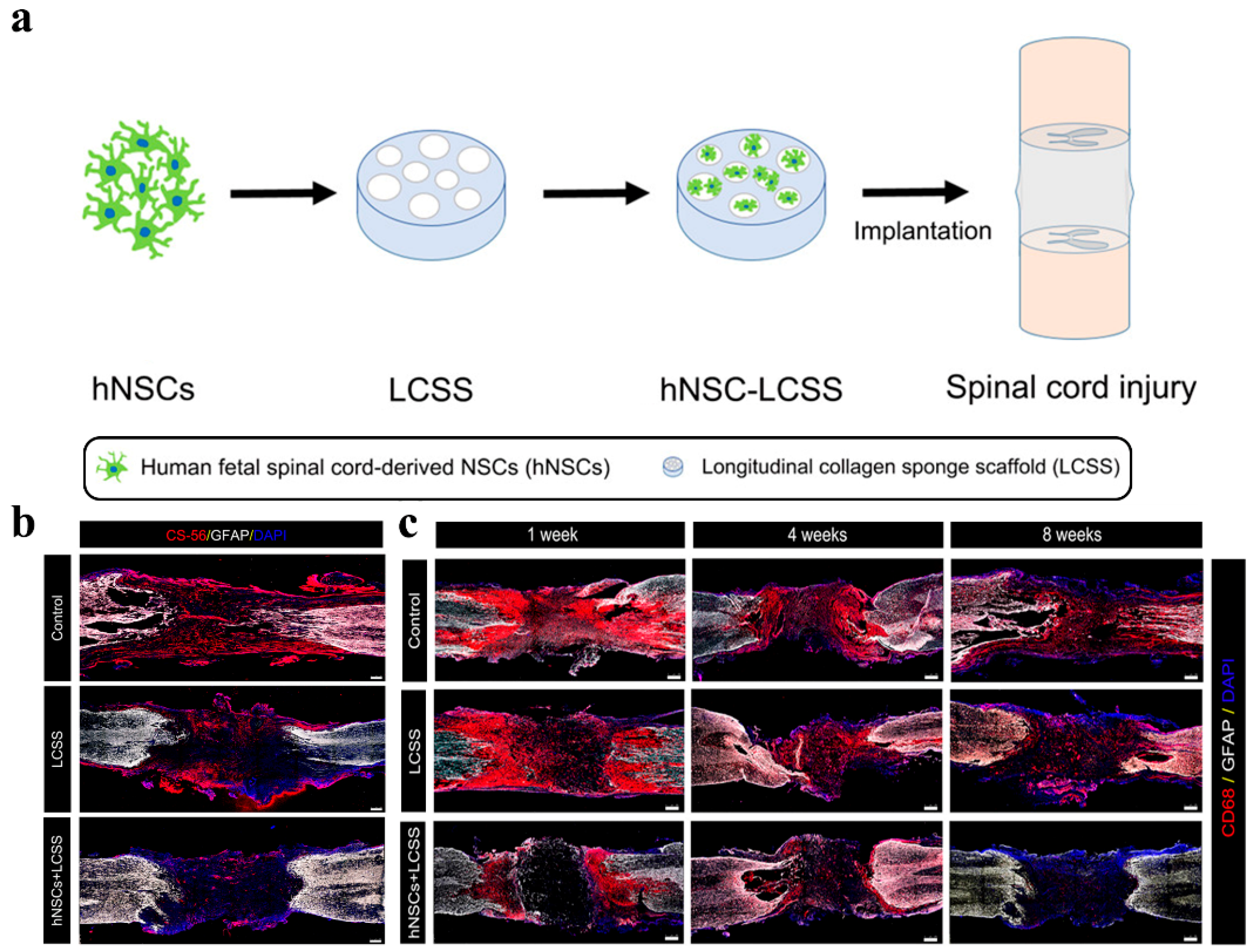
Figure 3. Schematic diagram and repair effect of neural stem cells in spinal cord repair. (a) Scheme of neural stem cells implantation for spinal cord repair; (b) Images of CS-56 immunostaining representing the formation of glial scars in the injury cavity at 8 weeks post-operation. The hNSCs implantation efficiently reduced glial scar forming around the injury cavity; (c) Representative immunofluorescence images of CD68 (activated microglia cell and macrophage marker)-positive signals in damaged sites. Grafted neural stem cells significantly suppress inflammation. (Adapted with permission from Ref. [106]. 2020, American Chemical Society).
Mesenchymal stem cells are also considered to be an important cell source for nerve repair after nervous system injury. Mesenchymal stem cells have strong proliferative ability, multi-directional differentiation potential and immunomodulatory ability [104]. They are also suitable for solving the physiological and pathological problems caused by nerve damage and have gradually become the dominant cells used to repair nerve function [109]. Mesenchymal stem cells include bone-marrow stromal cells [110], adipose-tissue-derived mesenchymal cells [111], human-umbilical-cord-blood-derived mesenchymal stem cells [112], etc., which can differentiate into Schwann-like cells to replace damaged cells and rebuild neural circuits, secrete cytokines to provide neuroprotection and nutritional support for injured nerve and axon regeneration, and promote the regeneration of neurons. Mesenchymal-stem-cell-derived factors can affect the maturation and function of all-immune cells and suppress both innate and adaptive immunity [113]. He found that mesenchymal-stem-cell-spheroids-loaded collagen hydrogels possessed both a superior anti-inflammatory efficacy and neurogenic activity. Mesenchymal stem cells spheroids promote cell–cell interactions and cell communication, accelerate the secretion of endogenous nutritional factors and the extracellular matrix, and facilitate the differentiation of neural stem cells into neuronal lines. In addition, the formation of the mesenchymal stem cells spheroids secreted more amounts and types of cytokines, as well as immunomodulatory paracrine factors to suppress lipopolysaccharide-induced inflammatory reactions [114]. Dental pulp stem cells are bone-marrow mesenchymal stem cells derived from dental pulp. Because they will not cause additional harm to the body or cause ethical problems due to the source of the cells, dental pulp stem cells have also attracted extensive attention. Luo selected the 10% formula (10% gelatin methacrylate hydrogel, recombinant human basic fibroblast growth factor and dental pulp stem cells) to fill a cellulose/soy protein isolate composite membrane tube to construct a nerve regeneration conduit. This conduit was applied to repair a 15 mm long defect of the sciatic nerve in a rat model. After 12-week post-implant surgery, it was found that almost all newly formed nerve tissues at the defect site originated from the direct differentiation of exogeneous dental pulp stem cells [115]. Dental pulp stem cells have a positive effect on nerve regeneration through glial differentiation and neuroprotective/neurotrophic effects on host tissues and significantly promote axon regeneration [116][117].
Schwann cells are the main glial cells of the peripheral nervous system and an important part of endogenous repair. Schwann cells form the myelin surrounding the nerve fibers and create a structural support to the axons. They also secrete growth factors essential for the maintenance of neuronal cells and provide directional clues to the regrowing axons [118]. Mosahebi encapsulated Schwann cells in different hydrogel materials. The viability and growth of Schwann cells were different, but there was no significant difference between these Schwann cells and those cultured on culture plates. Compared with alginate hydrogels without the encapsulation of Schwann cells, hydrogels-encapsulated Schwann cells significantly enhanced the mean neurite extension and increased the number of neurites sprouts of the chick embryo dorsal root ganglia [119]. In the collagen- and hyaluronic-acid-interpenetrating polymer network hydrogels prepared by Suri, Schwann cells with a high cell density in the hydrogel structure could actively secrete the nerve growth factor and brain-derived neurotrophic factor. In the co-culture of dissociated neurons and Schwann cells, some neurites were also observed to extend following the introduction of Schwann cells [120]. Compared with the use of gelatin sponge blocks to transplant Schwann cells suspension to the injury site, the transplantation of Schwann cells seeded in alginic acid hydrogel to the injury site of the rat spinal cord could inhibit cellular apoptosis, and the motor function of rats recovered better after 21 days [121]. However, it should be noted that, compared with homogeneous Schwann cells, heterologous Schwann cells elicit a strong immune reaction and hinder nerve regeneration when transplanted to the damaged site [122]. This suggests that researchers should pay attention to immunosuppression when using Schwann cells and other cells that can cause an immune response to nerve repair, or use autologous cells to obtain a better repair effect.

3.2. Bioactive Compounds

In order to avoid the problems of cell survival and a capacity decline, immune rejection, and limited cells, a number of studies have directly loaded cell components and cell secretions into hydrogels, which promote functional recovery from different aspects [123].
Exosomes are nano-scale extracellular vesicles and natural carriers. Their phospholipid bilayer structure can protect biologically active substances while loading growth factors, RNA, small-molecule drugs, etc., and achieve sustained release. Due to their nano size, exosomes can not only escape phagocytes and freely shuttle between cells or matrixes, but they also have a strong penetrating ability. Mesenchymal stem cells are the main source of exosomes for tissue repair. Exosomes contain specific substances from parental cells. The engineered preparation of exosomes can be achieved by the targeted modifications or genetic modifications of parental cells. As a cell-free biomaterial, exosomes can partially solve the problems encountered in the clinical application of regenerative medicine, such as the source, quantity and immune response of seed cells [124]. Li encapsulated exosomes derived from mesenchymal stem cells in a peptide-modified adhesive HA hydrogels, and the implanted exosomes exhibited an efficient retention and sustained release in the host nerve tissues. In the rat spinal cord injury model, hydrogels elicit significant nerve recovery and tissue preservation by effectively mitigating inflammation and oxidation [125]. Yang encapsulated Neurotrophin-3 mRNA in adipose-derived stem cell-derived exosomes. The exosomes were loaded into the alginate hydrogel and subsequently loaded into the nerve guide conduit to bridge sciatic nerve defects in rats. The exosomes could stably release Neurotrophin-3 mRNA for at least 2 weeks [126].
Some biologically active factors can regulate and promote nerve repair by interacting with corresponding receptors. At present, many studies have reported that various nutritional factors and growth factors can promote the survival and growth of neurons, and the therapeutic effect of active factors in nervous system injury has attracted more and more attention. However, active factors usually suffer from a reduced bioavailability, poor tissue distribution, short half-lives and so on. Higher doses may cause adverse immune reactions.
Neurotrophins are a family of growth factors that play important roles in the survival and function of neurons. The mammalian neurotrophic factor family has four known members: the brain-derived neurotrophic factor (BDNF), nerve growth factor (NGF), neurotrophin-3 (NT-3) and neurotrophin-4 (NT-4). Neurotrophins can regulate the development of the central and peripheral nervous system and inhibit apoptosis. Neurotrophins are key mediators that promote neuronal growth and survival after neuroplastic injury. Currently, the first two neurotrophic factors are under more intensive research as potential stroke treatments [127]. Cook utilized BDNF-loaded hyaluronic-acid hydrogels to locally release BDNF in the infarct cavity of a mouse stroke model for neural repair in the chronic phase after stroke. The experimental results showed that, compared with the direct injection of BDNF, the diffusion time of hydrogel-delivered BDNF from the stroke cavity to the surrounding infarct tissue was prolonged, and the recovery of motor function was promoted. Hydrogel–BDNF induces the initial migration of immature neurons into the peri-infarct cortex and enables long-term survival [128]. While loading NGF in the hydrogel to achieve a long-term effective release, the resulting nutritional factor gradient can stimulate the directional neurite extension of the ganglion in the hydrogel [129]. The hydrogel load with NGF exhibited a good morphology and stable bioactivity in vitro in Zhao’s research. The cell uptake efficiency of NGF was significantly improved. Tissue repairment in spinal gray matter was observed, including the recovery of nuclei and morphology, as well as a reduction in the organization cavity in the NGF–hydrogel group. Hydrogels could maximize NGF’s effects on neuron protection and spinal cord injury recovery [130].
The angiogenesis and reconstruction of a functional microvasculature is an important and necessary step to promote the recovery of nerve injury. The vascular endothelial growth factor (VEGF) has beneficial effects on motor neuron survival, neuronal viability and proliferation. VEGF is expressed in the nervous system after injury and participates in the process of nerve repair by regulating angiogenesis, neurogenesis, and neurite outgrowth through signal transduction [131]. Gelatin hydrogels loaded with VEGF can induce capillary-like tube formations and axonal outgrowth in vitro [132]. When the 15 mm rat sciatic nerve defects were repaired by a chitosan hydrogel loaded with a neurotrophic factor and VEGF at the same time, it can be observed that the hydrogel not only promoted nerve regeneration, but also promoted blood vessel penetration in the early stage of injury [133].
In addition, fibroblast growth factors (FGF) are also beneficial for neural repair, some members of which are produced by neurons and glial cells, in turn targeting other cells. Basic fibroblast growth factor (bFGF), a widely studied member of the FGF family, is abundant in the central nervous system. bFGF is closely related to neuroprotection, injury repair, and neurogenesis, and is an important contributor to the plasticity of hippocampal neurons. Fujimaki repaired a 15 mm sciatic nerve defect in a rat using a bFGF-loaded oriented collagen hydrogel tubes. The results indicated that bFGF could promote the regeneration of more myelinated fibers and the improvement of motor function [134]. Zhang used bFGF gene-modified neural stem cells to improve neurological deficits after transient middle cerebral artery occlusion in rats. The transplanted modified cells improved survival, migration, proliferation and differentiation in the brain. Moreover, bFGF promoted the differentiation of modified cells into mature neurons within the infarcted area [135]. Similar to bFGF, the acidic fibroblast growth factor (aFGF) can also regulate cell proliferation, migration, differentiation and survival, and down-regulate known axonal regeneration inhibitors, which play an important role in the regeneration of nerve fibers. After filling the collagen hydrogel with aFGF, it showed an effect equivalent to that of autologous transplantation, which had a significantly better regenerative ability than other groups (the empty conduit and the nerve conduit containing hydrogel only) in repairing a 10 mm defect of the rat sciatic nerve [136].
In addition, drugs can also be loaded into hydrogels to achieve long-term release. For example, when anti-inflammatory drugs and growth/nutrition factors are simultaneously loaded in hydrogels, the coordinated release of multiple components can effectively protect spare tissues/axons from secondary damage, reduce the expression of proinflammatory cytokines, cystic cavitation and glial scar formation and improve the survival rate of neurons [137][138]. The active ingredient of traditional Chinese medicine is also loaded into a hydrogel for nerve repair. Berberine is a plant alkaloid-based substrate that can promote the growth, development and reconstruction of axons in damaged peripheral nerves and the central nervous system. It has a neuroprotective effect on damaged neurons. Sadeghi loaded berberine in chitosan nanoparticles within a hybrid of alginate and chitosan hydrogel. The scaffolds with endometrial stem cells, as well as scaffolds alone, were transplanted into hemisected spinal cord injury rats. The data from the immunostaining of a neurofilament, as a neuronal growth marker, showed that all scaffolds had a certain immunoreactivity [139]. Curcumin is a diketone compound with an antioxidant activity and anti-inflammatory effect. Qian injected curcumin-loaded hydrogel under the endocranium into the traumatic brain injury mice. The slow and sustained released of curcumin can further reduce the level of reactive oxygen species in traumatic brain injury tissues and brain edema [140]. In Luo’s research, Curcumin encapsulated in the hybrid hydrogel was released in a sustained pattern over a 7-day period. The presented hybrid hydrogel could easily reassemble the extracellular matrix locally at the lesion site of rat spinal cord, and further modulate local inflammatory reaction by regulating the phenotypes of infiltrated inflammatory cells [141].

References

  1. Mietto, B.S.; Mostacada, K.; Martinez, A.M.B. Neurotrauma and Inflammation: CNS and PNS Responses. Mediat. Inflamm. 2015, 2015, 251204.
  2. Echternacht, S.R.; Chacon, M.A.; Leckenby, J.I. Central versus peripheral nervous system regeneration: Is there an exception for cranial nerves? Regen. Med. 2021, 16, 567–579.
  3. Wang, Q.; Capistrant, B.; Ehntholt, A.; Glymour, M.M. Long-Term Rate of Change in Memory Functioning before and after Stroke Onset. Stroke 2012, 43, 2561–2566.
  4. Maida, C.D.; Norrito, R.L.; Daidone, M.; Tuttolomondo, A.; Pinto, A. Neuroinflammatory Mechanisms in Ischemic Stroke: Focus on Cardioembolic Stroke, Background, and Therapeutic Approaches. Int. J. Mol. Sci. 2020, 21, 6454.
  5. Wei, K.; Wang, P.; Miao, C.-Y. A Double-Edged Sword with Therapeutic Potential: An Updated Role of Autophagy in Ischemic Cerebral Injury. CNS Neurosci. Ther. 2012, 18, 879–886.
  6. Sobesky, J. Akutversorgung des ischämischen Schlaganfalls. Der Internist 2009, 50, 1218–1226.
  7. Kumar, A.; Aakriti; Gupta, V. A review on animal models of stroke: An update. Brain Res. Bull. 2016, 122, 35–44.
  8. Liu, C.-L.; Liao, S.-J.; Zeng, J.-S.; Lin, J.-W.; Li, C.-X.; Xie, L.-C.; Shi, X.-G.; Huang, R.-X. dl-3n-butylphthalide prevents stroke via improvement of cerebral microvessels in RHRSP. J. Neurol. Sci. 2007, 260, 106–113.
  9. Yi, Y.Y.; Shin, H.J.; Choi, S.G.; Kang, J.W.; Song, H.-J.; Kim, S.K.; Kim, D.W. Preventive Effects of Neuroprotective Agents in a Neonatal Rat of Photothrombotic Stroke Model. Int. J. Mol. Sci. 2020, 21, 3703.
  10. Patel, P.; Yavagal, D.; Khandelwal, P. Hyperacute Management of Ischemic Strokes. J. Am. Coll. Cardiol. 2020, 75, 1844–1856.
  11. Ahmed, E.M. Hydrogel: Preparation, characterization, and applications: A review. J. Adv. Res. 2015, 6, 105–121.
  12. Carballo-Molina, O.A.; Velasco, I. Hydrogels as scaffolds and delivery systems to enhance axonal regeneration after injuries. Front. Cell. Neurosci. 2015, 9, 13.
  13. Russo, T.; Tunesi, M.; Giordano, C.; Gloria, A.; Ambrosio, L. Hydrogels for central nervous system therapeutic strategies. Proc. Inst. Mech. Eng. Part H J. Eng. Med. 2015, 229, 905–916.
  14. Gopalakrishnan, A.; Shankarappa, S.A.; Rajanikant, G.K. Hydrogel Scaffolds: Towards Restitution of Ischemic Stroke-Injured Brain. Transl. Stroke Res. 2019, 10, 1–18.
  15. González-Nieto, D.; Fernández-García, L.; Pérez-Rigueiro, J.; Guinea, G.V.; Panetsos, F. Hydrogels-Assisted Cell Engraftment for Repairing the Stroke-Damaged Brain: Chimera or Reality. Polymers 2018, 10, 184.
  16. Fornasari, B.E.; Carta, G.; Gambarotta, G.; Raimondo, S. Natural-Based Biomaterials for Peripheral Nerve Injury Repair. Front. Bioeng. Biotechnol. 2020, 8, 554257.
  17. Abatangelo, G.; Vindigni, V.; Avruscio, G.; Pandis, L.; Brun, P. Hyaluronic Acid: Redefining Its Role. Cells 2020, 9, 1743.
  18. Horn, E.M.; Beaumont, M.; Shu, X.Z.; Harvey, A.; Prestwich, G.D.; Horn, K.M.; Gibson, A.R.; Preul, M.C.; Panitch, A. Influence of cross-linked hyaluronic acid hydrogels on neurite outgrowth and recovery from spinal cord injury. J. Neurosurg. Spine 2007, 6, 133–140.
  19. Pan, L.; Ren, Y.; Cui, F.; Xu, Q. Viability and differentiation of neural precursors on hyaluronic acid hydrogel scaffold. J. Neurosci. Res. 2009, 87, 3207–3220.
  20. Seidlits, S.K.; Khaing, Z.; Petersen, R.R.; Nickels, J.; Vanscoy, J.E.; Shear, J.B.; Schmidt, C.E. The effects of hyaluronic acid hydrogels with tunable mechanical properties on neural progenitor cell differentiation. Biomaterials 2010, 31, 3930–3940.
  21. Liang, Y.; Walczak, P.; Bulte, J.W. The survival of engrafted neural stem cells within hyaluronic acid hydrogels. Biomaterials 2013, 34, 5521–5529.
  22. Wang, M.-D.; Zhai, P.; Schreyer, D.J.; Zheng, R.-S.; Sun, X.-D.; Cui, F.-Z.; Chen, X.-B. Novel crosslinked alginate/hyaluronic acid hydrogels for nerve tissue engineering. Front. Mater. Sci. 2013, 7, 269–284.
  23. Hou, S.; Xu, Q.; Tian, W.; Cui, F.; Cai, Q.; Ma, J.; Lee, I.-S. The repair of brain lesion by implantation of hyaluronic acid hydrogels modified with laminin. J. Neurosci. Methods 2005, 148, 60–70.
  24. Broguiere, N.; Isenmann, L.; Zenobi-Wong, M. Novel enzymatically cross-linked hyaluronan hydrogels support the formation of 3D neuronal networks. Biomaterials 2016, 99, 47–55.
  25. Jensen, G.; Holloway, J.L.; Stabenfeldt, S.E. Hyaluronic Acid Biomaterials for Central Nervous System Regenerative Medicine. Cells 2020, 9, 2113.
  26. Yoo, J.; Park, J.H.; Kwon, Y.W.; Chung, J.J.; Choi, I.C.; Nam, J.J.; Lee, H.S.; Jeon, E.Y.; Lee, K.; Kim, S.H.; et al. Augmented peripheral nerve regeneration through elastic nerve guidance conduits prepared using a porous PLCL membrane with a 3D printed collagen hydrogel. Biomater. Sci. 2020, 8, 6261–6271.
  27. Salehi, M.; Naseri-Nosar, M.; Ebrahimibarough, S.; Nourani, M.; Vaez, A.; Farzamfar, S.; Ai, J. Regeneration of sciatic nerve crush injury by a hydroxyapatite nanoparticle-containing collagen type I hydrogel. J. Physiol. Sci. 2018, 68, 579–587.
  28. Lu, D.; Mahmood, A.; Qu, C.; Hong, X.; Kaplan, D.; Chopp, M. Collagen Scaffolds Populated with Human Marrow Stromal Cells Reduce Lesion Volume and Improve Functional Outcome after Traumatic Brain Injury. Neurosurgery 2007, 61, 596–603.
  29. Deng, W.-S.; Ma, K.; Liang, B.; Liu, X.-Y.; Xu, H.-Y.; Zhang, J.; Shi, H.-Y.; Sun, H.-T.; Chen, X.-Y.; Zhang, S. Collagen scaffold combined with human umbilical cord-mesenchymal stem cells transplantation for acute complete spinal cord injury. Neural Regen. Res. 2020, 15, 1686–1700.
  30. Wang, P.; Zhao, H.; Yao, Y.; Lu, C.; Ma, J.; Chen, R.; Pan, J. Repair of facial nerve crush injury in rabbits using collagen plus basic fibroblast growth factor. J. Biomed. Mater. Res. Part A 2020, 108, 1329–1337.
  31. Hu, Q.; Sun, C.; Zhang, H.; Li, S.; Shen, X.; Liu, S. Design and Fabrication of Tissue-engineering Multiscale and Multichannel Nerve Conduits for Rabbits’ Sciatic Nerve Regeneration. J. Biomater. Tissue Eng. 2019, 9, 10–23.
  32. Matsumine, H.; Sasaki, R.; Tabata, Y.; Matsui, M.; Yamato, M.; Okano, T.; Sakurai, H. Facial nerve regeneration using basic fibroblast growth factor-impregnated gelatin microspheres in a rat model. J. Tissue Eng. Regen. Med. 2016, 10, E559–E567.
  33. Takagi, T.; Kimura, Y.; Shibata, S.; Saito, H.; Ishii, K.; Okano, H.J.; Toyama, Y.; Okano, H.; Tabata, Y.; Nakamura, M. Sustained bFGF-Release Tubes for Peripheral Nerve Regeneration. Plast. Reconstr. Surg. 2012, 130, 866–876.
  34. Tonda-Turo, C.; Gnavi, S.; Ruini, F.; Gambarotta, G.; Gioffredi, E.; Chiono, V.; Perroteau, I.; Ciardelli, G. Development and characterization of novel agar and gelatin injectable hydrogel as filler for peripheral nerve guidance channels. J. Tissue Eng. Regen. Med. 2017, 11, 197–208.
  35. Lin, C.-C.; Chang, J.-J.; Yung, M.-C.; Huang, W.-C.; Chen, S.-Y. Spontaneously Micropatterned Silk/Gelatin Scaffolds with Topographical, Biological, and Electrical Stimuli for Neuronal Regulation. ACS Biomater. Sci. Eng. 2020, 6, 1144–1153.
  36. Ye, W.; Li, H.; Yu, K.; Xie, C.; Wang, P.; Zheng, Y.; Zhang, P.; Xiu, J.; Yang, Y.; He, Y.; et al. 3D printing of gelatin methacrylate-based nerve guidance conduits with multiple channels. Mater. Des. 2020, 192, 108757.
  37. Yu, Z.; Li, H.; Xia, P.; Kong, W.; Chang, Y.; Fu, C.; Wang, K.; Yang, X.; Qi, Z. Application of fibrin-based hydrogels for nerve protection and regeneration after spinal cord injury. J. Biol. Eng. 2020, 14, 22.
  38. Johnson, P.; Tatara, A.; McCreedy, D.A.; Shiu, A.; Sakiyama-Elbert, S.E. Tissue-engineered fibrin scaffolds containing neural progenitors enhance functional recovery in a subacute model of SCI. Soft Matter 2010, 6, 5127–5137.
  39. Mooney, R.; Tawil, B.; Mahoney, M. Specific Fibrinogen and Thrombin Concentrations Promote Neuronal Rather Than Glial Growth When Primary Neural Cells Are Seeded within Plasma-Derived Fibrin Gels. Tissue Eng. Part A 2010, 16, 1607.
  40. Yao, S.; Liu, X.; Yu, S.; Wang, X.; Zhang, S.; Wu, Q.; Sun, X.; Mao, H. Co-effects of matrix low elasticity and aligned topography on stem cell neurogenic differentiation and rapid neurite outgrowth. Nanoscale 2016, 8, 10252–10265.
  41. Yang, W.; Wang, Z.; Zhang, J.; Yang, K.; Lu, C.; Cui, X.; Lu, H.; Cao, S.; Chen, Q.; Lu, X.; et al. Fibrin scaffolds embedded with sonic hedgehog/chitosan microspheres for recovery of spinal cord injury in rats. Mater. Express 2020, 10, 437–445.
  42. Robinson, M.; Douglas, S.; Willerth, S.M. Mechanically stable fibrin scaffolds promote viability and induce neurite outgrowth in neural aggregates derived from human induced pluripotent stem cells. Sci. Rep. 2017, 7, 6250.
  43. Rajabi, M.; Ali, A.; McConnell, M.; Cabral, J. Keratinous materials: Structures and functions in biomedical applications. Mater. Sci. Eng. C 2020, 110, 110612.
  44. Ozaki, Y.; Takagi, Y.; Mori, H.; Hara, M. Porous hydrogel of wool keratin prepared by a novel method: An extraction with guani-dine/2-mercaptoethanol solution followed by a dialysis. Mater. Sci. Eng. C 2014, 42, 146–154.
  45. Wang, S.; Taraballi, F.; Tan, L.P.; Ng, K.W. Human keratin hydrogels support fibroblast attachment and proliferation in vitro. Cell Tissue Res. 2012, 347, 795–802.
  46. Wang, S.; Wang, Z.; Foo, S.E.M.; Tan, N.S.; Yuan, Y.; Lin, W.; Zhang, Z.; Ng, K.W. Culturing Fibroblasts in 3D Human Hair Keratin Hydrogels. ACS Appl. Mater. Interfaces 2015, 7, 5187–5198.
  47. Sierpinski, P.; Garrett, J.; Ma, J.; Apel, P.; Klorig, D.; Smith, T.; Koman, L.A.; Atala, A.; Van Dyke, M. The use of keratin biomaterials derived from human hair for the promotion of rapid regeneration of peripheral nerves. Biomaterials 2008, 29, 118–128.
  48. Pace, L.A.; Plate, J.F.; Mannava, S.; Barnwell, J.C.; Koman, L.A.; Li, Z.; Smith, T.L.; Van Dyke, M. A Human Hair Keratin Hydrogel Scaffold Enhances Median Nerve Regeneration in Nonhuman Primates: An Electrophysiological and Histological Study. Tissue Eng. Part A 2014, 20, 507–517.
  49. Apel, P.J.; Garrett, J.P.; Sierpinski, P.; Ma, J.; Atala, A.; Smith, T.L.; Koman, L.A.; van Dyke, M.E. Peripheral Nerve Regeneration Using a Keratin-Based Scaffold: Long-Term Functional and Histological Outcomes in a Mouse Model. J. Hand Surg. 2008, 33, 1541–1547.
  50. Pace, L.A.; Plate, J.F.; Smith, T.L.; Van Dyke, M.E. The effect of human hair keratin hydrogel on early cellular response to sciatic nerve injury in a rat model. Biomaterials 2013, 34, 5907–5914.
  51. Zheng, K.; Feng, G.; Zhang, J.; Xing, J.; Huang, D.; Lian, M.; Zhang, W.; Wu, W.; Hu, Y.; Lu, X.; et al. Basic fibroblast growth factor promotes human dental pulp stem cells cultured in 3D porous chitosan scaffolds to neural differentiation. Int. J. Neurosci. 2021, 131, 625–633.
  52. Yuan, Y.; Zhang, P.; Yang, Y.; Wang, X.; Gu, X. The interaction of Schwann cells with chitosan membranes and fibers in vitro. Biomaterials 2004, 25, 4273–4278.
  53. Scanga, V.I.; Goraltchouk, A.; Nussaiba, N.; Shoichet, M.S.; Morshead, C.M. Biomaterials for neural-tissue engineering—Chitosan supports the survival, migration, and differentiation of adult-derived neural stem and progenitor cells. Rev. Can. Chim. 2010, 88, 277–287.
  54. Boecker, A.; Daeschler, S.; Kneser, U.; Harhaus, L. Relevance and Recent Developments of Chitosan in Peripheral Nerve Surgery. Front. Cell. Neurosci. 2019, 13, 104.
  55. Haastert-Talini, K.; Geuna, S.; Dahlin, L.B.; Meyer, C.; Stenberg, L.; Freier, T.; Heimann, C.; Barwig, C.; Pinto, L.F.; Raimondo, S.; et al. Chitosan tubes of varying degrees of acetylation for bridging peripheral nerve defects. Biomaterials 2013, 34, 9886–9904.
  56. Ai, J.; Farzin, A.; Zamiri, S.; Hadjighassem, M.; Ebrahimi-Barough, S.; Ai, A.; Mohandesnezhad, S.; Karampour, A.; Farahani, M.S.; Goodarzi, A. Repair of injured spinal cord using platelet-rich plasma- and endometrial stem cells-loaded chitosan scaffolds. Int. J. Polym. Mater. Polym. Biomater. 2021, 70, 1002–1011.
  57. Zhou, S.; Yang, Y.; Gu, X.; Ding, F. Chitooligosaccharides protect cultured hippocampal neurons against glutamate-induced neurotoxicity. Neurosci. Lett. 2008, 444, 270–274.
  58. Huang, H.-C.; Hong, L.; Chang, P.; Zhang, J.; Lu, S.-Y.; Zheng, B.-W.; Jiang, Z.-F. Chitooligosaccharides Attenuate Cu2+-Induced Cellular Oxidative Damage and Cell Apoptosis Involving Nrf2 Activation. Neurotox. Res. 2015, 27, 411–420.
  59. Wang, Y.; Zhao, Y.; Sun, C.; Hu, W.; Zhao, J.; Li, G.; Zhang, L.; Liu, M.; Liu, Y.; Ding, F.; et al. Chitosan Degradation Products Promote Nerve Regeneration by Stimulating Schwann Cell Proliferation via miR-27a/FOXO1 Axis. Mol. Neurobiol. 2016, 53, 28–39.
  60. Eftekharzadeh, B.; Khodagholi, F.; Abdi, A.; Maghsoudi, N. Alginate protects NT2 neurons against H2O2-induced neurotoxicity. Carbohydr. Polym. 2010, 79, 1063–1072.
  61. Matyash, M.; Despang, F.; Mandal, R.; Fiore, D.; Gelinsky, M.; Ikonomidou, C. Novel Soft Alginate Hydrogel Strongly Supports Neurite Growth and Protects Neurons Against Oxidative Stress. Tissue Eng. Part A 2012, 18, 55–66.
  62. Askarzadeh, N.; Nazarpak, M.H.; Mansoori, K.; Farokhi, M.; Gholami, M.; Mohammadi, J.; Mottaghitalab, F. Bilayer Cylindrical Conduit Consisting of Electrospun Polycaprolactone Nanofibers and DSC Cross-Linked Sodium Alginate Hydrogel to Bridge Peripheral Nerve Gaps. Macromol. Biosci. 2020, 20, e2000149.
  63. Bozza, A.; Coates, E.E.; Incitti, T.; Ferlin, K.M.; Messina, A.; Menna, E.; Bozzi, Y.; Fisher, J.P.; Casarosa, S. Neural differentiation of pluripotent cells in 3D alginate-based cultures. Biomaterials 2014, 35, 4636–4645.
  64. Suzuki, Y.; Kitaura, M.; Wu, S.; Kataoka, K.; Suzuki, K.; Endo, K.; Nishimura, Y.; Ide, C. Electrophysiological and horseradish peroxidase-tracing studies of nerve regeneration through alginate-filled gap in adult rat spinal cord. Neurosci. Lett. 2002, 318, 121–124.
  65. Ashton, R.S.; Banerjee, A.; Punyani, S.; Schaffer, D.V.; Kane, R.S. Scaffolds based on degradable alginate hydrogels and poly(lactide-co-glycolide) microspheres for stem cell culture. Biomaterials 2007, 28, 5518–5525.
  66. Novikova, L.N.; Mosahebi, A.; Wiberg, M.; Terenghi, G.; Kellerth, J.-O.; Novikov, L.N. Alginate hydrogel and matrigel as potential cell carriers for neurotransplantation. J. Biomed. Mater. Res. Part A 2006, 77, 242–252.
  67. Wang, G.; Wang, X.; Huang, L. Feasibility of chitosan-alginate (Chi-Alg) hydrogel used as scaffold for neural tissue engineering: A pilot studyin vitro. Biotechnol. Biotechnol. Equip. 2017, 31, 766–773.
  68. Karimi, S.; Bagher, Z.; Najmoddin, N.; Simorgh, S.; Pezeshki-Modaress, M. Alginate-magnetic short nanofibers 3D composite hydrogel enhances the encapsulated human olfactory mucosa stem cells bioactivity for potential nerve regeneration application. Int. J. Biol. Macromol. 2021, 167, 796–806.
  69. Wang, Z.; Wang, J.; Jin, Y.; Luo, Z.; Yang, W.; Xie, H.; Huang, K.; Wang, L. A Neuroprotective Sericin Hydrogel as an Effective Neuronal Cell Carrier for the Repair of Ischemic Stroke. ACS Appl. Mater. Interfaces 2015, 7, 24629–24640.
  70. Zhang, L.; Yang, W.; Tao, K.; Song, Y.; Xie, H.; Wang, J.; Li, X.; Shuai, X.; Gao, J.; Chang, P.; et al. Sustained Local Release of NGF from a Chitosan–Sericin Composite Scaffold for Treating Chronic Nerve Compression. ACS Appl. Mater. Interfaces 2017, 9, 3432–3444.
  71. Cheong, G.M.; Lim, K.; Jakubowicz, A.; Martens, P.; Poole-Warren, L.; Green, R.A. Conductive hydrogels with tailored bioactivity for implantable electrode coatings. Acta Biomater. 2014, 10, 1216–1226.
  72. Hernandes, M.S.; Troncone, L.R.P. Glycine as a neurotransmitter in the forebrain: A short review. J. Neural Transm. 2009, 116, 1551–1560.
  73. Zhao, Y.; Liang, Y.; Ding, S.; Zhang, K.; Mao, H.-Q.; Yang, Y. Application of conductive PPy/SF composite scaffold and electrical stimulation for neural tissue engineering. Biomaterials 2020, 255, 120164.
  74. You, R.; Zhang, Q.; Li, X.; Yan, S.; Luo, Z.; Qu, J.; Li, M. Multichannel Bioactive Silk Nanofiber Conduits Direct and Enhance Axonal Regeneration after Spinal Cord Injury. ACS Biomater. Sci. Eng. 2020, 6, 4677–4686.
  75. Carvalho, C.R.; Chang, W.; Silva-Correia, J.; Reis, R.L.; Oliveira, J.M.; Kohn, J. Engineering Silk Fibroin-Based Nerve Conduit with Neurotrophic Factors for Proximal Protection after Peripheral Nerve Injury. Adv. Health Mater. 2021, 10, e2000753.
  76. Yang, Y.; Chen, X.; Ding, F.; Zhang, P.; Liu, J.; Gu, X. Biocompatibility evaluation of silk fibroin with peripheral nerve tissues and cells in vitro. Biomaterials 2007, 28, 1643–1652.
  77. Gu, X.; Chen, X.; Tang, X.; Zhou, Z.; Huang, T.; Yang, Y.; Ling, J. Pure-silk fibroin hydrogel with stable aligned micropattern toward peripheral nerve regeneration. Nanotechnol. Rev. 2021, 10, 10–19.
  78. Chen, X.-Y.; Tu, Y.; Sun, S.-Z.; Kong, X.-B.; Tang, Q.-Y.; Sun, Z. Polyethylene glycol as a promising synthetic material for repair of spinal cord injury. Neural Regen. Res. 2017, 12, 1003–1008.
  79. Lin, C.-C.; Anseth, K.S. PEG Hydrogels for the Controlled Release of Biomolecules in Regenerative Medicine. Pharm. Res. 2009, 26, 631–643.
  80. Isaacs, J.; Klumb, I.; McDaniel, C. Preliminary investigation of a polyethylene glycol hydrogel “nerve glue”. J. Brachial Plex. Peripher. Nerve Inj. 2009, 4, e98–e102.
  81. Estradaa, V.; Brazdaa, N.; Schmitza, C.; Hellera, S.; Blazycab, H.; Martinib, R.; Müller, H.W. Long-lasting significant functional improvement in chronic severe spinal cord injury following scar resection and polyethylene glycol implantation. Neurobiol. Dis. 2014, 67, 165–179.
  82. Goncharuk-Oleksii, O.; Savosko-Serhii, I.; Petriv-Taras, I.; Medvediev-Volodymyr, V.; Tsymbaliuk-Vitaly, I. Morphometric Study of Rat Sciatic Nerve Recovery after Three Nerve Repair Techniques: Epineural Suture, Polyethylene Glycol Hydrogel and Fibrin Sealant. Int. J. Morphol. 2021, 39, 677–682.
  83. Mahoney, M.J.; Anseth, K.S. Contrasting effects of collagen and bFGF-2 on neural cell function in degradable synthetic PEG hydrogels. J. Biomed. Mater. Res. Part A 2007, 81A, 269–278.
  84. Hynes, S.R.; McGregor, L.M.; Rauch, M.F.; Lavik, E.B. Photopolymerized poly(ethylene glycol)/poly(L-lysine) hydrogels for the delivery of neural progenitor cells. J. Biomater. Sci. Polym. Ed. 2007, 18, 1017–1030.
  85. Burdick, J.A.; Ward, M.; Liang, E.; Young, M.J.; Langer, R. Stimulation of neurite outgrowth by neurotrophins delivered from degradable hydrogels. Biomaterials 2006, 27, 452–459.
  86. Zhang, K.; Li, J.; Jin, J.; Dong, J.; Li, L.; Xue, B.; Wang, W.; Jiang, Q.; Cao, Y. Injectable, anti-inflammatory and conductive hydrogels based on graphene oxide and diacerein-terminated four-armed polyethylene glycol for spinal cord injury repair. Mater. Des. 2020, 196, 109092.
  87. Hejčl, A.; Lesný, P.; Přádný, M.; Michálek, J.; Jendelová, P.; Štulík, J.; Syková, E. Biocompatible hydrogels in spinal cord injury repair. Physiol. Res. 2008, 57 (Suppl. S3), S121–S132.
  88. Bakshi, A.; Fisher, O.; Dagci, T.; Himes, B.T.; Fischer, I.; Lowman, A. Mechanically engineered hydrogel scaffolds for axonal growth and angiogenesis after transplantation in spinal cord injury. J. Neurosurg. Spine 2004, 1, 322–329.
  89. Hejčl, A.; Urdzikova, L.M.; Sedy, J.; Lesny, P.; Pradny, M.; Michálek, J.; Burian, M.; Hajek, M.; Zamecnik, J.; Jendelova, P.; et al. Acute and delayed implantation of positively charged 2-hydroxyethyl methacrylate scaffolds in spinal cord injury in the rat. J. Neurosurg. Spine 2008, 8, 67–73.
  90. Li, H.Y.; Führmann, T.; Zhou, Y.; Dalton, P.D. Host reaction to poly(2-hydroxyethyl methacrylate) scaffolds in a small spinal cord injury model. J. Mater. Sci. Mater. Electron. 2013, 24, 2001–2011.
  91. Pertici, V.; Trimaille, T.; Laurin, J.; Felix, M.-S.; Marqueste, T.; Pettmann, B.; Chauvin, J.-P.; Gigmes, D.; Decherchi, P. Repair of the injured spinal cord by implantation of a synthetic degradable block copolymer in rat. Biomaterials 2014, 35, 6248–6258.
  92. Kubinová, Š.; Horák, D.; Kozubenko, N.; Vaněček, V.; Proks, V.; Price, J.; Cocks, G.; Syková, E. The use of superporous Ac-CGGASIKVAVS-OH-modified PHEMA scaffolds to promote cell adhesion and the differentiation of human fetal neural precursors. Biomaterials 2010, 31, 5966–5975.
  93. Jhaveri, S.J.; Hynd, M.R.; Dowell-Mesfin, N.; Turner, J.N.; Shain, W.; Ober, C.K. Release of Nerve Growth Factor from HEMA Hydrogel-Coated Substrates and Its Effect on the Differentiation of Neural Cells. Biomacromolecules 2009, 10, 174–183.
  94. Hejčl, A.; Růžička, J.; Kekulová, K.; Svobodová, B.; Proks, V.; Macková, H.; Jiránková, K.; Kárová, K.; Urdziková, L.M.; Kubinová, Š.; et al. Modified Methacrylate Hydrogels Improve Tissue Repair after Spinal Cord Injury. Int. J. Mol. Sci. 2018, 19, 2481.
  95. Pertici, V.; Amendola, J.; Laurin, J.; Gigmes, D.; Madaschi, L.; Carelli, S.; Marqueste, T.; Gorio, A.; Decherchi, P. The Use of Poly(N--Methacrylamide) Hydrogel to Repair a T10 Spinal Cord Hemisection in Rat: A Behavioural, Electrophysiological and Anatomical Examination. ASN Neuro 2013, 5, AN20120082.
  96. Woerly, S.; Doan, V.D.; Sosa, N.; de Vellis, J.; Espinosa-Jeffrey, A. Prevention of gliotic scar formation by NeuroGel™ allows partial endogenous repair of transected cat spinal cord. J. Neurosci. Res. 2004, 75, 262–272.
  97. Plant, G.W.; Woerly, S.; Harvey, A.R. Hydrogels Containing Peptide or Aminosugar Sequences Implanted into the Rat Brain: Influence on Cellular Migration and Axonal Growth. Exp. Neurol. 1997, 143, 287–299.
  98. Woerly, S.; Fort, S.; Pignot-Paintrand, I.; Cottet, C.; Carcenac, C.; Savasta, M. Development of a Sialic Acid-Containing Hydrogel of Poly: Characterization and Implantation Study. Biomacromolecules 2008, 9, 2329–2337.
  99. Syková, E.; Jendelova, P.; Urdzikova, L.M.; Lesný, P.; Hejčl, A. Bone Marrow Stem Cells and Polymer Hydrogels—Two Strategies for Spinal Cord Injury Repair. Cell. Mol. Neurobiol. 2006, 26, 1113–1129.
  100. Stocco, E.; Barbon, S.; Lora, L.; Grandi, F.; Sartore, L.; Tiengo, C.; Petrelli, L.; Dalzoppo, D.; Parnigotto, P.P.; Macchi, V.; et al. Partially oxidized polyvinyl alcohol conduitfor peripheral nerve regeneration. Sci. Rep. 2018, 8, 604.
  101. Matsuura, Y.; Hiraizumi, Y.; Fujimaki, E. Application of a Polyvinyl Alcohol Hydrogel (Pvah) Mem-Brane as an Antiadhesive Interposition after Spinal Surgery. Showa Igakkai Zasshi 1996, 56, 355–362.
  102. Oh, S.H.; An, D.B.; Kim, T.H.; Lee, J.H. Wide-range stiffness gradient PVA/HA hydrogel to investigate stem cell differentiation behavior. Acta Biomater. 2016, 35, 23–31.
  103. Assinck, P.; Duncan, G.J.; Hilton, B.; Plemel, J.R.; Tetzlaff, W. Cell transplantation therapy for spinal cord injury. Nat. Neurosci. 2017, 20, 637–647.
  104. Ottoboni, L.; de Feo, D.; Merlini, A.; Martino, G. Commonalities in immune modulation between mesenchymal stem cells (MSCs) and neural stem/precursor cells (NPCs). Immunol. Lett. 2015, 168, 228–239.
  105. Tseng, T.-C.; Tao, L.; Hsieh, F.-Y.; Wei, Y.; Chiu, I.-M.; Hsu, S.-H. An Injectable, Self-Healing Hydrogel to Repair the Central Nervous System. Adv. Mater. 2015, 27, 3518–3524.
  106. Zou, Y.; Zhao, Y.; Xiao, Z.; Chen, B.; Ma, D.; Shen, H.; Gu, R.; Dai, J. Comparison of Regenerative Effects of Transplanting Three-Dimensional Longitudinal Scaffold Loaded-Human Mesenchymal Stem Cells and Human Neural Stem Cells on Spinal Cord Completely Transected Rats. ACS Biomater. Sci. Eng. 2020, 6, 1671–1680.
  107. Zhang, S.; Burda, J.E.; Anderson, M.A.; Zhao, Z.; Ao, Y.; Cheng, Y.; Sun, Y.; Deming, T.J.; Sofroniew, M.V. Thermoresponsive Copolypeptide Hydrogel Vehicles for Central Nervous System Cell Delivery. ACS Biomater. Sci. Eng. 2015, 1, 705–717.
  108. Prang, P.; Mueller, R.; Eljaouhari, A.; Heckmann, K.; Kunz, W.; Weber, T.; Faber, C.; Vroemen, M.; Bogdahn, U.; Weidner, N. The promotion of oriented axonal regrowth in the injured spinal cord by alginate-based anisotropic capillary hydrogels. Biomaterials 2006, 27, 3560–3569.
  109. Dasari, V.R.; Veeravalli, K.K.; Dinh, D.H. Mesenchymal stem cells in the treatment of spinal cord injuries: A review. World J. Stem Cells 2014, 6, 120.
  110. Zhang, J.; Cheng, T.; Chen, Y.; Gao, F.; Guan, F.; Yao, M.-H. A chitosan-based thermosensitive scaffold loaded with bone marrow-derived mesenchymal stem cells promotes motor function recovery in spinal cord injured mice. Biomed. Mater. 2020, 15, 35020.
  111. Rhode, S.C.; Beier, J.P.; Ruhl, T. Adipose tissue stem cells in peripheral nerve regeneration—In vitro and in vivo. J. Neurosci. Res. 2021, 99, 545–560.
  112. Lin, Y.-J.; Lee, Y.-W.; Chang, C.-W.; Huang, C.-C. 3D Spheroids of Umbilical Cord Blood MSC-Derived Schwann Cells Promote Peripheral Nerve Regeneration. Front. Cell Dev. Biol. 2020, 8, 1632.
  113. Volarevic, V.; Gazdic, M.; Markovic, B.S.; Jovicic, N.; Djonov, V.; Arsenijevic, N. Mesenchymal stem cell-derived factors: Immuno-modulatory effects and therapeutic potential. BioFactors 2017, 43, 633–644.
  114. He, J.; Zhang, N.; Zhu, Y.; Jin, R.; Wu, F. MSC spheroids-loaded collagen hydrogels simultaneously promote neuronal differentiation and suppress inflammatory reaction through PI3K-Akt signaling pathway. Biomaterials 2021, 265, 120448.
  115. Luo, L.; He, Y.; Jin, L.; Zhang, Y.; Guastaldi, F.P.; Albashari, A.A.; Hu, F.; Wang, X.; Wang, L.; Xiao, J.; et al. Application of bioactive hydrogels combined with dental pulp stem cells for the repair of large gap peripheral nerve injuries. Bioact. Mater. 2021, 6, 638–654.
  116. Kolar, M.K.; Itte, V.N.; Kingham, P.J.; Novikov, L.; Wiberg, M.; Kelk, P. The neurotrophic effects of different human dental mesenchymal stem cells. Sci. Rep. 2017, 7, 12605.
  117. Pisciotta, A.; Bertoni, L.; Vallarola, A.; Bertani, G.; Mecugni, D.; Carnevale, G. Neural crest derived stem cells from dental pulp and tooth-associated stem cells for peripheral nerve regeneration. Neural Regen. Res. 2020, 15, 373–381.
  118. Terenghi, G.; Wiberg, M.; Kingham, P.J. Chapter 21 Use of Stem Cells for Improving Nerve Regeneration. Int. Rev. Neurobiol. 2009, 87, 393–403.
  119. Mosahebi, A.; Simon, M.; Wiberg, M.; Terenghi, G. A Novel Use of Alginate Hydrogel as Schwann Cell Matrix. Tissue Eng. 2001, 7, 525–534.
  120. Suri, S.; Schmidt, C.E. Cell-Laden Hydrogel Constructs of Hyaluronic Acid, Collagen, and Laminin for Neural Tissue Engineering. Tissue Eng. Part A 2010, 16, 1703–1716.
  121. Wang, H.; Liu, C.; Ma, X. Alginic acid sodium hydrogel co-transplantation with Schwann cells for rat spinal cord repair. Arch. Med. Sci. 2012, 8, 563–568.
  122. Guenard, V.; Kleitman, N.; Morrissey, T.; Bunge, R.; Aebischer, P. Syngeneic Schwann cells derived from adult nerves seeded in semipermeable guidance channels enhance peripheral nerve regeneration. J. Neurosci. 1992, 12, 3310–3320.
  123. Tsai, E.; Dalton, P.D.; Shoichet, M.S.; Tator, C.H. Matrix inclusion within synthetic hydrogel guidance channels improves specific supraspinal and local axonal regeneration after complete spinal cord transection. Biomaterials 2006, 27, 519–533.
  124. Huang, J.; Xiong, J.; Yang, L.; Zhang, J.; Sun, S.; Liang, Y. Cell-free exosome-laden scaffolds for tissue repair. Nanoscale 2021, 13, 874–875.
  125. Li, L.; Zhang, Y.; Mu, J.; Chen, J.; Zhang, C.; Cao, H.; Gao, J. Transplantation of Human Mesenchymal Stem-Cell-Derived Exosomes Immobilized in an Adhesive Hydrogel for Effective Treatment of Spinal Cord Injury. Nano Lett. 2020, 20, 4298–4305.
  126. Yang, Z.; Yang, Y.; Xu, Y.; Jiang, W.; Shao, Y.; Xing, J.; Chen, Y.; Han, Y. Biomimetic nerve guidance conduit containing engineered exosomes of adipose-derived stem cells promotes peripheral nerve regeneration. Stem Cell Res. Ther. 2021, 12, 442.
  127. Sims, S.-K.; Wilken-Resman, B.; Smith, C.J.; Mitchell, A.; McGonegal, L.; Sims-Robinson, C. Brain-Derived Neurotrophic Factor and Nerve Growth Factor Therapeutics for Brain Injury: The Current Translational Challenges in Preclinical and Clinical Research. Neural Plast. 2022, 2022, 3889300.
  128. Cook, D.J.; Nguyen, C.; Chun, H.N.; Llorente, I.L.; Chiu, A.S.; Machnicki, M.; I Zarembinski, T.; Carmichael, S.T. Hydrogel-delivered brain-derived neurotrophic factor promotes tissue repair and recovery after stroke. J. Cereb. Blood Flow Metab. 2017, 37, 1030–1045.
  129. Yu, X.; Dillon, G.P.; Bellamkonda, R.V. A Laminin and Nerve Growth Factor-Laden Three-Dimensional Scaffold for Enhanced Neurite Extension. Tissue Eng. 1999, 5, 291–304.
  130. Zhao, Y.-Z.; Jiang, X.; Xiao, J.; Lin, Q.; Yu, W.-Z.; Tian, F.-R.; Mao, K.-L.; Yang, W.; Wong, H.L.; Lu, C.-T. Using NGF heparin-poloxamer thermosensitive hydrogels to enhance the nerve regeneration for spinal cord injury. Acta Biomater. 2016, 29, 71–80.
  131. Tong, Y.; Guo, H.; Zhou, H.; Lu, J.; Qu, Y.; Yu, D. Vascular endothelial growth factor: An attractive target in the treatment of hypoxic/ischemic brain injury. Neural Regen. Res. 2016, 11, 174–179.
  132. Gnavi, S.; di Blasio, L.; Tonda-Turo, C.; Mancardi, A.; Primo, L.; Ciardelli, G.; Gambarotta, G.; Geuna, S.; Perroteau, I. Gelatin-based hydrogel for vascular endothelial growth factor release in peripheral nerve tissue engineering. J. Tissue Eng. Regen. Med. 2014, 11, 459–470.
  133. Rao, F.; Wang, Y.; Zhang, D.; Lu, C.; Cao, Z.; Sui, J.; Wu, M.; Zhang, Y.; Pi, W.; Wang, B.; et al. Aligned chitosan nanofiber hydrogel grafted with peptides mimicking bioactive brain-derived neurotrophic factor and vascular endothelial growth factor repair long-distance sciatic nerve defects in rats. Theranostics 2020, 10, 1590–1603.
  134. Fujimaki, H.; Uchida, K.; Inoue, G.; Miyagi, M.; Nemoto, N.; Saku, T.; Isobe, Y.; Inage, K.; Matsushita, O.; Yagishita, S.; et al. Oriented collagen tubes combined with basic fibroblast growth factor promote peripheral nerve regeneration in a 15mm sciatic nerve defect rat model. J. Biomed. Mater. Res. Part A 2016, 105, 8.
  135. Zhang, J.J.; Zhu, J.J.; Hu, Y.B.; Xiang, G.H.; Xiao, J. Transplantation of bFGF-expressing neural stem cells promotes cell migration and functional recovery in rat brain after transient ischemic stroke. Oncotarget 2017, 8, 102067.
  136. Midha, R.; Munro, C.A.; Dalton, P.D.; Tator, C.H.; Shoichet, M.S. Growth factor enhancement of peripheral nerve regeneration through a novel synthetic hydrogel tube. J. Neurosurg. 2003, 99, 555.
  137. Ye, J.; Jin, S.; Cai, W.; Chen, X.; Zheng, H.; Zhang, T.; Lu, W.; Li, X.; Liang, C.; Chen, Q.; et al. Rationally Designed, Self-Assembling, Multifunctional Hydrogel Depot Repairs Severe Spinal Cord Injury. Adv. Health Mater. 2021, 10, 2100242.
  138. He, Z.; Zang, H.; Zhu, L.; Huang, K.; Yi, T.; Zhang, S.; Cheng, S. An anti-inflammatory peptide and brain-derived neurotrophic factor-modified hyaluronan-methylcellulose hydrogel promotes nerve regeneration in rats with spinal cord injury. Int. J. Nanomed. 2019, 14, 721–732.
  139. Mahya, S.; Ai, J.; Shojae, S.; Khonakdar, H.A.; Darbemamieh, G.; Shirian, S. Berberine loaded chitosan nanoparticles encapsulated in polysaccharide-based hydrogel for the repair of spinal cord. Int. J. Biol. Macromol. 2021, 182, 82–90.
  140. Qian, F.; Han, Y.; Hana, Z.; Zhanga, D.; Zhanga, L.; Zhaoa, G.; Lic, S.; Jind, G.; Yuabe, R.; Liuabe, H. In Situ implantable, post-trauma microenvironment-responsive, ROS Depletion Hydrogels for the treatment of Traumatic brain injury. Biomaterials 2021, 270, 120675.
  141. Luo, J.; Shi, X.; Li, L.; Tan, Z.; Feng, F.; Li, J.; Pang, M.; Wang, X.; He, L. An injectable and self-healing hydrogel with controlled release of curcumin to repair spinal cord injury. Bioact. Mater. 2021, 6, 4816–4829.
More
Information
Contributors MDPI registered users' name will be linked to their SciProfiles pages. To register with us, please refer to https://encyclopedia.pub/register : , , , , ,
View Times: 441
Entry Collection: Neurodegeneration
Revisions: 3 times (View History)
Update Date: 22 Apr 2022
1000/1000
Video Production Service