Submitted Successfully!
To reward your contribution, here is a gift for you: A free trial for our video production service.
Thank you for your contribution! You can also upload a video entry or images related to this topic.
Version Summary Created by Modification Content Size Created at Operation
1 -- 2822 2022-04-13 11:22:10 |
2 format change -3 word(s) 2819 2022-04-13 11:50:21 |

Video Upload Options

Do you have a full video?

Confirm

Are you sure to Delete?
Cite
If you have any further questions, please contact Encyclopedia Editorial Office.
Beretta, G.; Zaffaroni, N. Necroptosis and Prostate Cancer. Encyclopedia. Available online: https://encyclopedia.pub/entry/21703 (accessed on 26 October 2024).
Beretta G, Zaffaroni N. Necroptosis and Prostate Cancer. Encyclopedia. Available at: https://encyclopedia.pub/entry/21703. Accessed October 26, 2024.
Beretta, Giovanni, Nadia Zaffaroni. "Necroptosis and Prostate Cancer" Encyclopedia, https://encyclopedia.pub/entry/21703 (accessed October 26, 2024).
Beretta, G., & Zaffaroni, N. (2022, April 13). Necroptosis and Prostate Cancer. In Encyclopedia. https://encyclopedia.pub/entry/21703
Beretta, Giovanni and Nadia Zaffaroni. "Necroptosis and Prostate Cancer." Encyclopedia. Web. 13 April, 2022.
Necroptosis and Prostate Cancer
Edit

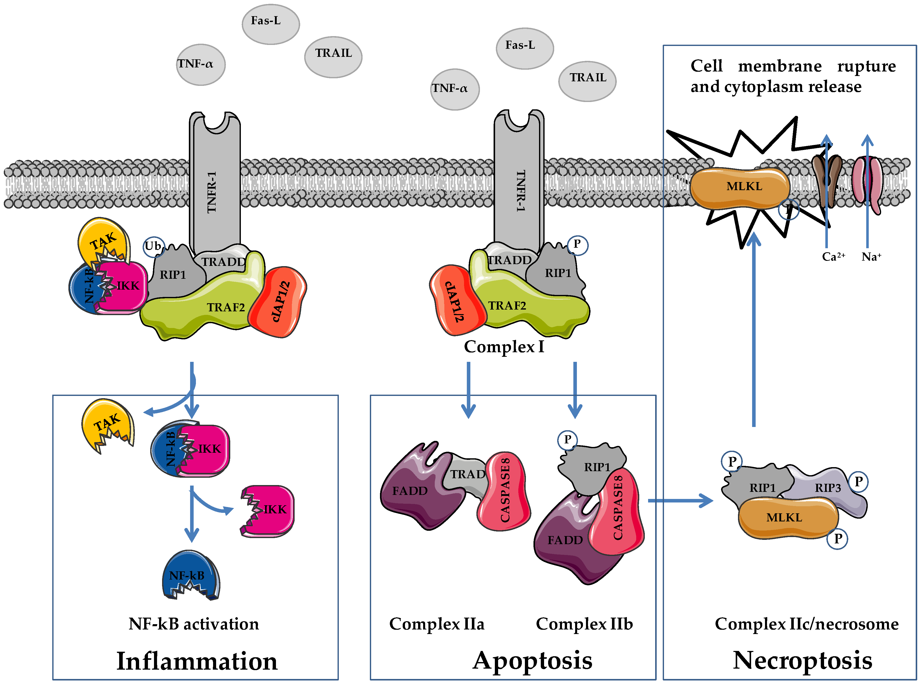
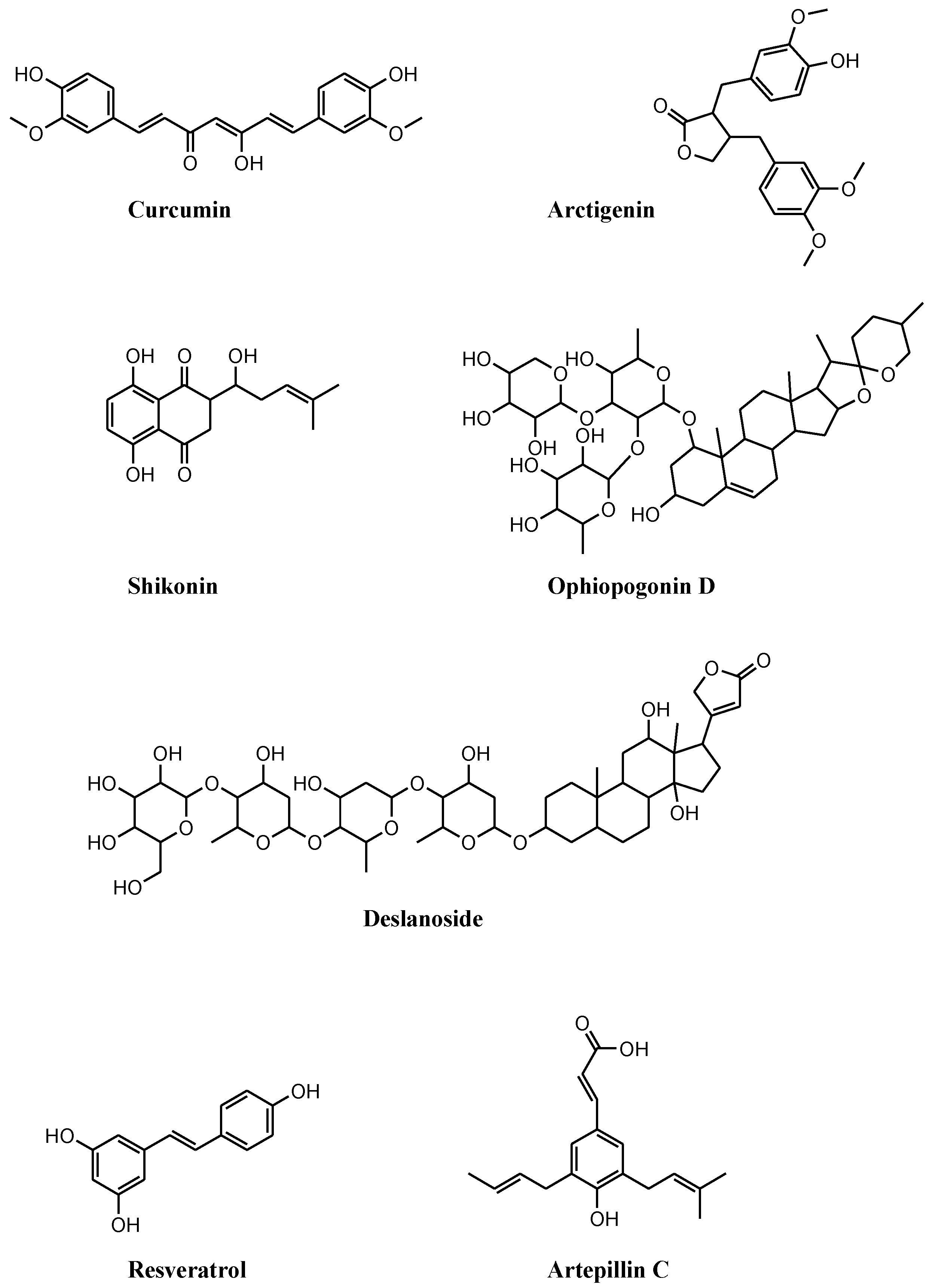
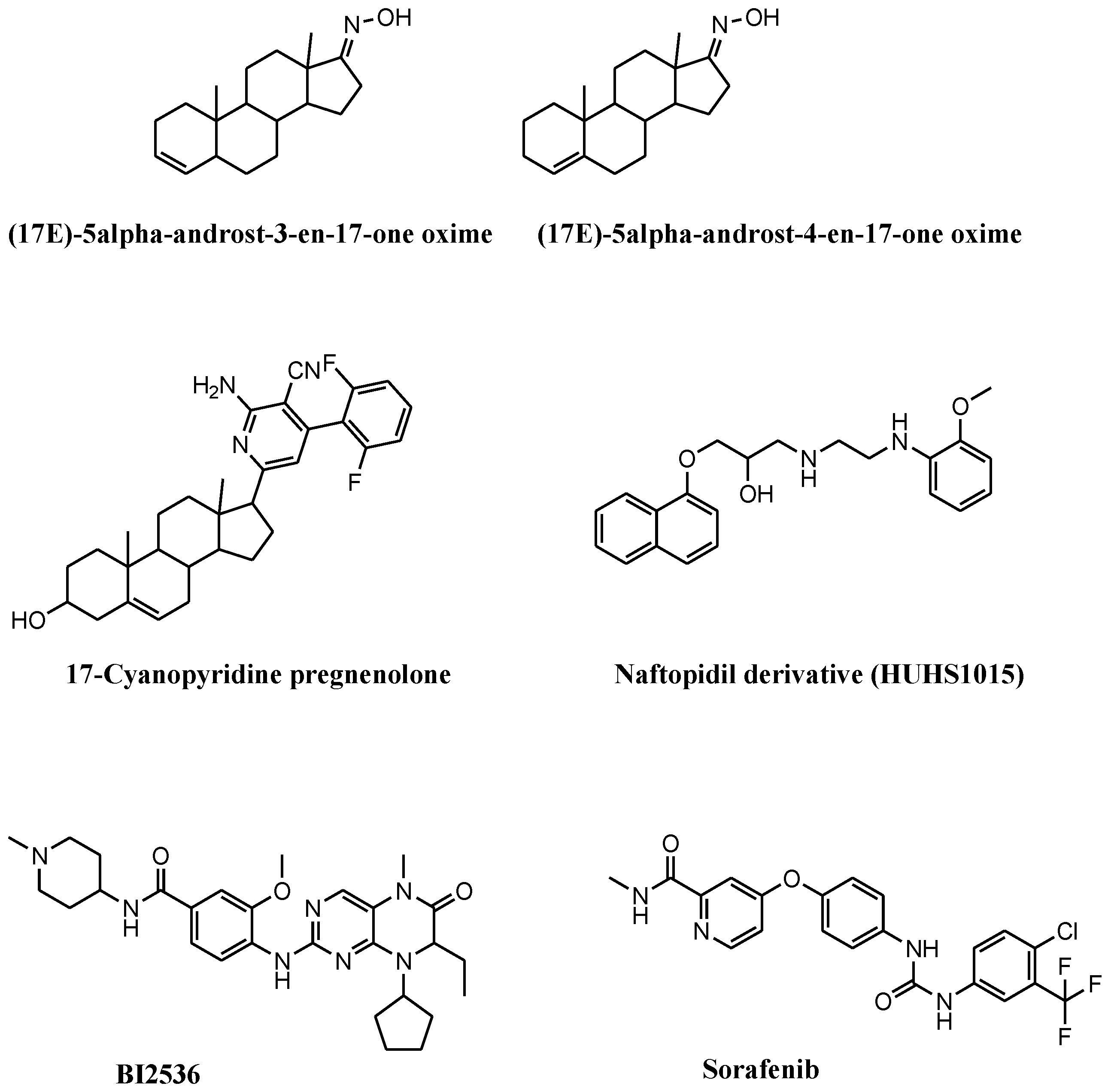
Necroptosis is a programmed form of necrosis characterized by mitochondrial alterations and plasma membrane permeabilization resulting in the release of cytoplasmic content into extracellular space, and leading to inflammatory reactions. Besides its critical role in viral defense mechanisms and inflammatory diseases, necroptosis plays pivotal functions in the drug response of tumors, including prostate cancer. Necroptosis is mainly governed by kinase enzymes, including RIP1, RIP3, and MLKL, and conversely to apoptosis, is a caspase-independent mechanism of cell death. Numerous compounds induce necroptosis in prostate cancer models, including (i) compounds of natural origin, (ii) synthetic and semisynthetic small molecules, and (iii) selenium and selenium-based nanoparticles.

necroptosis prostate cancer RIP1 RIP3 MLKL

1. Introduction

Cell death mechanisms include accidental cell death (ACD) and regulated cell death (RCD) [1]. External extreme temperature and pressure, chemical stress, and osmotic pressure exceeding the capability of the cell to restore a physiologic condition lead to ACD. This type of cell death is an uncontrolled process in which the hallmark is the cell membrane rupturing, causing the spillover of the cytoplasm into the extracellular environment. Alterations in the cell membrane and the release of cellular content are also typical features of RCD [1][2]. RCD is controlled by molecules of specific signal cascades governing biochemical, morphological, and immunological consequences in a regulated and programmed manner. RCD guides pivotal steps of physiological and pathological processes [1]. Besides the most studied apoptosis, RCD includes autophagy, pyroptosis, ferroptosis, and necroptosis (NEC) [3][4][5][6][7][8][9]. RCD is a concept in continuous evolution. Very recently, PANoptosis, which embraces pyroptosis, apoptosis, and NEC, has emerged as a new RCD mode controlled by the aggregation of specific effector molecules leading to the formation of the PANoptosome [10][11]. The role played by PANoptosis in tumors was reported in colorectal cancer [10].
Differences in cell morphology, gene expression, and biochemical properties classify diverse forms of RCD. Therefore, molecules governing different pathways of different RCDs can be considered to be biomarkers and potential therapeutic targets [12]. The pharmacological strategy to hit selective factors of specific RCD pathways showed efficacy for cancer therapy [13]. In this context, apoptosis is paradigmatic, and the development of drugs inducing apoptosis represented the major goal of medical research [14]. However, this strategy has demonstrated numerous limits, and the emergence of drug resistance rendering tumors insensitive to apoptosis is crucial [15]. In this scenario, the discovery of drugs that induce nonapoptotic RCD is an intriguing approach for anticancer therapy, and compounds inducing NEC in tumors, including prostate cancer (PCa), are interesting [16][17][18].
PCa is the second most diagnosed tumor (incidence of 14.1% over a total of 10.1 million new cases) and the fifth global cause of cancer death (mortality of 6.8% over a total of 5.5 million deaths) in men [19]. Men affected by localized or locally advanced PCa are treated by radical prostatectomy and radiotherapy. Conversely, metastatic androgen-sensitive PCa-suffering patients undergo androgen deprivation therapy (ADT). The disease invariably progresses towards lethal metastatic castration-resistant PCa (CRPC) that is insensitive or resistant to ADT. In spite of several efforts devised thus far in the development of new effective therapeutics on CRPC patients, including taxane-based chemotherapy (cabazitaxel and docetaxel), inhibitors of androgen synthesis (abiraterone) or signaling (enzalutamide), bone-targeting radiotherapy (radium-223) and immunotherapy (sipuleucel-T), the disease still remains lethal [20]. CRPC patients among all PCa patients, and their poor prognosis and survival reflect the need to develop other drugs. Thus, the discovery of new antitumor drugs and active medical interventions on CRPC patients is urgent, and compounds that induce NEC are expected to produce benefits [17][21].

2. Apoptosis and Necroptosis: Overview on Molecular Mechanisms

In 2005, Degterev and coworkers described NEC as a peculiar type of necrosis controlled by a specific signaling cascade that is inhibited by necrostatin 1 (Nec1) [22]. Taking advantage of a siRNA-mediated approach, receptor-interacting serine/threonine protein kinase 1 (RIP1) was identified as the target of Nec1. In 2018, such a type of programmed necrosis, not dependent on the activation of cysteine-aspartic proteases (caspases), and mainly governed by RIP1, receptor-interacting serine/threonine protein kinase 3 (RIP3) and substrate mixed lineage kinase domain like pseudokinase (MLKL) was called NEC [23]. NEC controls cell homeostasis, neurodegeneration, infectious diseases, inflammation, cardiovascular and skin diseases, acute kidney injury, and cancer [24].
Cells undergoing NEC and necrosis show similar behavior [25][26]. Although trauma, toxic stress, and infection are responsible for both necrosis and NEC, the latter differs from necrosis as a finely controlled type of RCD. Specific signaling pathways, including TNRF1 and RIP, govern NEC induction via the formation of the necroptosome complex. No important differences in morphology characterize cells undergoing NEC and necrosis (e.g., organelle and cell swelling, loss of membrane integrity, and the release of intracellular content). Moreover, both cell death mechanisms induce mitochondrial dysfunction with mitochondrial membrane collapse. However, only during NEC the production of reactive oxygen species (ROS) and the release of apoptosis-inducing factor (AIF) occur. Proinflammatory response characterizes both NEC and necrosis. Nec1 counteracts only NEC, while it is ineffective on necrosis.
Tumor necrosis factor-α (TNF-α), Fas ligand (FasL), and TNF-related apoptosis-inducing ligand (TRAIL) are death-inducing molecules acting on FasL/TNF receptor (TNFR)1, which can induce either apoptosis or NEC depending on caspase functionality (Figure 1) [27][28].
Figure 1. Schematic representation of cellular pathways involved in necroptosis. TNF-α, tumor necrosis factor α; FasL, Fas ligand; TRAIL, TNF-related apoptosis-inducing ligand; TNFR1, TNF receptor 1; RIP1/3, receptor-interacting serine/threonine-protein kinase 1/3; MLKL, mixed lineage linase domain like pseudokinase; TRADD, TNF receptor associated death domain protein; FADD, Fas-associated death domain; TRAF2, TNF receptor associated factor 2; cIAP1/2, cellular inhibitor of apoptosis proteins 1/2; TAK complex formed by TAK1, TAB2 and TAB3; IKK complex, formed by IKKα and IKKβ. Figure was prepared using tools from Servier Medical Art (http://www.servier.fr/servier-medical-art, accessed on 15 March 2022).
Cells undergoing apoptosis arrest their growth and division, and activate plasma membrane modifications, chromatin condensation, and DNA fragmentation (DNA ladder), and cell shrinkage favoring the formation of apoptotic bodies engulfing the surrounding environmental phagocytes (Table 1). During NEC, a cell increases its volume, and shows organelle shrinkage and plasma membrane disintegration. Cellular contents, including damage-related pattern molecules (DAMPs), high-mobility group box 1 (HMGB1), and mitochondrial DNA, are released into the extracellular environment. Conversely to apoptosis, which is characterized by limited DMAP release, the massive cytoplasm spillover observed during NEC induces an important immune response.
Table 1. Comparison of necroptosis-, necrosis-, and apoptosis-regulated cell death properties.
 

Characteristic

Necroptosis

Necrosis

Apoptosis

Involved proteins

RIP3

+

/

/


MLKL

+

/

/


Caspase 3

/

/

+

Cell properties

Membrane perforation

+

+

/


Membrane blebbing

/

/

+


DNA fragmentation

+

+

+


Cell lysis and swelling

+

+

/


Inflammation

+

+

/

Both membrane death receptors activation (via TNF-α, TRAIL, FasL) and intracellular stimulation (via genetic damage, hypoxia, osmotic stress, or starvation) trigger apoptosis [29]. The latter, known as intrinsic (e.g., mitochondrial) apoptosis, depends on mitochondria-secreted factors including cytochrome c, SMAC/DIABLO, HrtA2/Omi, and AIF that stimulate apoptosis. Intracellular cytotoxic stimuli activate BH3-only proteins leading to BAX and BAK activation, inducing the formation of mitochondrial permeability transition pores on the outer mitochondrial membrane. This feature stimulates the release of cytochrome c into the cytoplasm and its interaction with apoptotic protease activating factor 1 (APAF1) favoring the formation of the apoptosome. This molecular complex in turn stimulates procaspase 9, 3, and 7 cleavage leading to the dysfunction or disruption of cellular components and apoptosis.
The interaction of membrane death receptors and death ligands initiates extrinsic (e.g., extracellular) apoptosis. The formation of death-included signaling complex (DISC), which contains TNFR1, Fas-FasL, death receptor 4 (DR4), death receptor 3 (DR3), and tumor necrosis factor superfamily 10 (known as TRAIL/Apo2L), is the first step of the pathway [30]. The interaction of Fas with FasL induces conformational changes of the complex that favors the exposure of Fas death effector domains, facilitating the recruitment of adaptor FADD, and the activation of procaspase 8 and 10. In turn, the cleavage or activation of procaspases 8 and 10 stimulates procaspase 3 and 7 cleavage leading to the enzymatic activation of target proteins and apoptosis [30].
Similar to apoptosis, NEC is triggered by the interaction of death receptors (Fas, TNFR1, and TNFR2) with death ligands (TNF-α, FasL, and TRAIL). In apoptosis, NEC induction requires the inhibition of caspase signaling pathways (Figure 1) [22][31]. The interaction of death ligands with membrane receptors provokes conformational changes that induce the recruitment of TNF receptor-associated death domain protein (TRADD), RIP1, tumor necrosis factor-related factor 2 (TRAF2), the cellular inhibitors of apoptosis cIAP1/2 and ubiquitination complex, favoring the formation of a TNFR complex (complex I) [32]. This protein complex is responsible for cell fate, i.e., death or survival, depending on the activation of downstream pathways via ubiquitination and phosphorylation activities. Through the activation of NF-kB, complex I stimulates cell survival and promotes inflammation [33][34][35]. The ubiquitination status of RIP1 favors the recruitment of transforming growth factor β kinase 1 (TAK1), TAK1-binding protein 2 (TAB2), and TAB3 (TAK complex), and the IKK complex (IKKα and IKKβ). In this condition, RIP1 functions as a scaffold protein that favors the activation of NF-kB (e.g., cIAP2-mediated degradation of NF-kB inhibitory protein Ikβ of the IKK complex), which, following nuclear translocation, stimulates cell survival (e.g., activation of antiapoptotic genes including cFLIP) and inflammation. Apart from enzymatic activity, cFLIP is highly homologous to caspase 8, and interacting with caspase 8 inhibits its activation, favoring cell survival [36][37][38][39].
The ubiquitination status of complex I elements stabilizes its localization on the cell membrane, impeding the formation of complex II and favoring cell survival [38]. Reduced RIP1 ubiquitination favors the formation and the cytoplasmic localization of complex II (IIa, IIb, and IIc/necrosome) leading to apoptosis or NEC [37][38][39]. The formation of complex IIa, which contains procaspase 8, TRADD, and FADD, allows for caspase 8 and subsequently caspase 3 activation, leading to apoptosis. The inhibition of RIP1 polyubiquitination in complex I favors its cytoplasmic localization and the interaction with FADD and procaspase 8, leading to the formation of complex IIb (RIP1/FADD/procaspase 8). Regarding complex IIa, the activation of caspase 8 induces caspase 8-dependent apoptosis as well.
Phosphorylated RIP1 (p-RIP1) interacts with RIP3 and favors its phosphorylation (p-RIP3). Following the inhibition of caspase 8 activation (e.g., via cFLIP), the p-RIP1/p-RIP3 complex recruits MLKL favoring the formation of complex IIc/necrosome. Phosphorylation on threonine 357/serine 358 of MLKL provokes its translocation on the plasma membrane leading to the activation of sodium and calcium channels, and inducing membrane dysfunction, cell rupture, and NEC execution [40][41][42].
The levels of cFLIP influence the switch apoptosis/NEC. Elevated cFLIP expression inhibits caspase 8 by forming heteromeric complex caspase 8–cFLIP, thereby blocking apoptosis dependent on complexes IIa and IIb.
Besides plasma membrane localization, the RIP1/RIP3/MLKL complex translocates to the mitochondrial membrane provoking mitochondrial dysfunction (e.g., mitochondrial permeability transition), and inducing the production of ROS and the activation of mitochondrial phosphoglycerate mutase 5 (PGAM5). Activated PGAM5 recruits mitochondrial dynamin-related protein (DRP1) leading to mitochondrial fragmentation [43]. ROS are crucial for NEC, and some evidence links RIP3 to ROS levels. RIP3 stimulates mitochondrial enzymes, including pyruvate dehydrogenase complex, glutamine synthetase, and glutamate dehydrogenase, promoting ROS production and NEC [44]. However, the involvement of mitochondria in NEC is controversial. Indeed, cells depleted of mitochondria through forced mitophagy undergo NEC, implying that mitochondria or mitochondrial metabolism are not essential for this type of RCD [45]. This scenario reveals that the kinase-dependent NEC can be viewed as a rescue mechanism of cell death functioning when caspase-mediated apoptosis fails [26][40][46].

3. Necroptosis and Necroptosis Inducers in Prostate Cancer

Though reported since 2005, the induction of NEC for fighting PCa is still scarcely considered. RIP3 is decreased in PCa specimens and in cell lines (e.g., PC3, DU145, and 22Rv1). Moreover, in advanced PCa samples, RIP3 is significantly downregulated compared to normal tissue [47]. The reduced expression of RIP3 correlates with tumor size and prostate-specific antigen (PSA) levels [48]. High or normal levels of RIP3 counteract disease progression by favoring MLKL phosphorylation, leading to NEC. The overexpression of RIP3 in PC3 and 22Rv1 cell lines induces G2 cell-cycle arrest, reduces cell survival, proliferation, invasion (increased MMP2 and MMP9, vimentin, fibronectin, and N-cadherin), and favors NEC (phosphorylation of MLKL) [47]. In addition, compared to corresponding parental tumors, tumorigenesis and tumor volume are reduced in mice bearing RIP3-overexpressing PC3 and 22Rv1 tumors [47].
The seven members’ sirtuin (SIRT) family of proteins shows enzymatic activity implicated in numerous cellular functions, including DNA damage repair, senescence, metabolism, and tumor development and progression. Two enzymes of this family, SIRT3 and SIRT6, are involved in NEC induction in PCa. The increased expression of SIRT3 and SIRT6 is significantly associated with nodal metastasis and Gleason score, and negatively impacts on overall patient survival [49]. Therefore, the silencing of SIRT3 and SIRT6 in LNCaP, DU145, and PC3 cells significantly reduces cell growth, which is paralleled by RIP3-mediated NEC induction (e.g., increased phosphorylation of RIP3 and MLKL). These results were confirmed in vivo in SIRT3 and SIRT6 silenced LNCaP tumor xenografts that showed reduced tumor volume compared to the corresponding tumors expressing normal enzyme levels.
Compounds that induce NEC are receiving considerable attention, and several molecules, though not specific NEC inducers, showed antitumor activity on PCa cell lines (Figure 2 and Figure 3). Available compounds are: (i) of natural origin, (ii) synthetic and semisynthetic small molecules, and (iii) selenium and selenium-based nanoparticles (SeNPs).
Figure 2. Chemical structures of compounds of natural origin that induce necroptosis in prostate cancer cell lines. These compounds stimulate necroptosis by inducing (i) the dysfunction of mitochondrial membrane potential, curcumin, and arctigenin; (ii) cell-cycle arrest, shikonin; (iii) FADD stabilization, ophiopogonin D’; (iv) modulation of multiple necroptosis pathways, deslanoside; (v) reactive oxygen species, resveratrol; (vi) RIP1/3 activation following combination with autophagy inhibitors, artepillin C.
Figure 3. Chemical structures of synthetic and semisynthetic small molecules that induce necroptosis in prostate cancer cell lines. These compounds stimulate necroptosis by (i) inducing mitochondrial membrane permeability, (17E)-5α-androst-3-en-17-one oxime and (17E)-androst-4-en-17-one oxime; (ii) inducing reactive oxygen species and cell-cycle arrest, 17-cyanopyridine pregnenolone; (iii) inducing cell growth inhibition following combination with necrostatin, HUHS1015; (iv) inhibiting Polo-like kinase 1, BI2536; (v) inhibiting kinases, sorafenib.

4. Ongoing Clinical Trials Containing Necroptosis Inducers in Prostate Cancer

Some compounds reported here are contained in clinical trials enrolling patients suffering from PCa (Table 2, www.clinicaltrials.gov, accessed on 23 February 2022).
Table 2. Clinical trials containing necroptosis inducers ongoing in prostate cancer a.

Compound

NCT Number

Markers

Phase

Curcumin

NCT03769766

PSA

III


NCT03211104

PSA

na


NCT02064673

PSA

III

Curcumin and piperine

NCT04731844

nd

II

Curcumin and ursolic acid

NCT04403568

p65, NF-kB

I

Curcumin and Vitamin D, omega 3, turmeric

NCT03290417

PSA

na

Curcumin and taxotere

NCT02095717

PSA

III

Curcumin and radiotherapy

NCT01917890

TNF-α, NF-kB

na


NCT02724618

PSA

II


NCT03493997

nd

II

Polyphenon E

NCT00596011

PSA

II


NCT00676780

PSA, VEGF, HGF

II


NCT01340599

PSA, Ki67, Bcl2, Cyclin D, p27, VEGF, CD31, MMP2 and 9, IGF1

II


NCT00459407

MMP2, MMP9, IGF1

I


NCT00253643

FASN, Ki67

na


NCT04597359

PSA, Ki67

II

BI2536

NCT00706498

PSA

II

Sorafenib

NCT00090545

PSA

II


NCT00694291

PSA

II


NCT00466752

PSA, p-ERK, p-AKT, p-S6-kinase, caspase 3, Ki67

I


NCT00093457

PSA

II

Sorafenib and leuprolide or bicalutamide

NCT00924807

PSA

I-II

Sorafenib and docetaxel

NCT00589420

PSA

II


NCT00619996

PSA, p-ERK, VEGF-R2

II

Sorafenib and taxotere

NCT00405210

PSA

I

Sorafenib and mitoxantrone

NCT00452387

PSA

II

Sorafenib and gleevec

NCT00424385

PSA

I

Selenite and docetaxel

NCT01155791

PSA

I

Selenite and radiotherapy

NCT02184533

PSA

I

a Clinical data are from ClinicalTrials.gov, a service of the U.S. National Institutes of Health. www.clinicaltrial.gov (accessed on 23 February 2022). nd, not defined; na, not available.
Curcumin alone (NCT03769766, phase III; NCT03211104; NCT02064673, phase III) or in combination with piperine (NCT04731844, phase II), ursolic acid (NCT04403568, phase I), vitamin D, omega 3 and turmeric (NCT03290417), taxotere (NCT02095717, phase II) and radiotherapy (NCT01917890; NCT02724618, phase II; NCT03493997, phase II) is studied in PCa patients. Despite studies implying the measure of different biomarkers, mostly PSA, no specific markers of NEC are considered. Only trials NCT04403568 and NCT01917890 contemplate the evaluation of TNF-α and NF-kB. These two molecules are involved in NEC, though they are not specific biomarkers.
Polyphenon E was included in numerous studies as supplementary diet in subjects at high risk of developing PCa (prostatic hyperplasia) or in postsurgery patients (NCT00596011, phase II; NCT00676780, phase II; NCT01340599, phase II; NCT00459407, phase I; NCT00253643; NCT04597359, phase II). Polyphenon E-based studies contemplated the measure of numerous biological markers (e.g., PSA, Ki67, Bcl2, Cyclin D, p27, VEGF, CD31, MMP2 and 9, IGF1, HGF, FASN) but not specifically related to NEC.
Sorafenib alone (NCT00090545, phase II; NCT00694291, phase II; NCT00466752, phase I; NCT00093457, phase II) or in combination with leuprolide acetate or bicalutamide (NCT00924807, phase I-II), docetaxel (NCT00589420, phase II; NCT00619996, phase II), taxotere (NCT00405210, phase I) mitoxantrone (NCT00452387, phase II) or gleevec (NCT00424385, phase I) is investigated in PCa patients. In all these studies, PSA is used as biological marker. The trial NCT00619996 contemplates the measure of p-ERK and VEGF-R2 as well. Interestingly, in trial NCT00466752, samples from PCa patients collected before and after sorafenib administration were analyzed for gene and protein expression by microarray, Western blot, and immunohistochemistry. In this context, the levels of p-ERK, p-AKT, p-S6- kinase and caspase-3 expression, and the Ki67 index were considered. Though the study is completed, results are not yet available. In sorafenib-containing clinical trials, specific biomarkers of NEC are also not measured.
Selenite in combination with docetaxel (NCT01155791, phase I) or with radiotherapy (NCT02184533, phase I) is studied in PCa-affected patients. In these studies, PSA was the only considered biomarker.

References

  1. Tang, D.; Kang, R.; Berghe, T.V.; Vandenabeele, P.; Kroemer, G. The molecular machinery of regulated cell death. Cell Res. 2019, 29, 347–364.
  2. Choi, J.J.; Reich, C.F.; Pisetsky, D.S. Release of DNA from dead and dying lymphocyte and monocyte cell lines in vitro. Scand. J. Immunol. 2004, 60, 159–166.
  3. Kang, R.; Zeng, L.; Zhu, S.; Xie, Y.; Liu, J.; Wen, Q.; Cao, L.; Xie, M.; Ran, Q.; Kroemer, G.; et al. Lipid peroxidation drives gasdermin D-mediated pyroptosis in lethal polymicrobial sepsis. Cell Host. Microbe. 2018, 24, 97–108.e104.
  4. Canli, Ö.; Alankuş, Y.B.; Grootjans, S.; Vegi, N.; Hültner, L.; Hoppe, P.S.; Schroeder, T.; Vandenabeele, P.; Bornkamm, G.W.; Greten, F.R. Glutathione peroxidase 4 prevents necroptosis in mouse erythroid precursors. Blood J. Am. Soc. Hematol. 2016, 127, 139–148.
  5. Haberzettl, P.; Hill, B.G. Oxidized lipids activate autophagy in a JNK-dependent manner by stimulating the endoplasmic reticulum stress response. Redox Biol. 2013, 1, 56–64.
  6. Wang, Y.; Wu, N.; Jiang, N. Autophagy provides a conceptual therapeutic framework for bone metastasis from prostate cancer. Cell Death Dis. 2021, 12, 909.
  7. Zaffaroni, N.; Beretta, G.L. Nanoparticles for Ferroptosis Therapy in Cancer. Pharmaceutics 2021, 13, 1785.
  8. Zaffaroni, N.; Beretta, G.L. Ferroptosis inducers for prostate cancer therapy. Curr. Med. Chem. 2022, in press.
  9. Goodall, M.L.; Cramer, S.D.; Thorburn, A. Autophagy RIPs into cell death. Cell Cycle 2016, 15, 3014–3015.
  10. Karki, R.; Sharma, B.R.; Lee, E.; Banoth, B.; Malireddi, R.K.S.; Samir, P.; Tuladhar, S.; Mummareddy, H.; Burton, A.R.; Vogel, P.; et al. Interferon regulatory factor 1 regulates PANoptosis to prevent colorectal cancer. JCI Insight 2020, 5, e136720.
  11. Place, D.E.; Lee, S.; Kanneganti, T.D. PANoptosis in microbial infection. Curr. Opin. Microbiol. 2021, 59, 42–49.
  12. Heidaryan, F.; Bamehr, H.; Babaabasi, B.; Emamvirdizadeh, A.; Mohammadzadeh, N.; Khalili, A. The Trend of ripk1/ripk3 and mlkl Mediated Necroptosis Pathway in Patients with Different Stages of Prostate Cancer as Promising Progression Biomarkers. Clin. Lab. 2020, 66.
  13. Liu, W.; Jin, W.; Zhu, S.; Chen, Y.; Liu, B. Targeting regulated cell death (RCD) with small-molecule compounds in cancer therapy: A revisited review of apoptosis, autophagy-dependent cell death and necroptosis. Drug Discov. Today 2022, 27, 612–625.
  14. Carneiro, B.A.; El-Deiry, W.S. Targeting apoptosis in cancer therapy. Nat. Rev. Clin. Oncol. 2020, 17, 395–417.
  15. Fulda, S.; Vucic, D. Targeting IAP proteins for therapeutic intervention in cancer. Nat. Rev. Drug Discov. 2012, 11, 109–124.
  16. Yan, J.; Wan, P.; Choksi, S.; Liu, Z.G. Necroptosis and tumor progression. Trends Cancer 2022, 8, 21–27.
  17. Nie, Z.; Chen, M.; Gao, Y.; Huang, D.; Cao, H.; Peng, Y.; Guo, N.; Zhang, S. Regulated Cell Death in Urinary Malignancies. Front. Cell Dev. Biol. 2021, 9, 789004.
  18. Mohammad, R.M.; Muqbil, I.; Lowe, L.; Yedjou, C.; Hsu, H.Y.; Lin, L.T.; Siegelin, M.D.; Fimognari, C.; Kumar, N.B.; Dou, Q.P.; et al. Broad targeting of resistance to apoptosis in cancer. Semin. Cancer Biol. 2015, 35, S78–S103.
  19. Sung, H.; Ferlay, J.; Siegel, R.L.; Laversanne, M.; Soerjomataram, I.; Jemal, A.; Bray, F. Global Cancer Statistics 2020: GLOBOCAN Estimates of Incidence and Mortality Worldwide for 36 Cancers in 185 Countries. CA Cancer J. Clin. 2021, 71, 209–249.
  20. Nuhn, P.; De Bono, J.S.; Fizazi, K.; Freedland, S.J.; Grilli, M.; Kantoff, P.W.; Sonpavde, G.; Sternberg, C.N.; Yegnasubramanian, S.; Antonarakis, E.S. Update on systemic prostate cancer therapies: Management of metastatic castration resistant prostate cancer in the era of precision oncology. Eur. Urol. 2019, 75, 88–99.
  21. Martin-Sanchez, D.; Fontecha-Barriuso, M.; Sanchez-Niño, M.D.; Ramos, A.M.; Cabello, R.; Gonzalez-Enguita, C.; Linkermann, A.; Sanz, A.B.; Ortiz, A. Cell death-based approaches in treatment of the urinary tract-associated diseases: A fight for survival in the killing fields. Cell Death Dis. 2018, 9, 118.
  22. Degterev, A.; Huang, Z.; Boyce, M.; Li, Y.; Jagtap, P.; Mizushima, N.; Cuny, G.D.; Mitchison, T.J.; Moskowitz, M.A.; Yuan, J. Chemical inhibitor of nonapoptotic cell death with therapeutic potential for ischemic brain injury. Nat. Chem. Biol. 2005, 1, 112–119.
  23. Galluzzi, L.; Vitale, I.; Aaronson, S.A.; Abrams, J.M.; Adam, D.; Agostinis, P.; Alnemri, E.S.; Altucci, L.; Amelio, I.; Andrews, D.W.; et al. Molecular mechanisms of cell death: Recommendations of the Nomenclature Committee on Cell Death 2018. Cell Death Differ. 2018, 25, 486–541.
  24. Liu, X.; Xie, X.; Ren, Y.; Shao, Z.; Zhang, N.; Li, L.; Ding, X.; Zhang, L. The role of necroptosis in disease and treatment. MedComm 2021, 2, 730–755.
  25. Davidovich, P.; Kearney, C.J.; Martin, S.J. Inflammatory outcomes of apoptosis, necrosis and necroptosis. Biol. Chem. 2014, 395, 1163–1171.
  26. Vanden Berghe, T.; Linkermann, A.; Jouan-Lanhouet, S.; Walczak, H.; Vandenabeele, P. Regulated necrosis: The expanding network of non-apoptotic cell death pathways. Nat. Rev. Mol. Cell Biol. 2014, 15, 135–147.
  27. Laster, S.M.; Wood, J.G.; Gooding, L.R. Tumor necrosis factor can induce both apoptic and necrotic forms of cell lysis. J. Immunol. 1988, 141, 2629–2634.
  28. Vercammen, D.; Brouckaert, G.; Denecker, G.; Van de Craen, M.; Declercq, W.; Fiers, W.; Vandenabeele, P. Dual signaling of the Fas receptor: Initiation of both apoptotic and necrotic cell death pathways. J. Exp. Med. 1998, 188, 919–930.
  29. Mishra, A.P.; Salehi, B.; Sharifi-Rad, M.; Pezzani, R.; Kobarfard, F.; Sharifi-Rad, J.; Nigam, M. Programmed Cell Death, from a Cancer Perspective: An Overview. Mol. Diagn. Ther. 2018, 22, 281–295.
  30. D’Arcy, M.S. Cell death: A review of the major forms of apoptosis, necrosis and autophagy. Cell Biol. Int. 2019, 43, 582–592.
  31. Holler, N.; Zaru, R.; Micheau, O.; Thome, M.; Attinger, A.; Valitutti, S.; Bodmer, J.L.; Schneider, P.; Seed, B.; Tschopp, J. Fas triggers an alternative, caspase-8-independent cell death pathway using the kinase RIP as effector molecule. Nat. Immunol. 2000, 1, 489–495.
  32. Galluzzi, L.; Kepp, O.; Chan, F.K.; Kroemer, G. Necroptosis: Mechanisms and Relevance to Disease. Annu. Rev. Pathol. 2017, 12, 103–130.
  33. Dondelinger, Y.; Darding, M.; Bertrand, M.J.; Walczak, H. Poly-ubiquitination in TNFR1-mediated necroptosis. Cell Mol. Life Sci. 2016, 73, 2165–2176.
  34. Tortola, L.; Nitsch, R.; Bertrand, M.J.M.; Kogler, M.; Redouane, Y.; Kozieradzki, I.; Uribesalgo, I.; Fennell, L.M.; Daugaard, M.; Klug, H.; et al. The Tumor Suppressor Hace1 Is a Critical Regulator of TNFR1-Mediated Cell Fate. Cell Rep. 2016, 16, 3414.
  35. Zhang, K.X.; Firus, J.; Prieur, B.; Jia, W.; Rennie, P.S. To die or to survive, a fatal question for the destiny of prostate cancer cells after androgen deprivation therapy. Cancers 2011, 3, 1498–1512.
  36. Chan, F.K.; Luz, N.F.; Moriwaki, K. Programmed necrosis in the cross talk of cell death and inflammation. Annu. Rev. Immunol. 2015, 33, 79–106.
  37. Ting, A.T.; Bertrand, M.J.M. More to Life than NF-κB in TNFR1 Signaling. Trends Immunol. 2016, 37, 535–545.
  38. Dillon, C.P.; Oberst, A.; Weinlich, R.; Janke, L.J.; Kang, T.B.; Ben-Moshe, T.; Mak, T.W.; Wallach, D.; Green, D.R. Survival function of the FADD-CASPASE-8-cFLIP(L) complex. Cell Rep. 2012, 1, 401–407.
  39. Dondelinger, Y.; Declercq, W.; Montessuit, S.; Roelandt, R.; Goncalves, A.; Bruggeman, I.; Hulpiau, P.; Weber, K.; Sehon, C.A.; Marquis, R.W.; et al. MLKL compromises plasma membrane integrity by binding to phosphatidylinositol phosphates. Cell Rep. 2014, 22, 971–981.
  40. Quarato, G.; Guy, C.S.; Grace, C.R.; Llambi, F.; Nourse, A.; Rodriguez, D.A.; Wakefield, R.; Frase, S.; Moldoveanu, T.; Green, D.R. Sequential Engagement of Distinct MLKL Phosphatidylinositol-Binding Sites Executes Necroptosis. Mol. Cell 2016, 61, 589–601.
  41. Cho, Y.S.; Challa, S.; Moquin, D.; Genga, R.; Ray, T.D.; Guildford, M.; Chan, F.K. Phosphorylation-driven assembly of the RIP1-RIP3 complex regulates programmed necrosis and virus-induced inflammation. Cell 2009, 137, 1112–1123.
  42. Sun, L.; Wang, H.; Wang, Z.; He, S.; Chen, S.; Liao, D.; Wang, L.; Yan, J.; Liu, W.; Lei, X.; et al. Mixed lineage kinase domain-like protein mediates necrosis signaling downstream of RIP3 kinase. Cell 2012, 148, 213–227.
  43. Wang, Z.; Jiang, H.; Chen, S.; Du, F.; Wang, X. The mitochondrial phosphatase PGAM5 functions at the convergence point of multiple necrotic death pathways. Cell 2012, 148, 228–243.
  44. Yang, Z.; Wang, Y.; Zhang, Y.; He, X.; Zhong, C.Q.; Ni, H.; Chen, X.; Liang, Y.; Wu, J.; Zhao, S.; et al. RIP3 targets pyruvate dehydrogenase complex to increase aerobic respiration in TNF-induced necroptosis. Nat. Cell Biol. 2018, 20, 186–197.
  45. Tait, S.W.; Oberst, A.; Quarato, G.; Milasta, S.; Haller, M.; Wang, R.; Karvela, M.; Ichim, G.; Yatim, N.; Albert, M.L.; et al. Widespread mitochondrial depletion via mitophagy does not compromise necroptosis. Cell Rep. 2013, 5, 878–885.
  46. Zhou, W.; Yuan, J. Necroptosis in health and diseases. Semin. Cell Dev. Biol. 2014, 35, 14–23.
  47. Wang, K.J.; Wang, K.Y.; Zhang, H.Z.; Meng, X.Y.; Chen, J.F.; Wang, P.; Jiang, J.H.; Ma, Q. Up-Regulation of RIP3 Alleviates Prostate Cancer Progression by Activation of RIP3/MLKL Signaling Pathway and Induction of Necroptosis. Front. Oncol. 2020, 10, 1720.
  48. Lu, Z.; Wu, C.; Zhu, M.; Song, W.; Wang, H.; Wang, J.; Guo, J.; Li, N.; Liu, J.; Li, Y.; et al. Ophiopogonin D' induces RIPK1-dependent necroptosis in androgen-dependent LNCaP prostate cancer cells. Int J. Oncol. 2020, 56, 439–447.
  49. Fu, W.; Li, H.; Fu, H.; Zhao, S.; Shi, W.; Sun, M.; Li, Y. The SIRT3 and SIRT6 Promote Prostate Cancer Progression by Inhibiting Necroptosis-Mediated Innate Immune Response. J. Immunol. Res. 2020, 2020, 8820355.
More
Information
Subjects: Oncology
Contributors MDPI registered users' name will be linked to their SciProfiles pages. To register with us, please refer to https://encyclopedia.pub/register : ,
View Times: 618
Revisions: 2 times (View History)
Update Date: 13 Apr 2022
1000/1000
ScholarVision Creations