Submitted Successfully!
To reward your contribution, here is a gift for you: A free trial for our video production service.
Thank you for your contribution! You can also upload a video entry or images related to this topic.
Version Summary Created by Modification Content Size Created at Operation
1 -- 2201 2022-04-12 09:42:52 |
2 format correct -5 word(s) 2196 2022-04-12 11:13:57 | |
3 format correct Meta information modification 2196 2022-04-12 11:14:49 | |
4 format correct Meta information modification 2196 2022-04-13 04:18:22 |

Video Upload Options

Do you have a full video?

Confirm

Are you sure to Delete?
Cite
If you have any further questions, please contact Encyclopedia Editorial Office.
Cichocki, M.; Konecki, T.; , . Carotenoids Reduce Urological Cancers Risk. Encyclopedia. Available online: https://encyclopedia.pub/entry/21621 (accessed on 27 July 2024).
Cichocki M, Konecki T,  . Carotenoids Reduce Urological Cancers Risk. Encyclopedia. Available at: https://encyclopedia.pub/entry/21621. Accessed July 27, 2024.
Cichocki, Marcin, Tomasz Konecki,  . "Carotenoids Reduce Urological Cancers Risk" Encyclopedia, https://encyclopedia.pub/entry/21621 (accessed July 27, 2024).
Cichocki, M., Konecki, T., & , . (2022, April 12). Carotenoids Reduce Urological Cancers Risk. In Encyclopedia. https://encyclopedia.pub/entry/21621
Cichocki, Marcin, et al. "Carotenoids Reduce Urological Cancers Risk." Encyclopedia. Web. 12 April, 2022.
Carotenoids Reduce Urological Cancers Risk
Edit

Urological cancers, namely prostate, bladder, kidney, testicular, and penile cancers, are common conditions that constitute almost one-quarter of all malignant diseases in men. Urological cancers tend to affect older individuals, and their development is influenced by modifiable metabolic, behavioral, and environmental risk factors. Carotenoids may have cancer-fighting properties and protect against cancer development, slow its spread, and reduce the risk of cancer deaths in humans.

prostate cancer carotenoids

1. Prostate Cancer

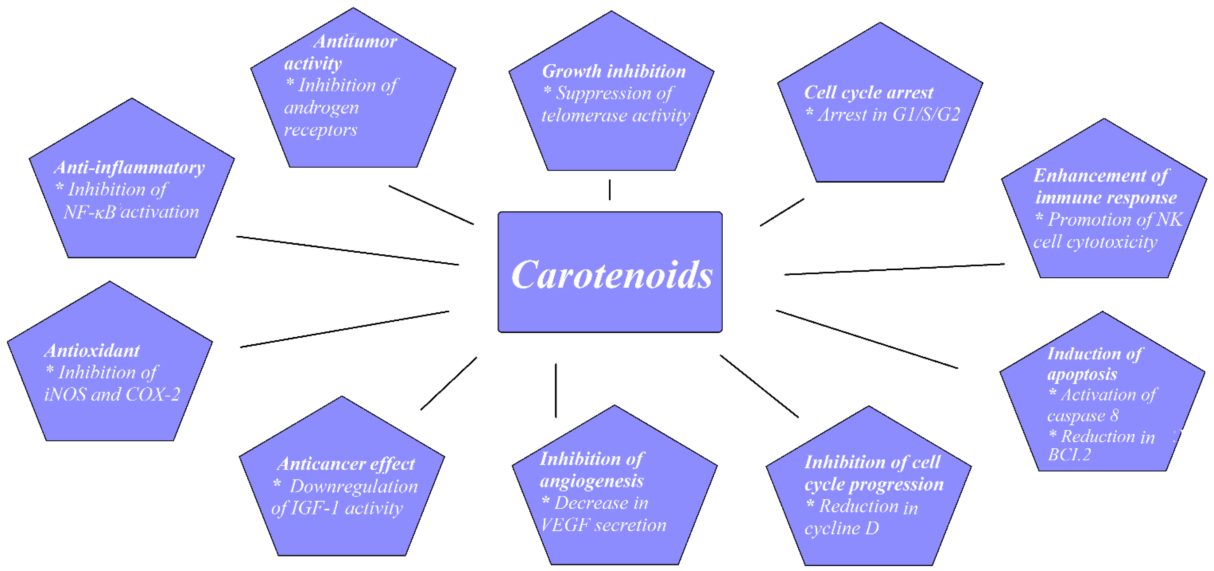
Prostate cancer (PCa) is a common malignancy among men worldwide. In 2018, PCa was reported as the second most common noncutaneous cancer in men worldwide (an estimated 1.3 million new cases) and the fifth most common cause of cancer death in men. There is wide geographical diversity in its prevalence, with age-standardized rates (ASR) per 100,000 of 97.2 in the United States and less than 20 in Asia and developing countries [1]. The most common pathological type of PCa is acinar adenocarcinoma, which accounts for more than 99% of all prostate tumors [2]. Family history of PCa, older age, and race are the only undisputed risk factors for PCa development. Asian men have low PCa risk compared with men from the Western world. However, when Japanese men moved from Japan to California, their risk of PCa rose, approaching that of American men, which implied a role of dietary or environmental factors, possibly including dietary carotenoid intake as a contributing factor [3]. There have been a few proposed mechanisms through which carotenoids may act to reduce cancer development, as shown in Figure 1.

Figure 1. Proposed mechanisms for carotenoids preventing cancer development.

1.1. Experimental Studies

The inhibition of PCa cell growth has been the subject of a number of experimental studies performed on the PCa cell cultures LNCap, DU145, and PC-3. The LNCaP cell line comes from a lymph node metastasis of human PCa; DU145 cells are derived from a brain metastasis of primary PCa; and the PC-3 cell line was established from a PCa bone metastasis from a 62-year-old male Caucasian. The growth-inhibitory effect of lycopene was compared with that of one of the main chemotherapeutic agents used to treat metastatic PCa, docetaxel. PCa cell growth was inhibited by 19–54% with docetaxel and by 5–24% with lycopene. A synergic effect of 78% growth reduction was also shown when combining docetaxel with lycopene [1][2]. A dose-dependent reduction in PCa cellular growth was shown with rising lycopene concentrations of up to 10 µM [3][4]. It may be suggested that the efficacy of lycopene depends on its source of origin. Lycopene extracts from algae (marine Chlorella) and from tomatoes were compared at concentrations of 20 and 50 µM. PCa cells’ viability was 10–18% lower after treatment with algae lycopene than after treatment with tomato lycopene at the same concentration, showing the better antiproliferative and apoptotic effect of algal lycopene [5]. Similarly, extracts from tomato paste, tomato sauce, ketchup, and tomato extract were compared on PCa cells that were obtained and cultured from prostates of patients submitted to radical prostatectomy. Cell growth reduction was highest for tomato paste, and then for ketchup, tomato sauce, and extract, with reductions in cellular growth of 54%, 51%, 47%, and 44%, respectively [6]. One must be cautious when interpreting cell-culture-based studies because there are differences between the concentrations of carotenoids in the body and those used in cell culture, with the latter usually being significantly higher. Concentrations of 1 µM and 0.5 µM of lycopene reduced cell proliferation of LNCaP cells; however, there was no effect observed at 0.25 µM [7]. Other studies have suggested that higher lycopene concentrations of around 1–1.25 µM lycopene are necessary for the antiproliferative effect [8]. Postmortem human studies showed that carotenoid concentrations in serum and individual organs were similar and that there was a correlation between intake and tissue/blood levels of carotenoids. The serum concentration of lycopene in plasma was around 740 nmol/kg, and that in prostate tissue was around 700 nmol/kg. These physiological concentrations may be at the edge of those that showed an antiproliferative effect in experimental studies.
Palozza et al. showed in an in vitro study on PCa cells that β-carotene acted as a growth-inhibitory agent by inhibiting AKT phosphorylation, which led to the increased expression of c-MYC and the activity of caspases. In the end, apoptosis was stimulated [9].
Furthermore, in another experimental animal study, Yang et al. subcutaneously implanted androgen-independent PCa (PC-3) cells in mice. Both lycopene and β-carotene, when supplemented to the mice, significantly inhibited tumor growth [10].
There have been few interesting studies, however, that have examined the impact of lycopene on PCa cells’ ability to spread and metastasize. Using PC3 and DU145 PCa cell lines, Elgass et al. showed that the adhesion of PCa cells to a basement membrane was significantly reduced at lycopene concentrations higher than 1.15 µM. There was also a reduction in PCa cellular motility and migration of 40% and 58% for PC3 cells and DU145 cells, respectively [11]. Some proteins known to be responsible for controlling the adhesion and migration of PC (namely intercellular adherence molecule 1 (ICAM1) and MMP9) were downregulated in PC-3 cell lines treated with tomato extract. These results indicate the ability of LC to diminish PCa’s metastatic abilities to some degree. The expression of proteins such as intracellular ICAM 1 and MMP9 was influenced by tomato extract and downregulated in PC3 cell lines [12]. This may suggest the potential ability of lycopene to reduce PCa spread.

1.2. Epidemiological Studies

It has been suggested that saturated fat consumption is linked to not only PCa incidence but PCa progression and mortality risk [13]. One of the key oncogenes in PCa tumorigenesis is c-MYC. It has been shown that increased fat consumption results in amplifying the MYC-dependent transcriptional cascade [14]. Blood lipids, and especially high lipoprotein A concentrations, seem to be associated with increased PCa risk and advanced disease [15], and lipoprotein A is known to promote inflammation. On the other hand, high levels of chronic inflammatory markers are associated with high-risk, aggressive PCa [16]. The well-established role of carotenoids as scavengers of reactive oxygen species and anti-inflammatory agents might contribute to PCa risk reduction [17].
High concentrations of carotenoids were identified in some vegetables consumed as part of a traditional Mediterranean diet [18]. There have been a few prospective cohort studies that found no protective association between Mediterranean diet prior to diagnosis and incidence of lethal or advanced PCa. However, an interesting observation was made that greater adherence to Mediterranean diet after the diagnosis of nonmetastatic PCa lead to 22% post-treatment reduction in overall mortality [19].
Hoang et al., in a case–control study with 652 participants, showed that PCa patients consumed significantly lower levels of carotenoids than controls (p < 0.05). In comparison with the highest versus the lowest tertile of lycopene or carrot and tomato intake, the odds ratios for PCa were 0.46 and 0.39, respectively. These correlations were true for low-, medium-, and high-grade PCa [20].
Antwi et al. found that higher dietary lycopene intake was associated with a decreased risk of having aggressive PCa. The odds ratio (OR) was 0.55 in the highest versus the lowest tertile after adjustment for multiple covariates. There were three groups of PCa defined in the study: highly aggressive PCa (Gleason sum ≥ 8 or PSA > 20 ng/mL or Gleason sum ≥ 7 and clinical stages T3–T4), low aggressive (Gleason sum < 7 and clinical stages T1–T2 and PSA < 10 ng/mL), and intermediate aggressive (all others). They also found that African-Americans, but not European-Americans, with higher β-cryptoxanthin dietary intake had 45% lower odds of aggressive PCa [21].
Because up to 80% of people develop some form of indolent PCa in their lives, it is more important for the urological community to look for factors associated with high-grade, clinically significant disease than to concentrate on any PCa. Cases are dominated by low-grade Gleason 6, mostly clinically nonsignificant disease. It seems that studies that included PCa patients diagnosed before the PSA-era with less indolent cancers reported more pronounced associations between carotenoid intake and PCa risk.
There seems to be more consistent evidence for a modest protective role for carotenoids against PCa progression than general incidence or initiation of the disease itself [22][23][24].
In general, the most robust evidence in the literature regarding the role of carotenoids in PCa has been associated with lycopene as a potent antioxidant. There have been three meta-analyses published in the literature on this topic, which are summarized in Table 1.
Table 1. Characteristics of meta-analyses assessing carotenoids’ effect on prostate cancer risk.
Author Number of Studies Measure Results
Chen et al., 2013 [25] 17 studies; 6 cohort, 11 NCC Effect of:
Lycopene intake on risk of PCa
Serum lycopene on risk of PCa
Lycopene intake on risk of advanced PCa
Serum lycopene on risk of advanced PCa
Not significant
Not significant
Not significant
Not significant
Wang et al., 2015 [26] 34 studies;
10 cohort
11 NCC
13 CC
Effect of:
α-carotene, β-carotene, lycopene intake on PCa
α-carotene, β-carotene, lycopene blood concentrations on PCa
Significant inverse association between dietary α-carotene intake and PCa
(RR: 0.81; CI: 0.76–0.99). No difference for β-carotene and lycopene intake.
Only blood levels of lycopene were
significantly associated with reduced PCa risk (RR: 0.81; CI: 0.69–0.96)
Rowles et al., 2017 [27] 42 studies;
19 CC
13 NCC
8 cohort
2 case-cohort
Effect of:
Lycopene intake on PCa
Lycopene circulating concentrations on PCa
Dietary intake (RR = 0.88, CI: 0.78–0.98) and circulating concentrations (RR = 0.88, CI: 0.79–0.98) of lycopene were significantly associated with reduced PCa risk
CC, case–control study; NCC, nested case–control study; CI, confidence interval; RR, relative risk.

2. Kidney Cancer

Kidney cancer is a broad term used to describe a heterogeneous group of tumors from a histological point of view. Kidney cancer can develop from renal parenchyma or the renal collecting system. Renal cell carcinoma (RCC) arises from renal parenchyma epithelial cells and accounts for approximately 90% of kidney cancer cases. RCC accounts for around 3% of all cancers, with the highest incidence reported in Western countries [28]. The most common histological type of RCC is clear cell RCC, which represents ~80% of RCCs. Among the other histological subtypes of kidney cancer, papillary (~10%) and chromophobe (~5%) are the most common. Clear cell and papillary RCCs stem from the epithelial cells of the proximal tubule. Chromophobe RCC is thought to arise from the epithelium of the collecting tubule [29]. Well-established risk factors include smoking, obesity, hypertension, and diabetes [30]. Carotenoids, as potent antioxidants, may influence RCC risk.
Recently, Sahin et al. conducted a very interesting experimental study to examine the role of a lycopene-rich diet in the development of RCC in the tuberous sclerosis 2 (TSC2) mutant Eker rat model. Eker rats develop spontaneous renal tumors and leiomyoma, which may be due to tuberous sclerosis 2 (TSC2) mutation resulting in the activation of the mammalian target of the rapamycin (mTOR) pathway. Eker rats received 0, 100, or 200 mg/kg of lycopene as part of their diet. After 18 months, the mean numbers of renal carcinomas were statistically significantly decreased in the lycopene-treated rats (p < 0.008) when compared with the untreated controls. Tumor numbers decreased linearly as the daily lycopene increased from 0 to 200, suggesting a role of lycopene in the prevention of RCC [31].
Bock et al. recently published the results of the U.S. Kidney Cancer Study, which was a population-based case–control study. Clear, significant inverse associations with RCC risk were found for α-carotene, β-carotene, lutein, zeaxanthin, and lycopene after adjusting for all variables [32].
In another case–control study conducted on 1138 histologically confirmed cases of RCC, Hu et al. showed a significant reduction in RCC risk, with ORs of 0.74 and 0.77 for the highest versus the lowest quartiles for β-carotene and lutein/zeaxanthin. This relationship was more pronounced in women, obese, and ever smoking individuals [33]. Two other case–control studies supported a significant protective influence of carotenoids on RCC risk [34][35]. However, a case–control study by Bosetti et al. did not show any protective impact of carotenoid intake and RCC risk [36].
The Women’s Health Initiative (WHI) was a prospective cohort study that included 96,196 postmenopausal women who were followed for up to 12 years.
There were 240 cases of RCC diagnosed during follow-up. Lycopene intake was inversely correlated with RCC risk. Individuals with the highest quartile of intake had a 39% lower risk of RCC compared with those in the lowest quartile. Interestingly, none of the other evaluated carotenoids (dietary β-carotene, α-carotene, β-cryptoxanthin, and lutein plus zeaxanthin) showed any significant association [37].
On the other hand, a prospective cohort study with 88,759 women and 47,828 men did not show a significant influence of lycopene on RCC risk, although it did find significant correlations for β-carotene, α-carotene, β-cryptoxanthin, and lutein plus zeaxanthin [38]. It was shown in the ATBC prospective study that neither lycopene nor other carotenoid intake was correlated with RCC risk [39], and this confirmed other cohort studies that showed null associations between carotenoids and RCC [40][41].
In 2009, Lee published a pooled cohort analysis of 13 prospective studies that included 1478 cases of RCC among 530,469 women and 244,483 men, who were followed for up to 7 to 20 years. It showed that increased vegetable and fruit consumption was associated with a decreased risk of RCC. Among the specific carotenoids they observed, there was an 18% lower risk of RCC when comparing the highest and lowest quintiles of β-carotene intake. There was a similar trend for other carotenoids (α-carotene, β-cryptoxanthin, and lutein/zeaxanthin) [42].
Zhang et al. recently published a meta-analysis that encompassed 19 observational studies (5 cohort and 14 case–control) with 10,215 RCC cases, summarizing current evidence from epidemiological data on the topic of fruit and vegetables and RCC risk. Although they did not examine carotenoid intake in their work, they found significant reductions in RCC risk for the highest versus the lowest intake of vegetables and fruit (RR = 0.73 and 0.86 respectively) [43]. It can only be speculated that carotenoids were at least partially responsible for the observed effect.

References

  1. World Health Organization. Regional Office for Europe. In World Cancer Report: Cancer Research for Cancer Development; IARC: Lyon, France, 2020; ISBN 9789283204473.
  2. Marcus, D.M.; Goodman, M.; Jani, A.B.; Osunkoya, A.O.; Rossi, P.J. A Comprehensive Review of Incidence and Survival in Patients with Rare Histological Variants of Prostate Cancer in the United States from 1973 to 2008. Prostate Cancer Prostatic Dis. 2012, 15, 283–288.
  3. Kimura, T.; Sato, S.; Takahashi, H.; Egawa, S. Global Trends of Latent Prostate Cancer in Autopsy Studies. Cancers 2021, 13, 359.
  4. Tang, Y.; Parmakhtiar, B.; Simoneau, A.R.; Xie, J.; Fruehauf, J.; Lilly, M.; Zi, X. Lycopene Enhances Docetaxel’s Effect in Castration-Resistant Prostate Cancer Associated with Insulin-like Growth Factor I Receptor Levels. Neoplasia 2011, 13, 108–119.
  5. Gong, X.; Marisiddaiah, R.; Zaripheh, S.; Wiener, D.; Rubin, L.P. Mitochondrial β-Carotene 9, 10 Oxygenase Modulates Prostate Cancer Growth via NF-ΚB Inhibition: A Lycopene-Independent Function Running Title: BCO2 Inhibits NF-ΚB Signaling in Prostate Cancer. Mol. Cancer Res. 2016, 14, 966–975.
  6. Yang, C.M.; Lu, Y.L.; Chen, H.Y.; Hu, M.L. Lycopene and the LXRα Agonist T0901317 Synergistically Inhibit the Proliferation of Androgen-Independent Prostate Cancer Cells via the PPARγ-LXRα-ABCA1 Pathway. J. Nutr. Biochem. 2012, 23, 1155–1162.
  7. Yang, C.M.; Lu, I.H.; Chen, H.Y.; Hu, M.L. Lycopene Inhibits the Proliferation of Androgen-Dependent Human Prostate Tumor Cells through Activation of PPARγ-LXRα-ABCA1 Pathway. J. Nutr. Biochem. 2012, 23, 8–17.
  8. Renju, G.L.; Muraleedhara Kurup, G.; Bandugula, V.R. Effect of Lycopene Isolated from Chlorella Marina on Proliferation and Apoptosis in Human Prostate Cancer Cell Line PC-3. Tumor Biol. 2014, 35, 10747–10758.
  9. Soares, N.d.C.P.; Machado, C.L.; Trindade, B.B.; Lima, I.C.d.C.; Gimba, E.R.P.; Teodoro, A.J.; Takiya, C.; Borojevic, R. Lycopene Extracts from Different Tomato-Based Food Products Induce Apoptosis in Cultured Human Primary Prostate Cancer Cells and Regulate TP53, Bax and Bcl-2 Transcript Expression. Asian Pac. J. Cancer Prev. 2017, 18, 339–345.
  10. Ivanov, N.I.; Cowell, S.P.; Brown, P.; Rennie, P.S.; Guns, E.S.; Cox, M.E. Lycopene Differentially Induces Quiescence and Apoptosis in Androgen-Responsive and -Independent Prostate Cancer Cell Lines. Clin. Nutr. 2007, 26, 252–263.
  11. Assar, E.A.; Vidalle, M.C.; Chopra, M.; Hafizi, S. Lycopene Acts through Inhibition of IκB Kinase to Suppress NF-ΚB Signaling in Human Prostate and Breast Cancer Cells. Tumor Biol. 2016, 37, 9375–9385.
  12. Palozza, P.; Sestito, R.; Picci, N.; Lanza, P.; Monego, G.; Ranelletti, F.O. The Sensitivity to β-Carotene Growth-Inhibitory and Proapoptotic Effects Is Regulated by Caveolin-1 Expression in Human Colon and Prostate Cancer Cells. Carcinogenesis 2008, 29, 2153–2161.
  13. Yang, C.M.; Yen, Y.T.; Huang, C.S.; Hu, M.L. Growth Inhibitory Efficacy of Lycopene and β-Carotene against Androgen-Independent Prostate Tumor Cells Xenografted in Nude Mice. Mol. Nutr. Food Res. 2011, 55, 606–612.
  14. Elgass, S.; Cooper, A.; Chopra, M. Lycopene Treatment of Prostate Cancer Cell Lines Inhibits Adhesion and Migration Properties of the Cells. Int. J. Med. Sci. 2014, 11, 948–954.
  15. Kolberg, M.; Pedersen, S.; Bastani, N.E.; Carlsen, H.; Blomhoff, R.; Paur, I. Tomato Paste Alters NF-B and Cancer-Related MRNA Expression in Prostate Cancer Cells, Xenografts, and Xenograft Microenvironment. Nutr. Cancer 2015, 67, 305–315.
  16. van Blarigan, E.L.; Kenfield, S.A.; Yang, M.; Sesso, H.D.; Ma, J.; Stampfer, M.J.; Chan, J.M.; Chavarro, J.E. Fat Intake after Prostate Cancer Diagnosis and Mortality in the Physicians’ Health Study. Cancer Causes Control 2015, 26, 1117–1126.
  17. Labbé, D.P.; Zadra, G.; Yang, M.; Reyes, J.M.; Lin, C.Y.; Cacciatore, S.; Ebot, E.M.; Creech, A.L.; Giunchi, F.; Fiorentino, M.; et al. High-Fat Diet Fuels Prostate Cancer Progression by Rewiring the Metabolome and Amplifying the MYC Program. Nat. Commun. 2019, 10, 4358.
  18. Ioannidou, A.; Watts, E.L.; Perez-Cornago, A.; Platz, E.A.; Mills, I.G.; Key, T.J.; Travis, R.C.; Tsilidis, K.K.; Zuber, V. The Relationship between Lipoprotein A and Other Lipids with Prostate Cancer Risk: A Multivariable Mendelian Randomisation Study. PLoS Med. 2022, 19, e1003859.
  19. Cacciatore, S.; Wium, M.; Licari, C.; Ajayi-Smith, A.; Masieri, L.; Anderson, C.; Salukazana, A.S.; Kaestner, L.; Carini, M.; Carbone, G.M.; et al. Inflammatory Metabolic Profile of South African Patients with Prostate Cancer. Cancer Metab. 2021, 9, 29.
  20. Kawata, A.; Murakami, Y.; Suzuki, S.; Fujisawa, S. Anti-Inflammatory Activity of β-Carotene, Lycopene and Tri-n-Butylborane, a Scavenger of Reactive Oxygen Species. Vivo 2018, 32, 255–264.
  21. Su, Q.; Rowley, K.G.; Itsiopoulos, C.; O’dea, K. ORIGINAL COMMUNICATION Identification and Quantitation of Major Carotenoids in Selected Components of the Mediterranean Diet: Green Leafy Vegetables, Figs and Olive Oil. Eur. J. Clin. Nutr. 2002, 56, 1149–1154.
  22. Kenfield, S.A.; Dupre, N.; Richman, E.L.; Stampfer, M.J.; Chan, J.M.; Giovannucci, E.L. Mediterranean Diet and Prostate Cancer Risk and Mortality in the Health Professionals Follow-up Study. Eur. Urol. 2014, 65, 887–894.
  23. van Hoang, D.; Pham, N.M.; Lee, A.H.; Tran, D.N.; Binns, C.W. Dietary Carotenoid Intakes and Prostate Cancer Risk: A Case-Control Study from Vietnam. Nutrients 2018, 10, 70.
  24. Antwi, S.O.; Steck, S.E.; Su, L.J.; Hebert, J.R.; Zhang, H.; Craft, N.E.; Fontham, E.T.H.; Smith, G.J.; Bensen, J.T.; Mohler, J.L.; et al. Carotenoid Intake and Adipose Tissue Carotenoid Levels in Relation to Prostate Cancer Aggressiveness among African-American and European-American Men in the North Carolina–Louisiana Prostate Cancer Project (PCaP). Prostate 2016, 76, 1053–1066.
  25. Zu, K.; Mucci, L.; Rosner, B.A.; Clinton, S.K.; Loda, M.; Stampfer, M.J.; Giovannucci, E. Dietary Lycopene, Angiogenesis, and Prostate Cancer: A Prospective Study in the Prostate-Specific Antigen Era. J. Natl. Cancer Inst. 2014, 106, djt430.
  26. Key, T.J.; Appleby, P.N.; Allen, N.E.; Travis, R.C.; Roddam, A.W.; Jenab, M.; Egevad, L.; Tjønneland, A.; Johnsen, N.F.; Overvad, K.; et al. Plasma Carotenoids, Retinol, and Tocopherols and the Risk of Prostate Cancer in the European Prospective Investigation into Cancer and Nutrition study. Am. J. Clin. Nutr. 2007, 86, 672–681.
  27. Giovannucci, E. Commentary: Serum Lycopene and Prostate Cancer Progression: A Re-Consideration of Findings from the Prostate Cancer Prevention Trial. Cancer Causes Control 2011, 22, 1055–1059.
  28. Chen, J.; Song, Y.; Zhang, L. Lycopene/Tomato Consumption and the Risk of Prostate Cancer: A Systemic Review and Meta-Analysis of Prospective Studies. J. Nutr. Sci. Vitaminol. (Tokyo) 2013, 59, 2013–2223.
  29. Wang, Y.; Cui, R.; Xiao, Y.; Fang, J.; Xu, Q. Effect of Carotene and Lycopene on the Risk of Prostate Cancer: A Systematic Review and Dose-Response Meta-Analysis of Observational Studies. PLoS ONE 2015, 10, e0137427.
  30. Rowles, J.L.; Ranard, K.M.; Smith, J.W.; An, R.; Erdman, J.W. Increased Dietary and Circulating Lycopene Are Associated with Reduced Prostate Cancer Risk: A Systematic Review and Meta-Analysis. Prostate Cancer Prostatic Dis. 2017, 20, 361–377.
  31. Cairns, P. Renal Cell Carcinoma. Cancer Biomark. 2011, 9, 461–473.
  32. Al-Bayati, O.; Hasan, A.; Pruthi, D.; Kaushik, D.; Liss, M.A. Systematic Review of Modifiable Risk Factors for Kidney Cancer. Urol. Oncol. Semin. Orig. Investig. 2019, 37, 359–371.
  33. Sahin, K.; Cross, B.; Sahin, N.; Ciccone, K.; Suleiman, S.; Osunkoya, A.O.; Master, V.; Harris, W.; Carthon, B.; Mohammad, R.; et al. Lycopene in the Prevention of Renal Cell Cancer in the TSC2 Mutant Eker Rat Model. Arch. Biochem. Biophys. 2015, 572, 36–39.
  34. Bock, C.H.; Ruterbusch, J.J.; Holowatyj, A.N.; Steck, S.E.; van Dyke, A.L.; Ho, W.J.; Cote, M.L.; Hofmann, J.N.; Davis, F.; Graubard, B.I.; et al. Renal Cell Carcinoma Risk Associated with Lower Intake of Micronutrients. Cancer Med. 2018, 7, 4087–4097.
  35. Hu, J.; la Vecchia, C.; Negri, E.; DesMeules, M.; Mery, L. Canadian Cancer Registries Epidemiology Research Group Dietary Vitamin C, E, and Carotenoid Intake and Risk of Renal Cell Carcinoma. Cancer Causes Control 2009, 20, 1451–1458.
  36. Yuan, J.-M.; Gago-Dominguez, M.; Castelao, J.E.; Hankin, J.H.; Ross, R.K.; Yu, M.C. Cruciferous Vegetables in Relation to Renal Cell Carcinoma.; Wiley-Liss, Inc.: New York, NY, USA, 1998; Volume 77.
  37. Brock, K.E.; Ke, L.; Gridley, G.; Chiu, B.C.H.; Ershow, A.G.; Lynch, C.F.; Graubard, B.I.; Cantor, K.P. Fruit, Vegetables, Fibre and Micronutrients and Risk of US Renal Cell Carcinoma. Br. J. Nutr. 2012, 108, 1077–1085.
  38. Bosetti, C.; Scotti, L.; Dal Maso, L.; Talamini, R.; Montella, M.; Negri, E.; Ramazzotti, V.; Franceschi, S.; la Vecchia, C. Micronutrients and the Risk of Renal Cell Cancer: A Case-Control Study from Italy. Int. J. Cancer 2007, 120, 892–896.
  39. Ho, W.J.; Simon, M.S.; Yildiz, V.O.; Shikany, J.M.; Kato, I.; Beebe-Dimmer, J.L.; Cetnar, J.P.; Bock, C.H. Antioxidant Micronutrients and the Risk of Renal Cell Carcinoma in the Women’s Health Initiative Cohort. Cancer 2015, 121, 580–588.
  40. Lee, J.E.; Giovannucci, E.; Smith-Warner, S.A.; Spiegelman, D.; Willett, W.C.; Curhan, G.C. Intakes of Fruits, Vegetables, Vitamins A, C, and E, and Carotenoids and Risk of Renal Cell Cancer. Cancer Epidemiol. Biomark. Prev. 2006, 15, 2445–2452.
  41. Bertoia, M.; Albanes, D.; Mayne, S.T.; Männistö, S.; Virtamo, J.; Wright, M.E. No Association between Fruit, Vegetables, Antioxidant Nutrients and Risk of Renal Cell Carcinoma. Int. J. Cancer 2010, 126, 1504–1512.
  42. Nicodemus, K.K.; Sweeney, C.; Folsom, A.R. Evaluation of Dietary, Medical and Lifestyle Risk Factors for Incident Kidney Cancer in Postmenopausal Women. Int. J. Cancer 2004, 108, 115–121.
  43. van Dijk, B.A.C.; Schouten, L.J.; Oosterwijk, E.; Hulsbergen-Van De Kaa, C.A.; Kiemeney, L.A.L.M.; Goldbohm, R.A.; Schalken, J.A.; van den Brandt, P.A. Carotenoid and Vitamin Intake, von Hippel-Lindau Gene Mutations and Sporadic Renal Cell Carcinoma. Cancer Causes Control 2008, 19, 125–134.
  44. Lee, J.E.; Männistö, S.; Spiegelman, D.; Hunter, D.J.; Bernstein, L.; van den Brandt, P.A.; Buring, J.E.; Cho, E.; English, D.R.; Flood, A.; et al. Intakes of Fruit, Vegetables, and Carotenoids and Renal Cell Cancer Risk: A Pooled Analysis of 13 Prospective Studies. Cancer Epidemiol. Biomark. Prev. 2009, 18, 1730–1739.
  45. Zhang, S.; Jia, Z.; Yan, Z.; Yang, J. Consumption of Fruits and Vegetables and Risk of Renal Cell Carcinoma: A Meta-Analysis of Observational Studies. Oncotarget 2017, 8, 27892.
More
Information
Contributors MDPI registered users' name will be linked to their SciProfiles pages. To register with us, please refer to https://encyclopedia.pub/register : , ,
View Times: 377
Revisions: 4 times (View History)
Update Date: 19 Apr 2022
1000/1000
Video Production Service