Submitted Successfully!
To reward your contribution, here is a gift for you: A free trial for our video production service.
Thank you for your contribution! You can also upload a video entry or images related to this topic.
Version Summary Created by Modification Content Size Created at Operation
1 -- 1706 2022-04-11 18:14:26 |
2 format correct + 4 word(s) 1710 2022-04-12 03:31:45 |

Video Upload Options

Do you have a full video?

Confirm

Are you sure to Delete?
Cite
If you have any further questions, please contact Encyclopedia Editorial Office.
Carotti, A.; , .; Borro, L.; Secinaro, A. Pulmonary Atresia with Ventricular Septal Defect. Encyclopedia. Available online: https://encyclopedia.pub/entry/21592 (accessed on 03 July 2024).
Carotti A,  , Borro L, Secinaro A. Pulmonary Atresia with Ventricular Septal Defect. Encyclopedia. Available at: https://encyclopedia.pub/entry/21592. Accessed July 03, 2024.
Carotti, Adriano, , Luca Borro, Aurelio Secinaro. "Pulmonary Atresia with Ventricular Septal Defect" Encyclopedia, https://encyclopedia.pub/entry/21592 (accessed July 03, 2024).
Carotti, A., , ., Borro, L., & Secinaro, A. (2022, April 11). Pulmonary Atresia with Ventricular Septal Defect. In Encyclopedia. https://encyclopedia.pub/entry/21592
Carotti, Adriano, et al. "Pulmonary Atresia with Ventricular Septal Defect." Encyclopedia. Web. 11 April, 2022.
Pulmonary Atresia with Ventricular Septal Defect
Edit

Pulmonary atresia with ventricular septal defect (PA/VSD) is the extreme form of Fallot's tetralogy in which pulmonary blood flow is ensured from systemic blood flow sources. In the presence of Major Aorto-Pulmonary Collateral Arteries (MAPCAs), the disease assumes the greatest complexity due to the variable pulmonary perfusion patterns, of which MAPCAs are an important, although not the only source. True pulmonary arteries may have varying degrees of hypoplasia, be absent and, more rarely, discontinuous, with unilateral pulmonary perfusion provided by the arterial duct and contralateral by MAPCAs. The variability of the pulmonary perfusion pattern is a determining factor in the complexity of patients with PA/VSD/MAPCAs and the consequent diversity of their surgical management.

Pulmonary Atresia with Ventricular Septal Defect Major Aortopulmonary Collateral Arteries Fallot's Tetralogy Unifocalization Pulmonary Artery Rehabilitation

1. Preoperative Imaging

1.1. Computed Tomography

CT is a reliable panoramic tomography modality for less invasive assessment of complex cardiothoracic anatomy in patients with PA/VSD/MAPCAs [1][2][3][4][5]. CT is particularly accurate in isolating true pulmonary arteries and detecting small collateral arteries. In particular, it is sometimes considered superior to cardiac catheterization in defining the mediastinal relationship between the great vessels, airways, and esophagus, and it has demonstrated accuracy in assessing complex anatomic overlaps that would necessarily be difficult to define with conventional two-dimensional imaging techniques. The researchers use it as the second imaging technique after 2D echocardiography to determine the type of surgical procedure the patient should undergo and as a primary source for 3D technologies.

1.2. Cardiac Catheterization

Cardiac catheterization is an essential step in the preoperative evaluation of patients with PA/VSD/MAPCAs and is usually performed in the vicinity of surgery. The purpose of cardiac catheterization is to identify the sources of pulmonary blood flow; the presence, size, and confluence of the central pulmonary arteries and their branches in the lungs; areas of stenosis in the lobar or segmental branches; and the origin and course of the MAPCAs and their connections to the true pulmonary arteries. In particular it should be emphasized the key role of the pulmonary vein wedge injection for the demonstration of native pulmonary arteries not detectabled with other methods and its consequent central role in the surgical planning of this peculiar disease.

1.3. Magnetic Resonance Imaging

CMR imaging provides also functional information for surgical decision making in patients with PA/VSD/MAPCAs. In practice, CMR is a particularly useful adjunct to cardiac catheterization, either in late referrals or in staging when it is critical to assess total pulmonary blood flow and Qp:Qs ratio in view of possible complete delayed repair of the disease. Indeed, in this disease, systemic blood flow (Qs) can be measured as the addition of superior venous caval flow and descending aortic flow (distal to the origin of MAPCAs). Pulmonary blood flow (Qp) can be measured either as total pulmonary venous return [6][7][8] or, in the presence of pulmonary atresia, by subtracting Qs from ascending aortic flow [9]. The preoperative Qp:Qs ratio calculated with CMR has been shown to correlate with RV systolic pressure after VSD closure, proving useful in selecting patients for complete repair [9]. Finally, coronal isotropic contrast-enhanced magnetic resonance angiography (CEMRA) with a gadolinium bolus tracking technique can be applied to obtain a 3-dimensional angiographic dataset.

1.4. 3D Reconstructions of CT or CMR Images

3D technology in cardiac surgery is now a consolidated clinical practice that has proven over the years that it can improve the surgical approach in complex clinical cases [10]. In the center, starting from the images of CT, but also from CMR sequences when available, the researchers routinely use 3D technology in patients with PA/VSD/MAPCAs. In addition to traditional 3D volume rendering reconstructions, the researchers increasingly use virtual anatomical models of the patient’s mediastinal and pulmonary anatomy [11]. From a purely technical point of view, the procedure for 3D reconstruction of an anatomical model of PA/VSD/ MAPCAs is divided into 3 main phases:
(1) Acquisition of CT images;
(2) 3D image segmentation using dedicated 3D medical software;
(3) Processing and editing of the virtual three-dimensional anatomical model.
Virtual models allow the surgeon to view and navigate the screen using a mouse, trackball, or remote control and can be used for surgical planning both preoperatively and intraoperatively. They allow the spatial relationship of MAPCAs to the aorta, airways, esophagus, and pulmonary veins to be identified. In addition, they allow determination of the exact origin and course of the collaterals in relation to the main bronchi and their branches and precise surgical planning of unifocalization preoperatively. Such preoperative planning can shorten surgical time and lead to improved clinical outcomes for the patient.

2. Surgical Treatment

2.1. Rehabilitation

Pulmonary artery rehabilitation aimed at inducing native pulmonary artery growth was the first surgical treatment described for PA/VSD/MAPCAs. Gates-Laks central shunt [12] is probably the most commonly performed procedure, although some groups still prefer RV connection to the pulmonary artery. The Melbourne group [13][14] advocates a strict rehabilitation policy supporting an early approach with the central shunt, followed by reassessment of pulmonary artery growth, conversion to an RV-to-pulmonary artery conduit, and hopefully complete repair with or without augmentation of the pulmonary artery branches. However, this approach relies almost exclusively on the presence of native pulmonary arteries. Another strategy for PA rehabilitation is stenting of the ductus arteriosus supplying a discontinuous pulmonary artery (usually the left).
The current technique of choice for rehabilitation is the central shunt. The researchers perform rehabilitation not only in patients with small confluent branch pulmonary arteries with normal arborization and dual-supply collaterals, but also in patients with prevalent nonexclusive distribution of native pulmonary arteries to the lungs (Figure 1), in whom unifocalization can be performed as a second step procedure [15]. In all patients who are clinically stable after rehabilitation, cardiac catheterization is performed several months after the procedure, and depending on the results, the researchers may proceed with unifocalization alone, in the case of associated terminal MAPCAs, unifocalization and repair, or just repair. The decision whether to perform a concomitant repair after unifocalization is based on the intraoperative pulmonary flow study.
Figure 1. Childrens’ Hospital Bambino Gesù algorithm for PA/VSD/MAPCAs treatment in infancy according to the presence, size and distribution of native pulmonary arteries.

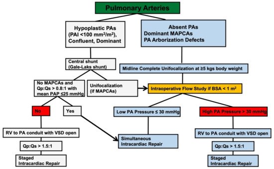
2.2. Unifocalization

The unifocalization technique, particularly in single-stage constitutes the approach of choice advocated by the Stanford group [16]. Access to the mediastinum during one-stage unifocalization is achieved via midline sternotomy. All collaterals originating from the descending thoracic aorta and/or from the epiaortic vessels are dissected, separated from their origin and anastomosed to each other and/or to the native pulmonary arteries to create a new pulmonary vascular tree that is adequately expanded through the use of homograft tissue. At this point, possibly after pulmonary pressure testing, the VSD is closed and the RV is connected to the unifocalized pulmonary vascular tree with a valved homograft conduit. In a subset of patients unsuitable for VSD closure, palliation is performed with shunt placement on the unifocalized pulmonary arteries [17]. Early removal of MAPCAs from the systemic circulation prevents both MAPCA degeneration and native pulmonary artery regression. Therefore, early unifocalization provides vascularization of all healthy pulmonary segments using both native pulmonary arteries and collaterals as source material.
The institutional approach for PA/VSD/MAPCAs is similar to that of the Stanford group. Pulmonary arterial rehabilitation is performed in all patients with confluent hypoplastic (i.e., pulmonary arterial index less than 100 mm2/m2) [18] but dominant (i.e., distributed over most pulmonary segments) pulmonary arteries with eventual MAPCAs supplying the areas not perfused by the true pulmonary arteries (Figure 1).
Unifocalization and rehabilitation focus on mobilization of collateral arteries and growth of native pulmonary vessels, respectively. Regardless of the strategy, the results have altered the natural history of the disease, with a complete cure rate of approximately 80% and low early and late mortality. In the experience to date, the ability to achieve definitive intracardiac repair is the critical factor for improved survival and adequate systolic RV performance at midterm follow-up [15]. Patients with the most unfavorable anatomy (absent central pulmonary arteries and hypoplastic MAPCAs) remain a challenge.

2.3. The Role of the Intraoperative Flow-Study in Assessing the Feasibility of Concomitant VSD Closure

The use of the intraoperative flow study as a functional measure of pulmonary vascular performance and suitability for VSD closure was first reported by Reddy and colleagues [19]. In October 1996 the researcher's group adopted the intraoperative flow study to assess the feasibility of VSD closure at the time of unifocalization and since then it has been performed according to an unchanged standardized protocol. The neopulmonary arterial confluence is cannulated to be perfused via a line derived from the arterial port of the oxygenator, and a pressure recording is placed simultaneously. The test is started with lungs deflated and the heart beating and fully vented. By a stepwise 25% increase, a maximum flow of oxygenated blood equal to 2.5 L · min−1 · m−2 is pumped into the reconstructed pulmonary bed and the mean pulmonary artery pressure (mPAP) is simultaneously recorded. An mPAP value of 30 mm Hg or less is selected for VSD closure, predicting adequate postoperative RV pressure. On the other hand, if mPAP rises above 30 mm Hg during the incremental steps of the flow study, the test is terminated, intracardiac repair is not performed and either a RV to PA conduit or a central shunt is placed based on individual patient assessment.
Proper performance of an intraoperative flow study can be problematic in patients with a body surface area greater than 1 m2 because excessive priming of the pump is required and the ability of the pump oxygenator to maintain adequate both systemic and pulmonary blood flow is compromised. Late presenting humanitarian referrals encompassing more than 1 m2 of body surface area are subject to preoperative CMR imaging with the Qp:Qs ratio calculation and VSD closure decided accordingly if the Qp:Qs value exceeds 1.5:1.

2.4. Criteria for Delayed VSD Closure

Patients who did not receive VSD closure in the first instance are re-evaluated, also on the basis of the individual clinical condition, 6–12 months after surgery by cardiac catheterization, possibly associated with CMR. Intracardiac repair is indicated in the presence of Qp:Qs greater than 1.5, if their source of pulmonary blood flow is an RV to PA connection, or in the presence of Qp:Qs greater than 0.8 and mPAP less than 25 mmHg, if their source of pulmonary blood flow is a systemic-to-pulmonary shunt.

References

  1. Ryan, J.R.; Moe, T.G.; Richardson, R.; Frakes, D.H.; Nigro, J.J.; Pophal, S. A Novel Approach to Neonatal Management of Tetralogy of Fallot, With Pulmonary Atresia, and Multiple Aortopulmonary Collaterals. JACC Cardiovasc. Imaging 2015, 8, 103–104.
  2. Liu, J.; Li, H.; Liu, Z.; Wu, Q.; Xu, Y. Complete Preoperative Evaluation of Pulmonary Atresia with Ventricular Septal Defect with Multi-Detector Computed Tomography. PLoS ONE 2016, 11, e0146380.
  3. Secinaro, A.; Curione, D. Congenital heart disease in children. In Medical Radiology—Diagnostic Imaging; Springer: Berlin/Heidelberg, Germany, 2019; pp. 987–1009. ISSN1 2197-4187. ISSN2 0942-5373.
  4. Ciancarella, P.; Ciliberti, P.; Santangelo, T.P.; Secchi, F.; Stagnaro, N.; Secinaro, A. Noninvasive imaging of congenital cardiovascular defects. Radiol. Med. 2020, 125, 1167–1185.
  5. Secinaro, A.; Curione, D.; Mortensen, K.H.; Santangelo, T.P.; Ciancarella, P.; Napolitano, C.; Del Pasqua, A.; Taylor, A.M.; Ciliberti, P. Dual-source computed tomography coronary artery imaging in children. Pediatr. Radiol. 2019, 49, 1823–1839.
  6. Malayeri, A.A.; Spevak, P.J.; Zimmerman, S.L. Utility of a High-Resolution 3D MRI Sequence (3D-SPACE) for Evaluation of Congenital Heart Disease. Pediatr. Cardiol. 2015, 36, 1510–1514.
  7. Niu, J.; Profirovic, J.; Pan, H.; Vaiskunaite, R.; Voyno-Yasenetskaya, T. G protein βγ subunits stimulate p114RhoGEF, a guanine nucleotide exchange factor for RhoA and Rac1: Regulation of cell shape and reactive oxygen species production. Circ. Res. 2003, 93, 848–856.
  8. Schicchi, N.; Secinaro, A.; Muscogiuri, G.; Ciliberti, P.; Leonardi, B.; Santangelo, T.P.; Napolitano, C.; Agliata, G.; Basile, M.C.; Guidi, F.; et al. Multicenter review: Role of cardiovascular magnetic resonance in diagnostic evaluation, pre-procedural planning and follow-up for patients with congenital heart disease. Radiol. Med. 2015, 121, 342–351.
  9. Grosse-Wortmann, L.; Yoo, S.-J.; Van Arsdell, G.; Chetan, D.; Macdonald, C.; Benson, L.; Honjo, O. Preoperative total pulmonary blood flow predicts right ventricular pressure in patients early after complete repair of tetralogy of Fallot and pulmonary atresia with major aortopulmonary collateral arteries. J. Thorac. Cardiovasc. Surg. 2013, 146, 1185–1190.
  10. Tack, P.; Victor, J.; Gemmel, P.; Annemans, L. 3D-printing techniques in a medical setting: A systematic literature review. Biomed. Eng. Online 2016, 15, 115.
  11. Byrne, N.; Forte, M.V.; Tandon, A.; Valverde, I.; Hussain, T. A systematic review of image segmentation methodology, used in the additive manufacture of patient-specific 3D printed models of the cardiovascular system. JRSM Cardiovasc. Dis. 2016, 5, 1691.
  12. Gates, R.N.; Laks, H.; Johnson, K. Side-to-Side Aorto–Gore-Tex Central Shunt. Ann. Thorac. Surg. 1998, 65, 515–516.
  13. Mumtaz, M.A.; Rosenthal, G.; Qureshi, A.; Prieto, L.; Preminger, T.; Lorber, R.; Latson, L.; Duncan, B.W. Melbourne Shunt Promotes Growth of Diminutive Central Pulmonary Arteries in Patients With Pulmonary Atresia, Ventricular Septal Defect, and Systemic-to-Pulmonary Collateral Arteries. Ann. Thorac. Surg. 2008, 85, 2079–2084.
  14. D’Udekem, Y.; Alphonso, N.; Nørgaard, M.A.; Cochrane, A.D.; Grigg, L.E.; Wilkinson, J.L.; Brizard, C.P. Pulmonary atresia with ventricular septal defects and major aortopulmonary collateral arteries: Unifocalization brings no long-term benefits. J. Thorac. Cardiovasc. Surg. 2005, 130, 1496–1502.
  15. Carotti, A.; Albanese, S.; Filippelli, S.; Ravà, L.; Guccione, P.; Pongiglione, G.; Di Donato, R.M. Determinants of outcome after surgical treatment of pulmonary atresia with ventricular septal defect and major aortopulmonary collateral arteries. J. Thorac. Cardiovasc. Surg. 2010, 140, 1092–1103.
  16. Reddy, V.; Liddicoat, J.R.; Hanley, F.L. Midline one-stage complete unifocalization and repair of pulmonary atresia with ventricular septal defect and major aortopulmonary collaterals. J. Thorac. Cardiovasc. Surg. 1995, 109, 832–845.
  17. Mainwaring, R.D.; Patrick, W.L.; Roth, S.J.; Kamra, K.; Wise-Faberowski, L.; Palmon, M.; Hanley, F.L. Surgical algorithm and results for repair of pulmonary atresia with ventricular septal defect and major aortopulmonary collaterals. J. Thorac. Cardiovasc. Surg. 2018, 156, 1194–1204.
  18. Itatani, K.; Miyaji, K.; Nakahata, Y.; Ohara, K.; Takamoto, S.; Ishii, M. The lower limit of the pulmonary artery index for the extracardiac Fontan circulation. J. Thorac. Cardiovasc. Surg. 2011, 142, 127–135.
  19. Reddy, V.; Petrossian, E.; McElhinney, D.B.; Moore, P.; Teitel, D.F.; Hanley, F.L. One-stage complete unifocalization in infants: When should the ventricular septal defect be closed? J. Thorac. Cardiovasc. Surg. 1997, 113, 858–868.
More
Information
Contributors MDPI registered users' name will be linked to their SciProfiles pages. To register with us, please refer to https://encyclopedia.pub/register : , , ,
View Times: 488
Revisions: 2 times (View History)
Update Date: 12 Apr 2022
1000/1000
Video Production Service