Submitted Successfully!
To reward your contribution, here is a gift for you: A free trial for our video production service.
Thank you for your contribution! You can also upload a video entry or images related to this topic.
Version Summary Created by Modification Content Size Created at Operation
1 -- 2097 2022-04-09 16:39:30 |
2 format -1 word(s) 2096 2022-04-11 05:11:38 |

Video Upload Options

Do you have a full video?

Confirm

Are you sure to Delete?
Cite
If you have any further questions, please contact Encyclopedia Editorial Office.
Yang, Y.; Guo, Y. Gap Junctions in Atrial Fibrillation. Encyclopedia. Available online: https://encyclopedia.pub/entry/21533 (accessed on 03 July 2024).
Yang Y, Guo Y. Gap Junctions in Atrial Fibrillation. Encyclopedia. Available at: https://encyclopedia.pub/entry/21533. Accessed July 03, 2024.
Yang, Yi-Qing, Yu-Han Guo. "Gap Junctions in Atrial Fibrillation" Encyclopedia, https://encyclopedia.pub/entry/21533 (accessed July 03, 2024).
Yang, Y., & Guo, Y. (2022, April 09). Gap Junctions in Atrial Fibrillation. In Encyclopedia. https://encyclopedia.pub/entry/21533
Yang, Yi-Qing and Yu-Han Guo. "Gap Junctions in Atrial Fibrillation." Encyclopedia. Web. 09 April, 2022.
Gap Junctions in Atrial Fibrillation
Edit

Atrial fibrillation (AF) represents the most common type of clinical cardiac arrhythmia worldwide and contributes to substantial morbidity, mortality and socioeconomic burden. Aggregating evidence highlights the strong genetic basis of AF. In addition to chromosomal abnormalities, pathogenic mutations in over 50 genes have been causally linked to AF, of which the majority encode ion channels, cardiac structural proteins, transcription factors and gap junction channels. In the heart, gap junctions comprised of connexins (Cxs) form intercellular pathways responsible for electrical coupling and rapid coordinated action potential propagation between adjacent cardiomyocytes. Among the 21 isoforms of connexins already identified in the mammal genomes, 5 isoforms (Cx37, Cx40, Cx43, Cx45 and Cx46) are expressed in human heart. Abnormal electrical coupling between cardiomyocytes caused by structural remodeling of gap junction channels (alterations in connexin distribution and protein levels) has been associated with enhanced susceptibility to AF and recent studies have revealed multiple causative mutations or polymorphisms in 4 isoforms of connexins predisposing to AF.

gap junctions connexins atrial fibrillation

1. Introduction

Atrial fibrillation (AF) is the most common cardiac rhythm disturbance, currently afflicting approximately 46.3 million individuals worldwide [1]. The prevalence of AF exponentially increases with age and reaches up to roughly 9% in individuals older than 80 years of age [2]. Comorbidities of AF substantially contribute to morbidity, mortality and medical care expenditure. The disease leads to severe major adverse cardiovascular events [3] and roughly 15% of all strokes [4]. AF is also associated with a three-fold increased risk for heart failure and a two-fold increased risk for all-cause mortality that is independent of comorbidities [5]. Nevertheless, the mechanisms causing and sustaining AF are multifactorial and incompletely understood. At present, it is still a great challenge to terminate AF.
AF is characterized by rapid and erratic atrial electrical activity, resulted from dynamic interplays among multiple electrophysiological, structural, inflammatory and genetic factors [6]. The disease is frequently secondary to diverse cardiovascular and systemic diseases such as hypertension, myocardial infarction, valvular dysfunction, heart failure and diabetes [7]. However, 2~16% of overall AF are defined as idiopathic AF for not having any identifiable underlying conditions [8]. Furthermore, approximately 30% of AF cases are of familial patterns [9], implying the genetic basis of AF. All these findings have provided substantial new insights into the mechanisms of AF. Up to now, mutations in over 50 genes have been causally linked to AF, most of which encode ion channels, structural proteins, transcription factors and gap junction channels [10].
Cardiac gap junctions are located at the intercalated disks between cardiomyocytes, forming intercellular pathways for the orderly propagation of electrical activation responsible for synchronized myocardial contraction [11]. The normal cardiac rhythm fundamentally depends on the cell-to-cell electrical coupling of cardiomyocytes through gap junctions. Therefore, it is possible that dysfunction or impairment of gap junctions my lead to cardiac arrhythmias. Indeed, a large amount of research has associated gap junctions and their structural components, connexins, with AF susceptibility.

2. Structure and Function of Gap Junctions

Gap junctions are clusters of transmembrane channels that enable the direct cytoplasmic exchange of ions or small metabolites (<1 kDa in size) between neighboring cells. They are constructed by the juxtaposition of a pair of hemichannels (connexons) from adjoining cells. Under pathological conditions or specific circumstances such as paracrine signaling [12], hemichannels that span the entire depth of the plasma membrane may also function as transmembrane channels in unopposed cells, allowing the permeation of ions and small metabolites [13] (Figure 1a). Each hemichannel is formed by oligomerization of six connexins (Cxs) surrounding a central aqueous pore [14]. Connexins are tetra-span transmembrane domain proteins with four highly conserved transmembrane domains (M1–M4) and intracellular N- and C-terminus (NT and CT), linked by two extracellular loops (E1 and E2) and one cytoplasmic loop (CL) (Figure 1b).
Figure 1. The schematic diagrams of gap junctions, hemichannels and connexins. (a) Two connexons from neighboring cells, which are formed by the oligomerization of six connexin subunits, can assemble into a gap junction. Connexons may also function as hemichannels under specific conditions; (b) Connexins are transmembrane proteins constituted by four transmembrane domains (M1–4) and intracellular N- and C-terminus (NT and CT), linked by two extracellular loops (E1 and E2) and one cytoplasmic loop (CL).
Practically all cells in solid tissues are linked by gap junctions and the majority of cells co-express more than one type of connexin [15]. Currently, 21 isoforms of connexin have been identified in the human genome and 20 in the murine genome. They are divided into five subfamilies (α, β, γ, δ and ε or GJA, GJB, GJC, GJD and GJE) according to sequence homology [16]. Identical Cxs in both docked hemichannels comprise homotypic gap junction channels while mixed Cxs constitute heteromeric gap junction channels. In addition, two different homotypic hemichannels make up a heterotypic gap junction channel. Particular connexin types or combination of connexin types in hemichannels leads to distinction in the physiological properties of gap junction channels [17].
The main functions of gap junctions are to share small nutrients or signaling molecules among groups of cells and permeate ions across electrically excitable cells to coordinate electrical and mechanical actions in tissues such as heart, neurons and smooth muscle [12]. Gating of gap junction intercellular channels is dynamically regulated by multiple factors including transjunctional voltage, intracellular calcium concentration, pH, phosphorylation and other post-translational modifications [15].

3. Subtypes of Cardiac Connexins

To date, six principal connexins are found expressed in human heart (Cx37, Cx40, Cx43, Cx45 and Cx46) [18][19]. Cx43 is the predominant cardiac connexin which is mainly distributed in the atrial and ventricular cardiomyocytes [20]. It is less expressed in the conduction system and not expressed in the sinoatrial or atrioventricular node [20][21]. Cx40, another significant cardiac connexin, is restricted primarily to the atrial tissue and the ventricular conduction system and is 2~3 fold higher in the right atrium than the left [18][21]. Cx45 is the first connexin expressed during early stages of cardiovascular development [20]. However, in the adult heart, it is expressed predominantly in the conduction system while expressed in low quantities in both ventricles and atria with a slightly higher level in the atrium than the ventricle [22]. Cx37 is expressed mainly in the vascular endothelium [21]. In addition, the less studied cardiac connexin Cx46 has been found in the atrial and ventricular myocytes in the human heart [23].
The biologic functions of these cardiac connexins are currently being elucidated. In addition to allowing rapid propagation of action potentials mediating coordinated myocardial contraction in the adult heart, these connexins are crucial for heart development through mediating the exchange of critical factors between cells. In neonatal mice, Cx43 knockout leads to death at birth because of right ventricular outflow tract malformations [24]. It was also reported that cardiac malformations are prevalent in Cx40-deficient mice [25]. Likewise, lacking Cx45 in mice predisposes to embryonic death for sinus node dysfunction and atrial arrhythmia [26]. In addition, several recent studies have linked Cx37 deficiency to both venous and lymphatic valve malformation [27][28].

4. Changes of Gap Junctions/Connexins in the Pathogenesis of AF

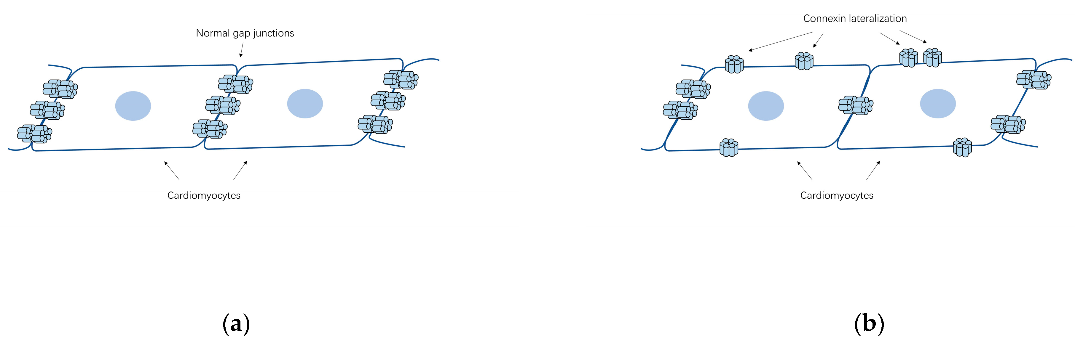
4.1. Gap Junction Remodeling

The process of gap junction remodeling, characterized by alterations in gap junction channel abundance, subcellular distribution, permeability (determined by the phosphorylation status of the constituent connexins) and conductance [29], is an important portion of the heart adaptive remodeling during cardiac diseased states. It is associated closely with cardiac electrical remodeling which leads to alterations in conduction [30]. The impaired gap junction intercellular communication is increasingly observed in human cardiovascular diseases such as heart failure, ischemic heart disease and cardiac arrythmias [17]. Nevertheless, the explicit relationship between gap junction remodeling and the pathogenesis of these diseases still needs further clarifications.
As mentioned above, cardiac gap junction channels play crucial roles in direct intercellular communication and myocardial synchronization in adult heart. Accordingly, any form of cardiac gap junction remodeling may disturb these functions and thus contribute to arrhythmia vulnerability. In fact, the onset of arrhythmias involves the interaction of gap junction intercellular communication, cell membrane excitability and the structures of cell and tissue [17]. Ventricular myocytes contain predominantly Cx43 isoform except for rather low levels of Cx45 and Cx46 while multiple isoforms of connexins (Cx40, Cx43 and Cx45) are expressed in the atria, the most connexin-heterogeneous tissue of the heart [18], causing diversity in types and physiological characteristics of atrial gap junctions. That is to say, it is much more difficult to figure out the patterns of gap junction remodeling of atrial arrhythmias than ventricular arrythmias.

4.2. Abundance and Distribution of Connexins Associated with AF

Ever since Spach et al. [31] first highlighted alterations in gap junction structure and function as potential therapeutic targets for AF, a large number of studies have been conducted on the role of connexins in the pathogenesis of AF [32]. Most of these studies focused on changes in the abundance and distribution of connexins in the AF models or patients.
Changes in Cx40/Cx43 quantity caused by AF are uncertain because of the inconsistency of the results. The abundance of Cx43 seems to be dependent on types of AF while the amount of Cx40 caused by AF may increase with Cx40 lateralization, or reduce significantly, or be indistinguishable from sinus rhythm, or be dependent on extracellular Ca2+ level [14]. Wetzel and his colleagues [33] found increases in both Cx40 and Cx43 concentration in left atrial tissue of lone AF patients and AF patients with mitral valve disease, when compared with sinus rhythm. In another study aiming at chronic AF [34], researchers observed no significant change in Cx43 content in patients’ atrial tissues while Cx40 was enhanced. Moreover, Kanagaratnam et al. [35] observed a reduction of Cx40 in chronic AF with complex activation.
Apart from changes in their quantity, the lateralization of Cx40/Cx43 from cell poles to lateral margins (Figure 2b) appears to be a general AF-associated alteration. In Polontchouk and his colleagues’ research [34], an increase in both Cx40 and Cx43 at the lateral membrane of human and rat atrial cells was found, indicating that AF might be accompanied by the spatial remodeling of gap junctions. Similar results were observed by Kostin et al. afterwards [36]. In their research, lateralization of Cx43, Cx40 and N-cadherin, reduction of Cx43 level and heterogeneous distribution of Cx40 together with augmentation of fibrosis were found in AF patients, constituting the anatomic substrates of AF [36]. In addition, Dhein et al. detected AF induced Cx40/Cx43 lateralization together with enhanced lateral conduction velocity in the left atrial [37]. These redistributed connexins lost their characteristic to assemble into gap junctions coupling with adjacent cells but may function as hemichannels which allow ionic currents and small metabolites [38][39]. It remains unclear what are the main factors that lead to the spatial remodeling of the gap junctions. However, AF causes a rather complicated intracellular remodeling which may constitute the basis for the redistribution of gap junctions and the intracellular remodeling of the Golgi-microtubular apparatus, where the connexins oligomerize, is also included [40]. In fact, the fragmentation and a decreased fragment size of the Golgi apparatus were observed in patients with chronic AF [41].
Figure 2. The schematic diagrams of the lateralization of cardiac connexins in AF. (a) Schema of the cell-to-cell connections by gap junctions in normal heart; (b) The lateralization of connexins from cell poles to lateral margins, which can be related to AF.
In brief, the pathogenesis of AF may be associated with the lateralization of cardiac connexins but the role of connexin abundance remains indistinct.

4.3. Changes in Atrial Connexin Expression Regulated by Transcription Factors

Abnormal transcriptional regulation of gene expression may be a characteristic feature in the occurrence of heart diseases. Transcription factors regulating cardiac connexins can be roughly classified into two categories based on the ubiquity or cardiac cell specificity of their binding sites. The former include Sp1/Sp3 and activator protein 1 (AP-1) while the latter include cardiac specific transcription factors such as NKX2-5, Shox2, T-box transcription factors and GATA family [42]. Some of these transcription factors might contribute to AF through regulating the expression of atrial connexins.
Ma et al. [43] sequenced a series of candidate genes in 139 Chinese patients with early-onset AF and found four missense TBX5 mutations. Subsequent functional experiments indicated that these mutations increased the expression of Cx40 and NPPA without altering the expression of cardiac structural protein genes in rat atrial myocytes. In the zebrafish model, the overexpression of TBX5 p.R355C mutation caused paroxysmal AF. Later in 2019, a gain-of-function mutation of the paired-like homeodomain transcription factor 2 (PITX2) were identified by Mechakra et al. in 1 out of 60 unrelated idiopathic AF patients [44]. The mutation was then introduced into HL-1 cells and increased mRNA level of GJA5 (coding for connexin 40, 3.1-fold increase) and GJA1 (coding for connexin 43, 2.1-fold) was observed.
In addition, phosphorylation of transcription factors can also affect connexin expression. For example, the activation of c-Jun (AP-1) N-terminal kinase has been reported to cause decreased Cx43 level in HL-1 cells [45]. Reduced conduction velocity and increased incidence of irregular rapid spontaneous activities that may contribute to AF were also observed.

References

  1. Benjamin, E.J.; Muntner, P.; Alonso, A.; Bittencourt, M.S.; Callaway, C.W.; Carson, A.P.; Chamberlain, A.M.; Chang, A.R.; Cheng, S.; Das, S.R.; et al. Heart Disease and Stroke Statistics-2019 Update: A Report From the American Heart Association. Circulation 2019, 139, e56–e528.
  2. Go, A.S.; Hylek, E.M.; Phillips, K.A.; Chang, Y.; Henault, L.E.; Selby, J.V.; Singer, D.E. Prevalence of diagnosed atrial fibrillation in adults: National implications for rhythm management and stroke prevention: The AnTicoagulation and Risk Factors in Atrial Fibrillation (ATRIA) Study. JAMA 2001, 285, 2370–2375.
  3. Wijesurendra, R.S.; Casadei, B. Mechanisms of atrial fibrillation. Heart 2019, 105, 1860–1867.
  4. Fuster, V.; Ryden, L.E.; Cannom, D.S.; Crijns, H.J.; Curtis, A.B.; Ellenbogen, K.A.; Halperin, J.L.; Kay, G.N.; Le Huezey, J.Y.; Lowe, J.E.; et al. American College of Cardiology Foundation/American Heart Association Task Force: 2011 ACCF/AHA/HRS focused updates incorporated into the ACC/AHA/ESC 2006 guidelines for the management of patients with atrial fibrillation: A report of the American College of Cardiology Foundation/American Heart Association Task Force on practice guidelines. Circulation 2011, 123, e269–e367.
  5. Zimetbaum, P. Atrial Fibrillation. Ann. Intern. Med. 2017, 166, ITC33–ITC48.
  6. Saffitz, J.E. Connexins, conduction, and atrial fibrillation. N. Engl. J. Med. 2006, 354, 2712–2714.
  7. January, C.T.; Wann, L.S.; Alpert, J.S.; Calkins, H.; Cigarroa, J.E.; Cleveland, J.C., Jr.; Conti, J.B.; Ellinor, P.T.; Ezekowitz, M.D.; Field, M.E.; et al. 2014 AHA/ACC/HRS guideline for the management of patients with atrial fibrillation: A report of the American College of Cardiology/American Heart Association Task Force on practice guidelines and the Heart Rhythm Society. Circulation 2014, 130, e199–e267.
  8. Campuzano, O.; Perez-Serra, A.; Iglesias, A.; Brugada, R. Genetic basis of atrial fibrillation. Genes Dis. 2016, 3, 257–262.
  9. Fox, C.S.; Parise, H.; D’Agostino, R.B.; Lloyd-Jones, D.M.; Vasan, R.S.; Wang, T.J.; Levy, D.; Wolf, P.A.; Benjamin, E.J. Parental atrial fibrillation as a risk factor for atrial fibrillation in offspring. J. Am. Med. Assoc. 2004, 291, 2851–2855.
  10. Guo, X.J.; Qiu, X.B.; Wang, J.; Guo, Y.H.; Yang, C.X.; Li, L.; Gao, R.F.; Ke, Z.P.; Di, R.M.; Sun, Y.M.; et al. PRRX1 Loss-of-Function Mutations Underlying Familial Atrial Fibrillation. J. Am. Heart Assoc. 2021, 10, e023517.
  11. Severs, N.J.; Dupont, E.; Thomas, N.; Kaba, R.; Rothery, S.; Jain, R.; Sharpey, K.; Fry, C.H. Alterations in cardiac connexin expression in cardiomyopathies. Adv. Cardiol. 2006, 42, 228–242.
  12. Goodenough, D.A.; Paul, D.L. Gap junctions. Cold Spring Harb. Perspect. Biol. 2009, 1, a002576.
  13. Saez, J.C.; Leybaert, L. Hunting for connexin hemichannels. FEBS Lett. 2014, 588, 1205–1211.
  14. Chaldoupi, S.M.; Loh, P.; Hauer, R.N.; de Bakker, J.M.; van Rijen, H.V. The role of connexin40 in atrial fibrillation. Cardiovasc. Res. 2009, 84, 15–23.
  15. Nielsen, M.S.; Axelsen, L.N.; Sorgen, P.L.; Verma, V.; Delmar, M.; Holstein-Rathlou, N.H. Gap junctions. Compr. Physiol. 2012, 2, 1981–2035.
  16. Beyer, E.C.; Berthoud, V.M. Gap junction gene and protein families: Connexins, innexins, and pannexins. Biochim. Biophys. Acta Biomembr. 2018, 1860, 5–8.
  17. Severs, N.J.; Bruce, A.F.; Dupont, E.; Rothery, S. Remodelling of gap junctions and connexin expression in diseased myocardium. Cardiovasc. Res. 2008, 80, 9–19.
  18. Duffy, H.S.; Wit, A.L. Is there a role for remodeled connexins in AF? No simple answers. J. Mol. Cell. Cardiol. 2008, 44, 4–13.
  19. Ortega, A.; Tarazon, E.; Gil-Cayuela, C.; Garcia-Manzanares, M.; Martinez-Dolz, L.; Lago, F.; Gonzalez-Juanatey, J.R.; Cinca, J.; Jorge, E.; Portoles, M.; et al. Intercalated disc in failing hearts from patients with dilated cardiomyopathy: Its role in the depressed left ventricular function. PLoS ONE 2017, 12, e0185062.
  20. Desplantez, T. Cardiac Cx43, Cx40 and Cx45 co-assembling: Involvement of connexins epitopes in formation of hemichannels and Gap junction channels. BMC Cell. Biol. 2017, 18, 3.
  21. van der Velden, H.M.; Jongsma, H.J. Cardiac gap junctions and connexins: Their role in atrial fibrillation and potential as therapeutic targets. Cardiovasc. Res. 2002, 54, 270–279.
  22. Severs, N.J.; Coppen, S.R.; Dupont, E.; Yeh, H.I.; Ko, Y.S.; Matsushita, T. Gap junction alterations in human cardiac disease. Cardiovasc. Res. 2004, 62, 368–377.
  23. Cheng, S.H.; Shakespeare, T.; Mui, R.; White, T.W.; Valdimarsson, G. Connexin 48.5 is required for normal cardiovascular function and lens development in zebrafish embryos. J. Biol. Chem. 2004, 279, 36993–37003.
  24. Reaume, A.G.; Desousa, P.A.; Kulkarni, S.; Langille, B.L.; Zhu, D.G.; Davies, T.C.; Juneja, S.C.; Kidder, G.M.; Rossant, J. Cardiac Malformation in Neonatal Mice Lacking Connexin43. Science 1995, 267, 1831–1834.
  25. Gu, H.; Smith, F.C.; Taffet, S.M.; Delmar, M. High incidence of cardiac malformations in connexin40-deficient mice. Circ. Res. 2003, 93, 201–206.
  26. Seki, A.; Ishikawa, T.; Daumy, X.; Mishima, H.; Barc, J.; Sasaki, R.; Nishii, K.; Saito, K.; Urano, M.; Ohno, S.; et al. Progressive Atrial Conduction Defects Associated With Bone Malformation Caused by a Connexin-45 Mutation. J. Am. Coll. Cardiol. 2017, 70, 358–370.
  27. Munger, S.J.; Kanady, J.D.; Simon, A.M. Absence of venous valves in mice lacking Connexin37. Dev. Biol. 2013, 373, 338–348.
  28. Kanady, J.D.; Munger, S.J.; Witte, M.H.; Simon, A.M. Combining Foxc2 and Connexin37 deletions in mice leads to severe defects in lymphatic vascular growth and remodeling. Dev. Biol. 2015, 405, 33–46.
  29. Martins-Marques, T.; Catarino, S.; Goncalves, A.; Miranda-Silva, D.; Goncalves, L.; Antunes, P.; Coutinho, G.; Moreira, A.L.; Pires, I.F.; Girao, H. EHD1 Modulates Cx43 Gap Junction Remodeling Associated With Cardiac Diseases. Circ. Res. 2020, 126, E97–E113.
  30. Tribulova, N.; Knezl, V.; Okruhlicova, L.; Slezak, J. Myocardial Gap Junctions: Targets for Novel Approaches in the Prevention of Life-Threatening Cardiac Arrhythmias. Physiol Res 2008, 57, S1–S13.
  31. Spach, M.S.; Starmer, C.F. Altering the Topology of Gap-Junctions—A Major Therapeutic Target for Atrial-Fibrillation. Cardiovasc. Res. 1995, 30, 337–344.
  32. Kato, T.; Iwasaki, Y.K.; Nattel, S. Connexins and Atrial Fibrillation Filling in the Gaps. Circulation 2012, 125, 203–206.
  33. Wetzel, U.; Boldt, A.; Lauschke, J.; Weigl, J.; Schirdewahn, P.; Dorszewski, A.; Doll, N.; Hindricks, G.; Dhein, S.; Kottkamp, H. Expression of connexins 40 and 43 in human left atrium in atrial fibrillation of different aetiologies. Heart 2005, 91, 166–170.
  34. Polontchouk, L.; Haefliger, J.A.; Ebelt, B.; Schaefer, T.; Stuhlmann, D.; Mehlhorn, U.; Kuhn-Regnier, F.; De Vivie, E.R.; Dhein, S. Effects of chronic atrial fibrillation on gap junction distribution in human and rat atria. J. Am. Coll. Cardiol. 2001, 38, 883–891.
  35. Kanagaratnam, P.; Cherian, A.; Stanbridge, R.D.L.; Glenville, B.; Severs, N.J.; Peters, N.S. Relationship between connexins and atrial activation during human atrial fibrillation. J. Cardiovasc. Electr. 2004, 15, 206–213.
  36. Kostin, S.; Klein, G.; Szalay, Z.; Hein, S.; Bauer, E.P.; Schaper, J. Structural correlate of atrial fibrillation in human patients. Cardiovasc. Res. 2002, 54, 361–379.
  37. Dhein, S.; Rothe, S.; Busch, A.; Rojas Gomez, D.M.; Boldt, A.; Reutemann, A.; Seidel, T.; Salameh, A.; Pfannmuller, B.; Rastan, A.; et al. Effects of metoprolol therapy on cardiac gap junction remodelling and conduction in human chronic atrial fibrillation. Br. J. Pharmacol. 2011, 164, 607–616.
  38. Rucker-Martin, C.; Milliez, P.; Tan, S.; Decrouy, X.; Recouvreur, M.; Vranckx, R.; Delcayre, C.; Renaud, J.F.; Dunia, I.; Segretain, D.; et al. Chronic hemodynamic overload of the atria is an important factor for gap junction remodeling in human and rat hearts. Cardiovasc. Res. 2006, 72, 69–79.
  39. Fakuade, F.E.; Tomsits, P.; Voigt, N. Connexin hemichannels in atrial fibrillation: Orphaned and irrelevant? Cardiovasc. Res. 2021, 117, 4–6.
  40. Dhein, S.; Salameh, A. Remodeling of Cardiac Gap Junctional Cell-Cell Coupling. Cells 2021, 10, 2422.
  41. Jungk, L.; Franke, H.; Salameh, A.; Dhein, S. Golgi Fragmentation in Human Patients with Chronic Atrial Fibrillation: A New Aspect of Remodeling. Thorac. Cardiov. Surg. 2019, 67, 98–106.
  42. Oyamada, M.; Takebe, K.; Oyamada, Y. Regulation of connexin expression by transcription factors and epigenetic mechanisms. Biochim. Biophys. Acta 2013, 1828, 118–133.
  43. Ma, J.F.; Yang, F.; Mahida, S.N.; Zhao, L.; Chen, X.; Zhang, M.L.; Sun, Z.; Yao, Y.; Zhang, Y.X.; Zheng, G.Y.; et al. TBX5 mutations contribute to early-onset atrial fibrillation in Chinese and Caucasians. Cardiovasc. Res. 2016, 109, 442–450.
  44. Mechakra, A.; Footz, T.; Walter, M.; Aranega, A.; Hernandez-Torres, F.; Morel, E.; Millat, G.; Yang, Y.Q.; Chahine, M.; Chevalier, P.; et al. A Novel PITX2c Gain-of-Function Mutation, p.Met207Val, in Patients With Familial Atrial Fibrillation. Am. J. Cardiol. 2019, 123, 787–793.
  45. Yan, J.; Kong, W.; Zhang, Q.; Beyer, E.C.; Walcott, G.; Fast, V.G.; Ai, X. c-Jun N-terminal kinase activation contributes to reduced connexin43 and development of atrial arrhythmias. Cardiovasc. Res. 2013, 97, 589–597.
More
Information
Contributors MDPI registered users' name will be linked to their SciProfiles pages. To register with us, please refer to https://encyclopedia.pub/register : ,
View Times: 494
Revisions: 2 times (View History)
Update Date: 11 Apr 2022
1000/1000
Video Production Service