 +1 credit
| Version | Summary | Created by | Modification | Content Size | Created at | Operation |
|---|---|---|---|---|---|---|
| 1 | Ramar Thangam | -- | 3489 | 2022-04-06 10:36:05 | | | |
| 2 | Peter Tang | -30 word(s) | 3459 | 2022-04-08 05:46:54 | | | | |
| 3 | Peter Tang | Meta information modification | 3459 | 2022-04-08 05:47:22 | | |
Video Upload Options
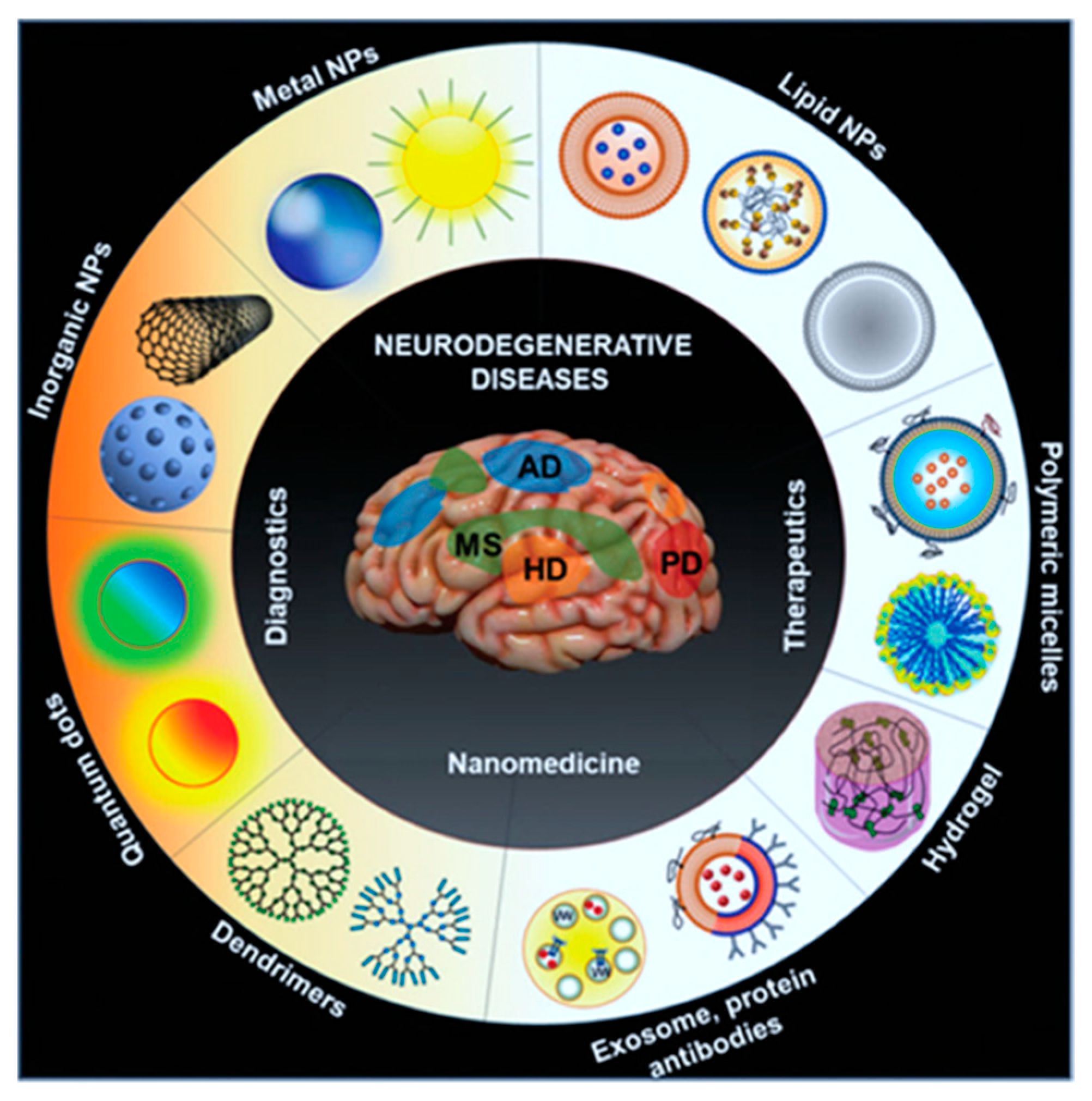
Theranostic nanoparticles in molecular imaging significantly impact non-invasive strategies to understand biological and biochemical events in intact cells within living subjects. It plays a prominent role in disease diagnosis and therapeutic monitoring outcomes in vivo. The theranostic application of nanomaterials can be classified into morphological and functional imaging based on their roles in image contrast abilities during applicable imaging methods.
1. Introduction

|
Target Disease |
Nanoformulation |
Model Organism |
Therapeutic Outcome |
Ref. No. |
|---|---|---|---|---|
|
Parkinsons |
Selegiline nanoemulsion |
Rat |
Intranasally administered selegiline nanoemulsion improved the behavioral activities in comparison to oral administration. |
[54] |
|
Parkinsons |
Resveratrol and curcumin nanoemulsion |
Sheep |
Intranasal delivery of hyaluronic acid-based lipidic nanoemulsion proven as a successful carrier to enhance the solubility, stability, and brain targetability of polyphenols. |
[55] |
|
Alzheimer’s disease |
Rivastigmine-loaded nanoemulsion |
Rat |
Achieved higher drug delivery to the brain with enhanced safety, non-toxic and non-irritating to the nasal mucosa. |
[56] |
|
Alzheimer’s disease |
Donepezil nanoemulsion |
Pig |
Effective strategy using polymers improved the adhesion and penetration of the drug through the nasal mucosa. |
[57] |
|
Alzheimer’s disease |
Cholera Toxin B subunit-based nanoparticles |
Mice |
Delivered nanosystem exhibited a notable performance in accumulating in the hippocampus that further showed an excellent magnetic resonance imaging (MRI) potential in vivo. |
[3] |
|
Epilepsy |
Letrozole loaded nanoemulsion |
Mice |
Intranasal administration of nanoemulsion improved the prolonged drug release profile in brain as compared to suspension. |
[58] |
|
Migraine |
Zolmitriptan mucoadhesive nanoemulsion |
Rat |
In vivo delivery showed higher permeability through the nasal mucosa. |
[59] |
|
Neuroprotective |
Kaempferol loaded chitosan nanoemulsion |
Rat |
In vivo delivery and biodistribution studies exhibited a higher drug concentration in the brain upon intranasal administration. |
[60] |
|
Glioblastoma |
Bevacizumab-PLGA NPs |
Mice |
Bevacizumab-loaded PLGA NPs showed effective tumor reductions as accompanied by higher anti-angiogenic potentials than free drug. |
[61] |
|
Glioma |
Ecto-50-nucleotidase (CD73 siRNA) nanoemulsion |
Rat |
Intranasal nasal administration of cationic nanoemulsion with CD73 siRNA delivery system improved glioblastoma therapy. |
[62] |
|
Glioma |
Temozolomide-Anti-EPHA3 PLGA NPs |
Rat |
Study results indicated that anti-EPHA3-decorated PLGA NPs targeted the Glioma via a nose-to-brain drug delivery approach. |
[63] |
|
Glioblastoma |
Farnesylthiosalicylicacid (FTA) loaded hybrid NPs |
Rat |
Intranasal delivery of FTA-NPs improved the glioblastoma therapy in vivo. |
[64] |
|
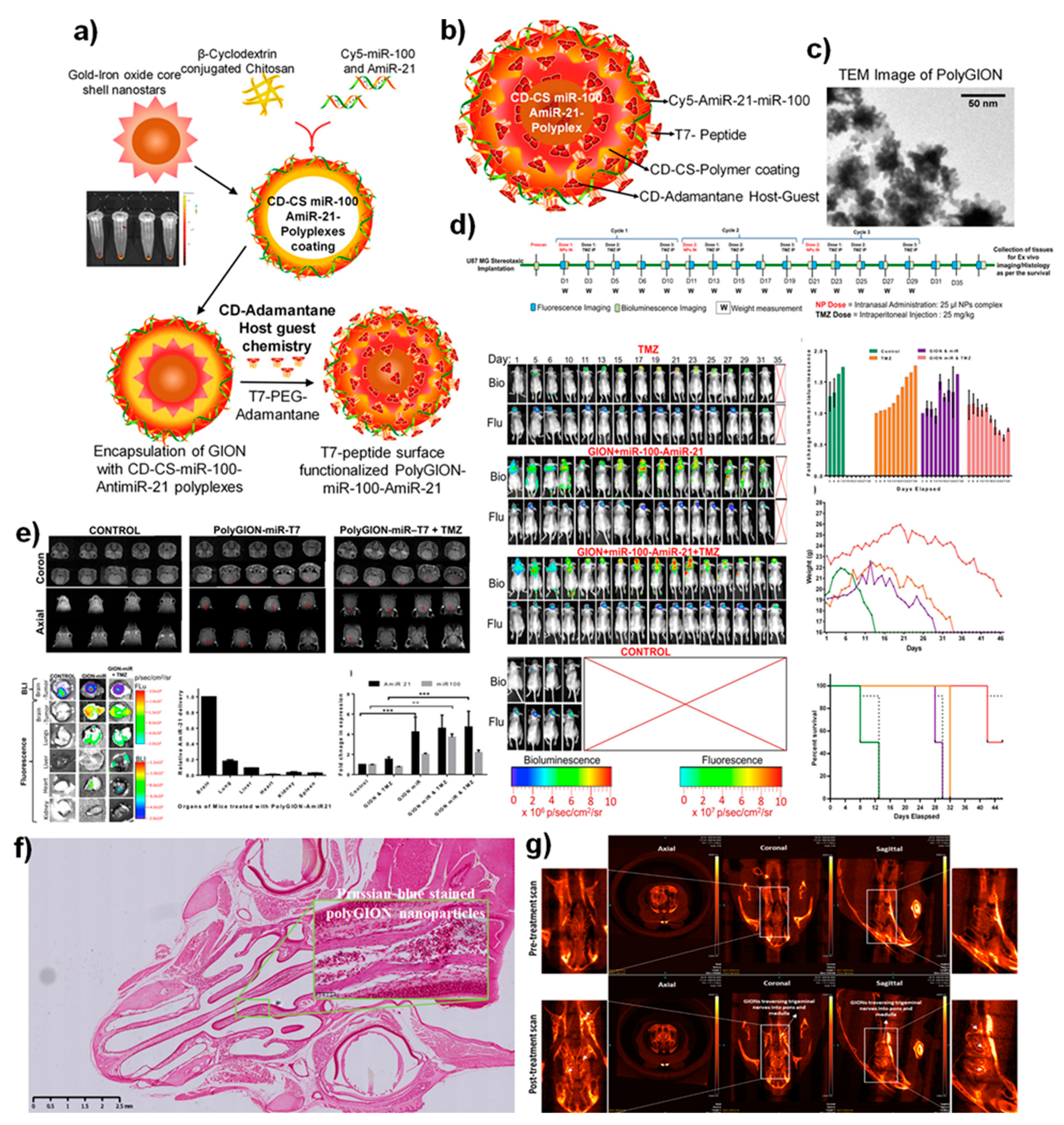
Glioblastoma |
miR-100 and antimiR-21 loaded PolyGIONS |
Mice |
Intranasal delivery of NPs strategy potentiated the nano-theranostic effects in vivo. |
[51] |
|
Glioblastoma |
siRNA + TMZ loaded chitosan NPs |
Mice |
Intranasal delivery of nanoparticle adjuvants increase the efficiency of immune-checkpoint blockade and chemotherapy in vivo. |
[65] |
|
Glioblastoma |
Self-assembled BMP4 plasmid DNA with poly(beta-amino ester) NPs |
Rat |
Intransally administered NPs could target brain tumors to enhance targeted therapies. |
[66] |
|
Gliobastoma |
Self-assembly of MPEG-PCL-Tat with siRaf-1/ Camptothecin |
Rat |
Nose-to-brain delivery proved the excellent therapeutic functions for treating glioblastoma. |
[67] |
|
Glioblastoma |
Extracellular vesicles (EVs) loaded with CXCR4 receptor, antimiRNA-21 and miRNA-100 biomaterials |
Mice |
Intranasally delivered EVs with miRNA sensitized the tumor cells to treat temozolomide, thereby improving mice’s survival rate. |
[68] |
|
Epilepsy |
Carbamazepine loaded carboxymethyl chitosan nanoparticles |
Mice |
Enhanced drug bioavailability and brain targeting was achieved via nasal administration. |
[69] |
|
Central nervous systems disorders |
Rabies Virus Glycoprotein (RVG29)-Modified PLGA Nanoparticles |
Mice |
Engineered nanoparticulate systems proved the viral delivery vectors to target and treat CNS via intranasal delivery. |
[70] |
|
Huntington’s disease |
Chitosan nanoparticles loaded with anti-HTT siRNA |
Mice |
Intranasal delivery proved the promising therapeutic alternative for safe and effective which further decreases the mutant HTT expression. |
[71] |
|
Ischemic stroke |
17β-estradiol (E2) loaded gelatin nanoparticles |
Mice |
The intranasally administered nanoparticles achieved higher delivery efficacy in vivo. |
[72] |
|
Newcastle disease and infectious bronchitis |
Chitosan nanoparticles loaded with the combined attenuated live vaccine |
Chicken |
Intranasal adjuvant and delivery carrier made a mucosal vaccine and delivery of drugs for enhanced immune functions. |
[73] |
|
SARS-CoV-2 |
Receptor-binding domain (RBD) of SARS-CoV-2 spike glycoprotein loaded chitosan nanoparticles |
Mice |
An alternative route of intranasal vaccination mimics the natural route of SARS-CoV-2 infection and stimulates both mucosal and systemic compartments of the immune responses. |
[74] |
|
SARS-CoV2 vaccine mucosal immunization |
Au-nanostar-chitosan loaded with SARS CoV-2 DNA vaccine |
Mice |
Intranasal administered SARS-CoV2 DNA vaccines encoded the spike protein antigen loaded nanomaterial achieved the humoral antibody responses and providing long-lasting immunity. |
[75] |
|
Respiratory infection |
Chitosan Nanoparticles–Adjuvanted Chlamydia Vaccine |
Mice |
Intranasal adjuvants induced the humoral, mucosal, cell-mediated immunity against bacterial infections in vivo by acting as nano vaccines. |
[76] |
2. Nanomaterials Improving Theranostic Imaging Modalities
References
- Menon, J.U.; Jadeja, P.; Tambe, P.; Vu, K.; Yuan, B.; Nguyen, K.T. Nanomaterials for photo-based diagnostic and therapeutic applications. Theranostics 2013, 3, 152.
- Burgos-Ravanal, R.; Campos, A.; Díaz-Vesga, M.C.; González, M.F.; León, D.; Lobos-González, L.; Leyton, L.; Kogan, M.J.; Quest, A.F. Extracellular vesicles as mediators of cancer disease and as nanosystems in theranostic applications. Cancers 2021, 13, 3324.
- Chen, Y.; Fan, H.; Xu, C.; Hu, W.; Yu, B. Efficient cholera toxin B subunit-based nanoparticles with MRI capability for drug delivery to the brain following intranasal administration. Macromol. Biosci. 2019, 19, 1800340.
- Gu, X.; Kwok, R.T.; Lam, J.W.; Tang, B.Z. AIEgens for biological process monitoring and disease theranostics. Biomaterials 2017, 146, 115–135.
- Weissleder, R.; Pittet, M.J. Imaging in the era of molecular oncology. Nature 2008, 452, 580–589.
- Pirovano, G.; Roberts, S.; Kossatz, S.; Reiner, T. Optical imaging modalities: Principles and applications in preclinical research and clinical settings. J. Nucl. Med. 2020, 61, 1419–1427.
- James, M.L.; Gambhir, S.S. A molecular imaging primer: Modalities, imaging agents, and applications. Physiol. Rev. 2012, 92, 897–965.
- Mukherjee, S.; Madamsetty, V.S.; Bhattacharya, D.; Roy Chowdhury, S.; Paul, M.K.; Mukherjee, A. Recent advancements of nanomedicine in neurodegenerative disorders theranostics. Adv. Funct. Mater. 2020, 30, 2003054.
- Kang, H.; Wong, S.H.D.; Pan, Q.; Li, G.; Bian, L. Anisotropic ligand nanogeometry modulates the adhesion and polarization state of macrophages. Nano Lett. 2019, 19, 1963–1975.
- Kang, H.; Jung, H.J.; Wong, D.S.H.; Kim, S.K.; Lin, S.; Chan, K.F.; Zhang, L.; Li, G.; Dravid, V.P.; Bian, L. Remote control of heterodimeric magnetic nanoswitch regulates the adhesion and differentiation of stem cells. J. Am. Chem. Soc. 2018, 140, 5909–5913.
- Min, S.; Jeon, Y.S.; Jung, H.J.; Khatua, C.; Li, N.; Bae, G.; Choi, H.; Hong, H.; Shin, J.E.; Ko, M.J. Independent tuning of nano-ligand frequency and sequences regulates the adhesion and differentiation of stem cells. Adv. Mater. 2020, 32, 2004300.
- Kang, H.; Jung, H.J.; Kim, S.K.; Wong, D.S.H.; Lin, S.; Li, G.; Dravid, V.P.; Bian, L. Magnetic manipulation of reversible nanocaging controls in vivo adhesion and polarization of macrophages. ACS Nano 2018, 12, 5978–5994.
- Thangam, R.; Kim, M.S.; Bae, G.; Kim, Y.; Kang, N.; Lee, S.; Jung, H.J.; Jang, J.; Choi, H.; Li, N. Remote switching of elastic movement of decorated ligand nanostructures controls the adhesion-regulated polarization of host macrophages. Adv. Funct. Mater. 2021, 31, 2008698.
- Hong, H.; Min, S.; Koo, S.; Lee, Y.; Yoon, J.; Jang, W.Y.; Kang, N.; Thangam, R.; Choi, H.; Jung, H.J. Dynamic ligand screening by magnetic nanoassembly modulates stem cell differentiation. Adv. Mater. 2021, 2105460.
- Kang, H.; Kim, S.; Wong, D.S.H.; Jung, H.J.; Lin, S.; Zou, K.; Li, R.; Li, G.; Dravid, V.P.; Bian, L. Remote manipulation of ligand nano-oscillations regulates adhesion and polarization of macrophages in vivo. Nano Lett. 2017, 17, 6415–6427.
- Kang, H.; Zhang, K.; Jung, H.J.; Yang, B.; Chen, X.; Pan, Q.; Li, R.; Xu, X.; Li, G.; Dravid, V.P. An in situ reversible heterodimeric nanoswitch controlled by metal-ion–ligand coordination regulates the mechanosensing and differentiation of stem cells. Adv. Mater. 2018, 30, 1803591.
- Kim, Y.; Choi, H.; Shin, J.E.; Bae, G.; Thangam, R.; Kang, H. Remote active control of nanoengineered materials for dynamic nanobiomedical engineering. View 2020, 1, 20200029.
- Cheng, Z.; Yan, X.; Sun, X.; Shen, B.; Gambhir, S.S. Tumor molecular imaging with nanoparticles. Engineering 2016, 2, 132–140.
- Cherry, S.R. Multimodality in vivo imaging systems: Twice the power or double the trouble? Annu. Rev. Biomed. Eng. 2006, 8, 35–62.
- Smith, B.R.; Gambhir, S.S. Nanomaterials for in vivo imaging. Chem. Rev. 2017, 117, 901–986.
- Fan, W.; Yung, B.; Huang, P.; Chen, X. Nanotechnology for multimodal synergistic cancer therapy. Chem. Rev. 2017, 117, 13566–13638.
- Bjornmalm, M.; Thurecht, K.J.; Michael, M.; Scott, A.M.; Caruso, F. Bridging bio–nano science and cancer nanomedicine. ACS Nano 2017, 11, 9594–9613.
- Hainfeld, J.; Slatkin, D.; Focella, T.; Smilowitz, H. Gold nanoparticles: A new X-ray contrast agent. Br. J. Radiol. Suppl. 2006, 79, 248–253.
- Zhao, W.; Yu, X.; Peng, S.; Luo, Y.; Li, J.; Lu, L. Construction of nanomaterials as contrast agents or probes for glioma imaging. J. Nanobiotechnol. 2021, 19, 1–31.
- Rangnekar, A.; Sarma, T.K.; Singh, A.K.; Deka, J.; Ramesh, A.; Chattopadhyay, A. Retention of enzymatic activity of α-amylase in the reductive synthesis of gold nanoparticles. Langmuir 2007, 23, 5700–5706.
- Liu, T.; Jin, R.; Yuan, P.; Bai, Y.; Cai, B.; Chen, X. Intracellular enzyme-triggered assembly of amino acid-modified gold nanoparticles for accurate cancer therapy with multimode. ACS Appl. Mater. Interfaces 2019, 11, 28621–28630.
- Hainfeld, J.F.; Smilowitz, H.M.; O’Connor, M.J.; Dilmanian, F.A.; Slatkin, D.N. Gold nanoparticle imaging and radiotherapy of brain tumors in mice. Nanomedicine 2013, 8, 1601–1609.
- Liu, Y.; Bhattarai, P.; Dai, Z.; Chen, X. Photothermal therapy and photoacoustic imaging via nanotheranostics in fighting cancer. Chem. Soc. Rev. 2019, 48, 2053–2108.
- Jia, X.; Han, Y.; Pei, M.; Zhao, X.; Tian, K.; Zhou, T.; Liu, P. Multi-functionalized hyaluronic acid nanogels crosslinked with carbon dots as dual receptor-mediated targeting tumor theranostics. Carbohydr. Polym. 2016, 152, 391–397.
- Mu, J.; Lin, J.; Huang, P.; Chen, X. Development of endogenous enzyme-responsive nanomaterials for theranostics. Chem. Soc. Rev. 2018, 47, 5554–5573.
- Song, N.; Zhang, Z.; Liu, P.; Yang, Y.W.; Wang, L.; Wang, D.; Tang, B.Z. Nanomaterials with supramolecular assembly based on AIE luminogens for theranostic applications. Adv. Mater. 2020, 32, 2004208.
- Kreuter, J. Drug delivery to the central nervous system by polymeric nanoparticles: What do we know? Adv. Drug Deliv. Rev. 2014, 71, 2–14.
- Rabanel, J.-M.; Delbreil, P.; Banquy, X.; Brambilla, D.; Ramassamy, C. Periphery-confined particulate systems for the management of neurodegenerative diseases and toxicity: Avoiding the blood-brain-barrier challenge. J. Control. Release 2020, 322, 286–299.
- Li, X.; Vemireddy, V.; Cai, Q.; Xiong, H.; Kang, P.; Li, X.; Giannotta, M.; Hayenga, H.N.; Pan, E.; Sirsi, S.R. Reversibly modulating the blood–brain barrier by laser stimulation of molecular-targeted nanoparticles. Nano Lett. 2021, 21, 9805–9815.
- Pandit, R.; Chen, L.; Götz, J. The blood-brain barrier: Physiology and strategies for drug delivery. Adv. Drug Deliv. Rev. 2020, 165, 1–14.
- Gao, H. Progress and perspectives on targeting nanoparticles for brain drug delivery. Acta Pharm. Sin. B 2016, 6, 268–286.
- Poulose, A.C.; Veeranarayanan, S.; Mohamed, M.S.; Nagaoka, Y.; Aburto, R.R.; Mitcham, T.; Ajayan, P.M.; Bouchard, R.R.; Sakamoto, Y.; Yoshida, Y. Multi-stimuli responsive Cu2S nanocrystals as trimodal imaging and synergistic chemo-photothermal therapy agents. Nanoscale 2015, 7, 8378–8388.
- Gai, S.; Yang, G.; Yang, P.; He, F.; Lin, J.; Jin, D.; Xing, B. Recent advances in functional nanomaterials for light–triggered cancer therapy. Nano Today 2018, 19, 146–187.
- Banerjee, R.; Katsenovich, Y.; Lagos, L.; McIintosh, M.; Zhang, X.; Li, C.-Z. Nanomedicine: Magnetic nanoparticles and their biomedical applications. Curr. Med. Chem. 2010, 17, 3120–3141.
- Gelmi, A.; Schutt, C.E. Stimuli-responsive biomaterials: Scaffolds for stem cell control. Adv. Healthc. Mater. 2021, 10, 2001125.
- Kim, J.; Piao, Y.; Hyeon, T. Multifunctional nanostructured materials for multimodal imaging, and simultaneous imaging and therapy. Chem. Soc. Rev. 2009, 38, 372–390.
- Na, H.B.; Song, I.C.; Hyeon, T. Inorganic nanoparticles for MRI contrast agents. Adv. Mater. 2009, 21, 2133–2148.
- Kircher, M.F.; De La Zerda, A.; Jokerst, J.V.; Zavaleta, C.L.; Kempen, P.J.; Mittra, E.; Pitter, K.; Huang, R.; Campos, C.; Habte, F. A brain tumor molecular imaging strategy using a new triple-modality MRI-photoacoustic-Raman nanoparticle. Nat. Med. 2012, 18, 829–834.
- Jeon, M.; Halbert, M.V.; Stephen, Z.R.; Zhang, M. Iron oxide nanoparticles as T1 contrast agents for magnetic resonance imaging: Fundamentals, challenges, applications, and prospectives. Adv. Mater. 2021, 33, 1906539.
- Zhang, J.; Ning, L.; Huang, J.; Zhang, C.; Pu, K. Activatable molecular agents for cancer theranostics. Chem. Sci. 2020, 11, 618–630.
- Medintz, I.L.; Uyeda, H.T.; Goldman, E.R.; Mattoussi, H. Quantum dot bioconjugates for imaging, labelling and sensing. Nat. Mater. 2005, 4, 435–446.
- Zhang, H.; Li, L.; Liu, X.L.; Jiao, J.; Ng, C.-T.; Yi, J.B.; Luo, Y.E.; Bay, B.-H.; Zhao, L.Y.; Peng, M.L. Ultrasmall ferrite nanoparticles synthesized via dynamic simultaneous thermal decomposition for high-performance and multifunctional T 1 magnetic resonance imaging contrast agent. ACS Nano 2017, 11, 3614–3631.
- Nowak, M.; Helgeson, M.E.; Mitragotri, S. Delivery of nanoparticles and macromolecules across the blood–brain barrier. Adv. Ther. 2020, 3, 1900073.
- Muthuraman, A.; Rishitha, N.; Mehdi, S. Role of nanoparticles in bioimaging, diagnosis and treatment of cancer disorder. In Design of Nanostructures for Theranostics Applications; Elsevier: Amsterdam, The Netherlands, 2018; pp. 529–562.
- Mukhtar, M.; Bilal, M.; Rahdar, A.; Barani, M.; Arshad, R.; Behl, T.; Brisc, C.; Banica, F.; Bungau, S. Nanomaterials for diagnosis and treatment of brain cancer: Recent updates. Chemosensors 2020, 8, 117.
- Sukumar, U.K.; Bose, R.J.; Malhotra, M.; Babikir, H.A.; Afjei, R.; Robinson, E.; Zeng, Y.; Chang, E.; Habte, F.; Sinclair, R. Intranasal delivery of targeted polyfunctional gold–iron oxide nanoparticles loaded with therapeutic microRNAs for combined theranostic multimodality imaging and presensitization of glioblastoma to temozolomide. Biomaterials 2019, 218, 119342.
- Zhou, Z.; Qutaish, M.; Han, Z.; Schur, R.M.; Liu, Y.; Wilson, D.L.; Lu, Z.-R. MRI detection of breast cancer micrometastases with a fibronectin-targeting contrast agent. Nat. Commun. 2015, 6, 1–11.
- Wu, C.; Muroski, M.E.; Miska, J.; Lee-Chang, C.; Shen, Y.; Rashidi, A.; Zhang, P.; Xiao, T.; Han, Y.; Lopez-Rosas, A. Repolarization of myeloid derived suppressor cells via magnetic nanoparticles to promote radiotherapy for glioma treatment. Nanomed. Nanotechnol. Biol. Med. 2019, 16, 126–137.
- Kumar, S.; Ali, J.; Baboota, S. Design Expert® supported optimization and predictive analysis of selegiline nanoemulsion via the olfactory region with enhanced behavioural performance in Parkinson’s disease. Nanotechnology 2016, 27, 435101.
- Nasr, M. Development of an optimized hyaluronic acid-based lipidic nanoemulsion co-encapsulating two polyphenols for nose to brain delivery. Drug Deliv. 2016, 23, 1444–1452.
- Haider, M.F.; Khan, S.; Gaba, B.; Alam, T.; Baboota, S.; Ali, J.; Ali, A. Optimization of rivastigmine nanoemulsion for enhanced brain delivery: In-vivo and toxicity evaluation. J. Mol. Liq. 2018, 255, 384–396.
- Espinoza, L.C.; Silva-Abreu, M.; Clares, B.; Rodríguez-Lagunas, M.J.; Halbaut, L.; Cañas, M.-A.; Calpena, A.C. Formulation strategies to improve nose-to-brain delivery of donepezil. Pharmaceutics 2019, 11, 64.
- Iqbal, R.; Ahmed, S.; Jain, G.K.; Vohora, D. Design and development of letrozole nanoemulsion: A comparative evaluation of brain targeted nanoemulsion with free letrozole against status epilepticus and neurodegeneration in mice. Int. J. Pharmaceut. 2019, 565, 20–32.
- Abdou, E.M.; Kandil, S.M.; El Miniawy, H.M. Brain targeting efficiency of antimigrain drug loaded mucoadhesive intranasal nanoemulsion. Int. J. Pharmaceut. 2017, 529, 667–677.
- Colombo, M.; de Lima Melchiades, G.; Figueiró, F.; Battastini, A.M.O.; Teixeira, H.F.; Koester, L.S. Validation of an HPLC-UV method for analysis of Kaempferol-loaded nanoemulsion and its application to in vitro and in vivo tests. J. Pharm. Biomed. Anal. 2017, 145, 831–837.
- Sousa, F.; Dhaliwal, H.K.; Gattacceca, F.; Sarmento, B.; Amiji, M.M. Enhanced anti-angiogenic effects of bevacizumab in glioblastoma treatment upon intranasal administration in polymeric nanoparticles. J. Control. Release 2019, 309, 37–47.
- Azambuja, J.; Schuh, R.; Michels, L.; Gelsleichter, N.; Beckenkamp, L.; Iser, I.; Lenz, G.; De Oliveira, F.; Venturin, G.; Greggio, S. Nasal administration of cationic nanoemulsions as CD73-siRNA delivery system for glioblastoma treatment: A new therapeutical approach. Mol. Neurobiol. 2020, 57, 635–649.
- Chu, L.; Wang, A.; Ni, L.; Yan, X.; Song, Y.; Zhao, M.; Sun, K.; Mu, H.; Liu, S.; Wu, Z. Nose-to-brain delivery of temozolomide-loaded PLGA nanoparticles functionalized with anti-EPHA3 for glioblastoma targeting. Drug Deliv. 2018, 25, 1634–1641.
- Sekerdag, E.; Lüle, S.; Pehlivan, S.B.; Öztürk, N.; Kara, A.; Kaffashi, A.; Vural, I.; Işıkay, I.; Yavuz, B.; Oguz, K.K. A potential non-invasive glioblastoma treatment: Nose-to-brain delivery of farnesylthiosalicylic acid incorporated hybrid nanoparticles. J. Control. Release 2017, 261, 187–198.
- Van Woensel, M.; Mathivet, T.; Wauthoz, N.; Rosière, R.; Garg, A.D.; Agostinis, P.; Mathieu, V.; Kiss, R.; Lefranc, F.; Boon, L. Sensitization of glioblastoma tumor micro-environment to chemo-and immunotherapy by Galectin-1 intranasal knock-down strategy. Sci. Rep. 2017, 7, 1–14.
- Mangraviti, A.; Tzeng, S.Y.; Gullotti, D.; Kozielski, K.L.; Kim, J.E.; Seng, M.; Abbadi, S.; Schiapparelli, P.; Sarabia-Estrada, R.; Vescovi, A. Non-virally engineered human adipose mesenchymal stem cells produce BMP4, target brain tumors, and extend survival. Biomaterials 2016, 100, 53–66.
- Kanazawa, T.; Morisaki, K.; Suzuki, S.; Takashima, Y. Prolongation of life in rats with malignant glioma by intranasal siRNA/drug codelivery to the brain with cell-penetrating peptide-modified micelles. Mol. Pharm. 2014, 11, 1471–1478.
- Wang, K.; Kumar, U.S.; Sadeghipour, N.; Massoud, T.F.; Paulmurugan, R. A microfluidics-based scalable approach to generate extracellular vesicles with enhanced therapeutic microRNA loading for intranasal delivery to mouse glioblastomas. ACS Nano 2021, 15, 18327–18346.
- Liu, S.; Yang, S.; Ho, P.C. Intranasal administration of carbamazepine-loaded carboxymethyl chitosan nanoparticles for drug delivery to the brain. Asian J. Pharm. Sci. 2018, 13, 72–81.
- Chung, E.P.; Cotter, J.D.; Prakapenka, A.V.; Cook, R.L.; DiPerna, D.M.; Sirianni, R.W. Targeting small molecule delivery to the brain and spinal cord via intranasal administration of rabies virus glycoprotein (RVG29)-modified PLGA nanoparticles. Pharmaceutics 2020, 12, 93.
- Sava, V.; Fihurka, O.; Khvorova, A.; Sanchez-Ramos, J. Enriched chitosan nanoparticles loaded with siRNA are effective in lowering Huntington’s disease gene expression following intranasal administration. Nanomed. Nanotechnol. Biol. Med. 2020, 24, 102119.
- Joachim, E.; Barakat, R.; Lew, B.; Kim, K.K.; Ko, C.; Choi, H. Single intranasal administration of 17β-estradiol loaded gelatin nanoparticles confers neuroprotection in the post-ischemic brain. Nanomed. Nanotechnol. Biol. Med. 2020, 29, 102246.
- Zhao, K.; Li, S.; Li, W.; Yu, L.; Duan, X.; Han, J.; Wang, X.; Jin, Z. Quaternized chitosan nanoparticles loaded with the combined attenuated live vaccine against Newcastle disease and infectious bronchitis elicit immune response in chicken after intranasal administration. Drug Deliv. 2017, 24, 1574–1586.
- Jearanaiwitayakul, T.; Seesen, M.; Chawengkirttikul, R.; Limthongkul, J.; Apichirapokey, S.; Sapsutthipas, S.; Phumiamorn, S.; Sunintaboon, P.; Ubol, S. Intranasal administration of RBD nanoparticles confers induction of mucosal and systemic immunity against SARS-CoV-2. Vaccines 2021, 9, 768.
- Kumar, U.S.; Afjei, R.; Ferrara, K.; Massoud, T.F.; Paulmurugan, R. Gold-nanostar-chitosan-mediated delivery of SARS-CoV-2 DNA vaccine for respiratory mucosal immunization: Development and proof-of-principle. ACS Nano 2021, 15, 17582–17601.
- Li, Y.; Wang, C.; Sun, Z.; Xiao, J.; Yan, X.; Chen, Y.; Yu, J.; Wu, Y. Simultaneous intramuscular and intranasal administration of chitosan nanoparticles–Adjuvanted Chlamydia vaccine elicits elevated protective responses in the lung. Int. J. Nanomed. 2019, 14, 8179.
- Cheng, L.; Wang, X.; Gong, F.; Liu, T.; Liu, Z. 2D nanomaterials for cancer theranostic applications. Adv. Mater. 2020, 32, 1902333.
- Li, S.; Su, W.; Wu, H.; Yuan, T.; Yuan, C.; Liu, J.; Deng, G.; Gao, X.; Chen, Z.; Bao, Y. Targeted tumour theranostics in mice via carbon quantum dots structurally mimicking large amino acids. Nat. Biomed. Eng. 2020, 4, 704–716.
- Molaei, M.J. Carbon quantum dots and their biomedical and therapeutic applications: A review. RSC Adv. 2019, 9, 6460–6481.
- Li, N.; Liang, X.; Wang, L.; Li, Z.; Li, P.; Zhu, Y.; Song, J. Biodistribution study of carbogenic dots in cells and in vivo for optical imaging. J. Nanopart. Res. 2012, 14, 1–9.
- Huang, N.; Cheng, S.; Zhang, X.; Tian, Q.; Pi, J.; Tang, J.; Huang, Q.; Wang, F.; Chen, J.; Xie, Z. Efficacy of NGR peptide-modified PEGylated quantum dots for crossing the blood–brain barrier and targeted fluorescence imaging of glioma and tumor vasculature. Nanomed. Nanotechnol. Biol. Med. 2017, 13, 83–93.
- Kabanov, A.V.; Batrakova, E.V. Polymer nanomaterials for drug delivery across the blood brain barrier. In Neuroimmune Pharmacology; Springer: Berlin/Heidelberg, Germany, 2017; pp. 847–868.
- Huang, H.; Lovell, J.F. Advanced functional nanomaterials for theranostics. Adv. Funct. Mater. 2017, 27, 1603524.
- Van der Meel, R.; Sulheim, E.; Shi, Y.; Kiessling, F.; Mulder, W.J.; Lammers, T. Smart cancer nanomedicine. Nat. Nanotechnol. 2019, 14, 1007–1017.
- Toy, R.; Bauer, L.; Hoimes, C.; Ghaghada, K.B.; Karathanasis, E. Targeted nanotechnology for cancer imaging. Adv. Drug Deliv. Rev. 2014, 76, 79–97.
- Kevadiya, B.D.; Ottemann, B.M.; Thomas, M.B.; Mukadam, I.; Nigam, S.; McMillan, J.; Gorantla, S.; Bronich, T.K.; Edagwa, B.; Gendelman, H.E. Neurotheranostics as personalized medicines. Adv. Drug Deliv. Rev. 2019, 148, 252–289.
- Ehlerding, E.B.; Grodzinski, P.; Cai, W.; Liu, C.H. Big potential from small agents: Nanoparticles for imaging-based companion diagnostics. ACS Nano 2018, 12, 2106–2121.
- Chen, J.; Li, J.; Zhou, J.; Lin, Z.; Cavalieri, F.; Czuba-Wojnilowicz, E.; Hu, Y.; Glab, A.; Ju, Y.; Richardson, J.J. Metal–phenolic coatings as a platform to trigger endosomal escape of nanoparticles. ACS Nano 2019, 13, 11653–11664.

