You're using an outdated browser. Please upgrade to a modern browser for the best experience.
Collagen D-Band Periodicity
Edit

The molecules follow a quarter-staggered fashion packing, which leads to the formation of the so call D-band periodicity. This D-band periodicity is a repeating banding pattern of about 67 nm (depending on the different tissue) and includes gap and overlap regions. Collagen fibrils form bundles and fibers by appropriate alignment.

collagen Atomic Force Microscopy (AFM) D-band D-periodicity D-spacing mechanical properties biomaterials

1. General

Collagens comprise about thirty percent (30%) of the whole protein in mammals and they have been recognized as very promising substances for complementing the structure of biomaterials so that they can better interact with biological systems [1][2][3]. Among the vertebrate collagen superfamily, which consist of more than 50 collagens and/or collagen-like proteins [4][5], the greatest interest is presented by collagen type I. This fibril-forming collagen is the most abundant in mammals, including humans, and fulfills unique characteristics, including self-assembly [6].
In order to enhance the understanding concerning collagen’s role in a wide range of functions or diseases and collagen possible applications, an extensive investigation at the nanoscale of the surface properties and structure is demanded. Atomic Force Microscopy (AFM) arises as a novel technique for the nano-characterization of biological samples and biomaterials [7][8]. AFM was invented in 1986 by Binning et al. [9], and the first commercial AFMs started to appear in the market in the beginning of 1990s [10]. Soon after its development, researchers worldwide started using it for nano-imaging and mechanical properties characterization of a wide range of specimens, including biomaterials and biological samples [11][12].

2. Collagen

2.1. Collagen Superfamily and Collagen-Related Pathological Conditions

The superfamily of vertebrate collagen includes more than fifty collagen and collagen-like proteins [4][5]. All the members of this superfamily are identified by the same structure of the collagen molecule, consisting by a triple-helix from 3 polypeptide chains [5][13]. The characteristic of each polypeptide chain is the repeating pattern of amino acids (Gly-X-Y), where X and Y can be any amino acid. So far, 28 different collagens have been identified in the human body (a group of the collagen superfamily) with different structures and functions. There are different subgroups, such as the fibril-forming collagens, among which the type I collagen is of most interest [13][14]. Other members of the fibril-forming collagens are the collagen types II, III, V, XI, XXIV, and XXVII.
Generally, collagen and collagen mutations are related to many diseases, such as osteogenesis imperfecta, chondrodysplasias, osteoporosis and a number of syndromes (for example, Ehler–Danlos, Alport, Knobloch). Furthermore, structural variations of collagen at the nanoscale are related to a number of pathological conditions [15][16][17]. In addition, collagen alterations in terms of structure, orientation and mechanical properties have been shown to have a significant role in desmoplastic solid tumors, such as breast and pancreatic cancers [18][19][20][21].

2.2. Collagen Type I

Collagen type I is member of the fibril-forming subgroup of the collagen superfamily. Over 90% of the collagen in humans is type I as it is the major protein in the extracellular matrix (ECM). Collagen type I can be found mainly in skin, tendon, vascular ligature, organs and it is the main part of the organic part of bone [22][23]. The molecules of type I collagen form rod-shaped triple helices that are assembled in order to form fibrils [4][24]. The molecules follow a quarter-staggered fashion packing, which leads to the formation of the so call D-band periodicity [1][5][25][26][27][28]. This D-band periodicity is a repeating banding pattern of about 67 nm (depending on the different tissue) and includes gap and overlap regions. Collagen fibrils form bundles and fibers by appropriate alignment [4][24][29]. The fibrils of collagen type I play the role of the elementary building blocks in a wide range of collagen-rich tissues [30][31].
Depending on the tissue, collagen type I fibrils present different morphologies, properties, and have a crucial role in different functions, such as scaffolding and mechanical strength [1][4][32]. For example, in the case of tendons, collagen fibrils have a lateral packing and present a uniform distribution of diameters [33]. On the other hand, in skin, the collagen type I fibrils are randomly oriented and present the form of loosely interwoven and wavy bundles. However, one unique characteristic for collagen type I fibrils is the presence of the so-called D-band periodicity.

2.3. Collagen D-Band Periodicity

The packing of collagen type I molecules follow a quarter-staggered fashion that leads to the formation of a unique banding pattern, the D-band periodicity (also known as D-band, D-periodicity, D-spacing) [1][5][25][26]. Generally, the collagen D-band periodicity is considered a unique nanocharacteristic of all fibrils-forming collagens. However, there is a debate whether the length of the D-band periodicity is identical for all fibrillar collagens [34][35]. For instance, some researchers state that there is not a common D-band periodicity among fibrillar collagens [36]. These arguments are based mainly on electron microscopy studies where different D-band patterns were found on different collagen types (collagen type I, II and III), both on positively [37][38][39][40] and negatively [41] stained specimens. In addition, different patterns have been found in studies that compared native collagen type I with type II [42], and reconstructed fibrils type I and V [43]. Furthermore, a D-period of 65 nm has been reported in tissues rich in collagen type III, such as dermis [44] and cornea [45]. It must also be noted that for type I collagen, a fibrillar variant the so-called Fibrous Long Spacing Collagen (FLS) has been reported [46][47][48][49]. These fibrils are characterized by significant larger values in D-band periodicity, typically 200–300 nm. FLS were first reported in in vitro experiments [50], but subsequently, it was found in a number of pathological and normal tissues [51][52][53]. Nevertheless, it was focused on collagen type I D-band periodicity. The D-band periodicity plays a significant role in the collagen type I fibrils mechanical properties and the cell-collagen crosstalk, while it has been associated with a wide range of pathological conditions [27][28][54][55].
The length of the D-band periodicity is tissue-dependent, but in all cases includes gap and overlapping regions [1][5][27][28]. Fibrillar collagens are generally characterized by a D-band periodicity of 64–67 nm depending on the tissue [1]. A number of the variations of the D-band periodicity that have been reported in the literature is presented in relevant articles [56]. For example, some of the values that have been reported using different microscopy techniques are: 64.6 ± 5.3 nm (55–80) for human skin [57], 67.7 ± 0.9 (central zone) and 71.3 ± 0.4 (distal zone) for vitrified predentin [58], and 54–75 nm demineralized dentin [59] (variations in D-band periodicity are also presented in the Results). Furthermore, it has been reported that a D-periodicity of 65 nm it can be found in corneal stroma. One explanation is that the fibrils are more hydrated and, as a result, the molecules are tilted by about 15o to the fibril axis, leading to a reduced axial periodicity [60]. In addition, as mentioned previously, some researchers state that tissues that are rich in collagen type III, such as cornea and dermis, present the 65-nm D-band periodicity [44][45]. In the literature, frequently, the D-band periodicity is introduced as a single value of 67 nm, but as it has been discussed this is not true [56]. Both the collagen type and the tissue play a role. So, it can be said that collagen D-band periodicity is collagen-type and tissue-dependent.
Collagen D-band periodicity has been correlated with fibrils’ mechanical properties, collagen–cell interactions and a number of pathological conditions [27][28][54][55]. It has been shown that cells respond to this periodic pattern and, for example, cell elongation along collagen fibrils/fibers major axis has been correlated with the D-band periodicity orientation [54]. In general, the so called “contact guidance mechanism” has been associated with the motion of the cell along the axis of fibrous features, such as the fibrous proteins (including collagen) of the ECM [61] and the cells’ morphodynamics respond to both surface characteristics and mechanical properties of the surrounding environment [61]. According to the literature, there is a limiting threshold regarding the response of the cells on the contact mechanism, while it has been shown that, at least for fibroblasts, this threshold is ~35 nm [62]. However, the exact mechanism of cell-collagen and/or cell-collagen-based biomaterials is not yet fully clarified.

2.4. Collagen-Based Biomaterials

Since collagen possesses unique properties, such as non-toxicity, bio-compatibility, bio-degradability and the ability for self-assembly [6], it has been identified as unique biomaterial for the development of novel biomaterials [1][2][3]. For example, controlling the self-assembly process of collagen can lead to the development of collagen-based biomaterials that can be used as in vitro models of collagen-rich tissues. In addition, it has been demonstrated that collagen-based surfaces with well-organized geometrical features, such as aligned fibers and or porous structures, can guide cell behavior towards a better performance [63][64]. The control of cells movement has attracted significant research interest as it has been associated with specific biological processes, such as wound-healing and metastasis.
Although the process of collagen self-assembly is entropy-driven and the basic principles are known [1], the exact mechanism is not well defined [31] and the development of collagen-based biomaterials with tunable and/or pre-determined characteristics and properties remains a challenging task. The formation of the D-band periodicity is a consequence of the self-assembly process. In vivo, the cells’ (mainly fibroblasts) membrane includes recesses, which appropriately arrange thin collagen fibrils. In the case of the in vitro formation of collagen fibers with D-band periodicity, the mechanism is modified as the self-assembly occurs in the absence of cellular control and a number of growth steps, both linear and lateral, take place [65].
As the majority of the biological reactions occur on interfaces and/or surfaces and specific biological processes, including cells’ proliferation and adhesion, are influenced by the nano-characteristics of the biomaterials, the features of the biomaterials’ surface are of pivotal importance in bioengineering and biomedicine [66][67]. Consequently, the nano-characterization of the surface properties of collagen and collagen-based biomaterials is of crucial importance. What is more, the formation of collagen-based biomaterials is not straightforward and that is why novel techniques are required for evaluating and studying the properties of the final material. One of the major tools for these purposes (the characterization of collagen and collagen-based biomaterials), is Atomic Force Microscopy (Figure 1) [10][68][69][70]. However, a number of other techniques can be used for characterizing and imaging collagen and collagen-based biomaterials.
Figure 1. An illustration of an Atomic Force Microscopy scanning a collagen fiber.

2.5. Imaging Collagen and Collagen-Based Biomaterials

Collagen and collagen-based samples can be imaged with a variety of imaging techniques, including optical and electron microscopy techniques and Atomic Force Microscopy (AFM). Optical microscopy possesses the advantages of the non-invasiveness and the minimum sample destruction due to light–collagen interactions. It must be noted that there are some specific types of optical microscopy that present selectivity on collagen, such as second harmonic generation (SHG) [71][72][73][74][75] and polarized microscopy, especially when combined with picrosirius red staining [76][77][78]. SHG is a nonlinear optical microscopy technique and no staining is demanded, while for picrosirius red staining polarization microscopy, fixation and staining is demanded. However, for all the light microscopy techniques, it must be taken into account that the size of the collagen fibrils is smaller or at the limit of optical microscopy. Of course, the D-band periodicity is much smaller of the resolution of optical microscopy. Furthermore, in tissues, fibrils are assembled into bundles and sheets and the imaging of individual fibrils is impossible with a light microscope. However, in cases that high resolution is demanded, optical microscopy techniques are used. In addition, in many cases, correlative microscopy or different microscopy techniques are combined, such as AFM and SHG [25][26]. Concerning, electron microscopy techniques, transmission electron microscopy (TEM) and scanning electron microscopy (SEM) were for many years the major imaging techniques of collagen and collagen-based biomaterials, especially TEM that presents higher resolution [79][80][81]. Electron microscopy techniques enable also the measurement of the fibrils’ length, the fibril volume fraction (FVF) and the 3D organization of the fibrils [79]. The D-band periodicity was originally observed in the early 1960s on electron micrographs that led the introduction of the D-band periodicity (D-stagger), and subsequently, the model that describe it with the gap and overlap regions [82]. Studies with TEM showed that the average D-band periodicity is in the range of 64–70 nm [83]. Unfortunately, electron microscopy techniques present some significant limitations. First of all, during sample preparation biomolecules such as collagen irreversibly denature as a consequence of the harsh chemical fixation and the staining procedure [84]. In addition, the specimen–electron beam interaction may destroy collagen ultrastructure. Some of these drawbacks may be overcome with cryo-electron microscopy techniques that protect biomolecules’ native ultrastructure and have the advantages of electron microscopy [85]. However, the fact that these techniques require a highly specialized equipment, complicated analysis strategies, complicated preparation protocols (for example, freezing tissues) and are characterized by poor signal-noise ratio that minimize their applications. Finally, AFM is a high-resolution surface technique that can combine both imaging and mechanical properties characterization of biological samples. Minimum sample preparation is demanded and it can be performed under different environmental conditions. As the focuses is the characterization of collagen D-band periodicity with AFM, detailed information about AFM and its applications concerning collagen are presented in the next sections.

3. Atomic Force Microscopy

3.1. General

Atomic Force Microscopy (AFM) was developed in 1980s [9] and is member of the scanning probe microscopies family. AFM measures the probe (a tip mounted on a cantilever) surface interactions in order to assess information concerning the topography and the mechanical properties of the sample at the nanoscale. Since its invention, AFM has arisen as a fundamental nano- and microscopy technique, as it possesses a number of advantages compared to other microscopes (such as optical microscopy, Scanning and Transmission Electron Microscopy). AFM can operate in different environmental conditions, with different modes, that can offer a wide range of information from surface topography to mechanical characterization of specimens that require minimum preparation [10][86][87]. These features make it very attractive for biological samples, including collagen- based tissues and biomaterials.

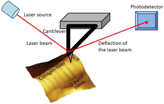
3.2. AFM Basic Principles

AFM is very different than conventional microscopes as it has no lenses of any kind. Actually, AFM “feels” rather than “looks” at the sample surface [10]. An AFM system uses a probe in order to collect the specimen information. The AFM probe is a tip (with different shapes depending on the application) mounted on a cantilever, which are attached on a chip. Piezoelectric elements are used for the accurate scanning of the tip over the sample. The movement of the tip is being monitored by using a laser beam. A laser source emits a laser beam in the backside of the cantilever. The deflection beam is then detected by a photodiode (a photodetector). During the scanning procedure the cantilever bends, due to the tip-sample forces, and the photodetector monitors the movement of the tip by the changes of the laser spot position. The photodetector, which is a position-sensitive detector, translates the laser deflection into an electric signal that is used for the formation of a 3-D image of the surface [9] or it is used for force spectroscopy purposes depending on the used mode.

3.3. AFM Modes

One of the more significant characteristics of AFM is that it can operate in a wide range of different modes, and as a result, it can offer both qualitative and quantitative information concerning bio-samples [70][87][88], such as nano-topography and mechanical properties characterization [7][89][90][91]. The most widely used AFM modes are the contact mode, the tapping mode, and the non-contact mode (where the tip does not come in contact with the sample surface). In contact mode, the tip is always in contact with the sample surface and topographical maps are provided. In addition, during contact mode, Lateral Force Microscopy (LFM) imaging can be performed. LFM offers information relevant to the surface friction which is a consequence of the material’s surface inhomogeneities. In tapping mode (also known as intermittent mode, dynamic contact mode and AC mode), the cantilever oscillates at its resonance frequency. Tapping mode can be used for acquiring topographical images, while it can simultaneously capture phase images [10]. Phase imaging use as contrast the phase lag between the cantilever’s driving signal and its output signal. Phase images are useful in order to understand variations in composition, friction, adhesion and viscoelastic properties of heterogeneous specimens [59][69]. The information from phase imaging goes beyond topographical features [59][69]. Tapping mode is preferred in the case of the biological specimens as they are quite soft, and this mode is less destructive as lateral and frictional forces are minimized [7][11]. Finally, AFM can also operate in non-contact mode. This mode is not frequently used in biological samples as it is quite difficult to perform. During this mode, the AFM tip does not come in contact with the sample surface and uses only attractive forces.
AFM can also be used for nanomechanical properties characterization of the samples with force spectroscopy. In this experimental procedure, the AFM probe acts as a force sensor [92] for assessing the mechanical properties of the sample with different modes [93]. For instance, the AFM nanoindentation procedure (where indentation–force curves are formed) [94][95][96] or the force scanning mode [97] can be used for acquiring information regarding the samples stiffness (in terms of Young’s modulus values or stiffness maps) [69][89][91][98][99]. For acquiring the specific values, mechanical models are used, such as the Hertz model, which is the most widely used, while commercial or home-built software are used for the fitting the mathematical expressions [100][101].

3.4. AFM Limitations

It must be noted that AFM, as the majority of the microscopes, is also characterized by a number of drawbacks [102]. First of all, AFM is a surface imaging and mechanical properties characterization technique. Although AFM can provide information concerning the z axis (is one of the few microscopes that provide 3D maps of the surface), this ability is limited to the length of the tip. So, it provides only characterization of the surface and not the whole specimen in the z axis. In the case of the collagen-based materials, AFM cannot measure the organization of the fibrils in the three dimensions or the fibril volume fraction (FMV) [79]. Furthermore, in the cases of the biological derived collagen-based specimens (such as tissue sections), the characterization of the surface is very challenging as the specimen collection procedure or preparation methodology (for example, needle biopsy collection, section with surgical blades) may destroy the collagen ultrastructure. However, the combination with cryosectioning techniques enables the visualization of the ultrastructure of unstained specimens [103], while more complicated specimen preparation techniques have been proposed in the literature [104][105]. One more limitation is that AFM systems can image a single nano-sized image at a time. The size of the maximum size of the image depends on the different AFM scanners, which are different depending on the manufacturer. As a result, information, such as the collagen fibril length, cannot be assessed, but it does not affect features such as the D-band periodicity. Furthermore, AFM imaging is characterized by low scanning time, which can cause thermal drift on the sample. This limitation is overcome by novel developments in AFM modes and techniques, such as the fast/high-speed scanning [106]. One more limitation, concerning collagen characterization is related to the sample preparation and the need for the specimen to be firmly attached to a substrate. As the AFM tip exerts forces on the specimens, loosely attached samples cannot be characterized. In the case of tissue specimens, biocompatible glues are used, but careful handling is demanded so as to protect the surface. On the other hand, for in vitro self-assembled collagen fibrils, physical adsorption of the collagen solutions on a flat substrate, such as mica, is usually used. Another source of drawback arises when collagen characterization is performed in air as a layer of water condensation and other contamination often covers both probe and sample. This leads to attractive forces that affect imaging. In order to overcome this limitation, AFM characterization under liquid conditions (for example, saline buffers) is usually used. Finally, a source of possible limitation arises from the size and shape of the AFM tip. Inappropriate, damaged or contaminated tips may influence topographical characteristics, such as the D-band periodicity and the relevant measurements. Currently, a number of manufactures offers a wide range of AFM tips, with different shapes, sizes and for different environmental conditions and modes, so as the appropriate ones to be selected. In addition, calibration gratings can be used for assessing tips’ performance. Overall, as the AFM technology matures, new advances and developments help researchers to overcome the majority of the limitations of this technique.

3.5. AFM and Collagen

One of the major advantages of AFM in biology and bioengineering ones is the fact that it does not demand significant sample preparation. For example, for the AFM characterization, it is not necessary to coat or label the specimen with dyes/antibodies, while depending on the sample, dehydration is not mandatory [11][12][21]. Furthermore, AFM can operate both in air and liquid [86], while also experiments under vacuum conditions have been performed. Concerning the application of AFM on collagen-based samples both imaging and mechanical properties characterization have been applied in a wide range of samples, from pure collagen to collagen rich-tissues and biomaterials. AFM scanning does not affect or destroy the collagen structure, while AFM resolution can provide information from molecules to individual fibrils/fibers [6][107]. AFM has been applied for investigating different properties of collagen, including collagen structure, the role of collagen in a number of pathological conditions and collagen–cell interactions. 

References

  1. Fratzl, P. Collagen Structure and Mechanics; Springer: New York, NY, USA, 2008.
  2. Tihan, G.T.; Rău, I.; Zgârian, R.G.; Ghica, M.V. Collagen-based biomaterials for ibuprofen delivery. Comptes Rendus Chim. 2016, 19, 389–393.
  3. Walters, B.D.; Stegemann, J.P. Strategies for directing the structure and function of three-dimensional collagen biomaterials across length scales. Acta Biomater. 2014, 10, 1488–1501.
  4. Ricard-Blum, S. The collagen family. Cold Spring Harb. Perspect. Biol. 2011, 3, a004978.
  5. Hulmes, D.J.S. Collagen diversity, synthesis and assembly. In Collagen: Structure and Mechanics; Springer: New York, NY, USA, 2008; pp. 15–47.
  6. Hasirci, V.; Vrana, E.; Zorlutuna, P.; Ndreu, A.; Yilgor, P.; Basmanav, F.B.; Aydin, E. Nanobiomaterials: A review of the existing science and technology, and new approaches. J. Biomater. Sci. Polym. Ed. 2006, 17, 1241–1268.
  7. Stylianou, A.; Yova, D. Surface nanoscale imaging of collagen thin films by Atomic Force Microscopy. Mater. Sci. Eng. C 2013, 33, 2947–2957.
  8. Stylianou, A.; Yova, D.; Politopoulos, K. Atomic force microscopy surface nanocharacterization of UV-irradiated collagen thin films. In Proceedings of the 12th IEEE International Conference on BioInformatics and BioEngineering, BIBE, Larnaca, Cyprus, 11–13 November 2012; pp. 602–607.
  9. Binnig, G.; Quate, C.F.; Gerber, C. Atomic force microscope. Phys. Rev. Lett. 1986, 56, 930–933.
  10. Morris, V.J.; Kirby, A.R.; Gunning, A.P. Atomic Force Microscopy for Biologists; Imperial College Press: London, UK, 2008.
  11. Gadegaard, N. Atomic force microscopy in biology: Technology and techniques. Biotech. Histochem. 2006, 81, 87–97.
  12. Stylianou, A.; Yova, D.; Alexandratou, E. Nanotopography of collagen thin films in correlation with fibroblast response. J. Nanophotonics 2013, 7, 073590.
  13. Engel, J.; Bächinger, H.P. Structure, stability and folding of the collagen triple helix. Top. Curr. Chem. 2005, 247, 7–33.
  14. Kadler, K.E.; Baldock, C.; Bella, J.; Boot-Handford, R.P. Collagens at a glance. J. Cell Sci. 2007, 120, 1955–1958.
  15. Abou Neel, E.A.; Bozec, L.; Knowles, J.C.; Syed, O.; Mudera, V.; Day, R.; Hyun, J.K. Collagen—Emerging collagen based therapies hit the patient. Adv. Drug Deliv. Rev. 2013, 65, 429–456.
  16. Åsling, B.; Jirholt, J.; Hammond, P.; Knutsson, M.; Walentinsson, A.; Davidson, G.; Agreus, L.; Lehmann, A.; Lagerström-Fermer, M. Collagen type III alpha I is a gastro-oesophageal reflux disease susceptibility gene and a male risk factor for hiatus hernia. Gut 2009, 58, 1063–1069.
  17. Fang, M.; Yuan, J.; Peng, C.; Li, Y. Collagen as a double-edged sword in tumor progression. Tumor Biol. 2014, 35, 2871–2882.
  18. Gkretsi, V.; Stylianou, A.; Papageorgis, P.; Polydorou, C.; Stylianopoulos, T. Remodeling components of the tumor microenvironment to enhance cancer therapy. Front. Oncol. 2015, 5, 214.
  19. Stylianou, A.; Gkretsi, V.; Louca, M.; Zacharia, L.; Stylianopoulos, T. Collagen Content and Extracellular Matrix Stiffness Remodels Pancreatic Fibroblasts Cytoskeleton. J. R. Soc. Interface 2019, 16, 20190226.
  20. Stylianou, A.; Gkretsi, V.; Stylianopoulos, T. Transforming Growth Factor-β modulates Pancreatic Cancer Associated Fibroblasts cell shape, stiffness and invasion. Biochim. Biophys. Acta 2018, 1862, 1537–1546.
  21. Stylianou, A.; Stylianopoulos, T. Atomic Force Microscopy Probing of Cancer Cells and Tumor Microenvironment Components. BioNanoScience 2016, 6, 33–46.
  22. Birk, D.E.; Bruckner, P. Collagen suprastructures. Top. Curr. Chem. 2005, 247, 185–205.
  23. Brinckmann, J. Collagens at a glance. In Collagen; Brinckmann, J., Mueller, P.K., Notbohm, H., Eds.; Springer: Berlin/Heidelberg, Germany, 2005; Volume 247, pp. 1–6.
  24. Ricard-Blum, S.; Ruggiero, F.; van der Rest, M. The collagen superfamily. Top. Curr. Chem. 2005, 247, 35–84.
  25. Bozec, L.; van der Heijden, G.; Horton, M. Collagen Fibrils: Nanoscale Ropes. Biophys. J. 2007, 92, 70–75.
  26. Petruska, J.A.; Hodge, A.J. A Subunit Model for the Tropocollagen Macromolecule. Proc. Natl. Acad. Sci. USA 1964, 51, 871–876.
  27. Wallace, J.M.; Orr, B.G.; Marini, J.C.; Holl, M.M.B. Nanoscale morphology of Type I collagen is altered in the Brtl mouse model of Osteogenesis Imperfecta. J. Struct. Biol. 2011, 173, 146–152.
  28. Grant, C.A.; Phillips, M.A.; Thomson, N.H. Dynamic mechanical analysis of collagen fibrils at the nanoscale. J. Mech. Behav. Biomed. Mater. 2012, 5, 165–170.
  29. Fratzl, P. Collagen: Structure and mechanics, an introduction. In Collagen: Structure and Mechanics; Springer: Boston, MA, USA, 2008; pp. 1–13.
  30. Ivanova, V.P.; Krivchenko, A.I. A current viewpoint on structure and evolution of collagens. I. Fibrillar collagens. J. Evol. Biochem. Physiol. 2012, 48, 127–139.
  31. Shoulders, M.D.; Raines, R.T. Collagen structure and stability. Annu. Rev. Biochem. 2009, 78, 929–958.
  32. Gordon, M.K.; Hahn, R.A. Collagens. Cell Tissue Res. 2010, 339, 247–257.
  33. Cen, L.; Liu, W.; Cui, L.; Zhang, W.; Cao, Y. Collagen tissue engineering: Development of novel biomaterials and applications. Pediatr. Res. 2008, 63, 492–496.
  34. Bender, E.; Silver, F.H.; Hayashi, K. Model conformations of the carboxyl telopeptides in vivo based on type I collagen fibral banding patterns. Coll. Relat. Res. 1983, 3, 407–418.
  35. Bruns, R.R.; Gross, J. High-resolution analysis of the modified quarter-stagger model of the collagen fibril. Biopolymers 1974, 13, 931–941.
  36. Ortolani, F.; Giordano, M.; Marchini, M. A model for type II collagen fibrils: Distinctive D-band patterns in native and reconstituted fibrils compared with sequence data for helix and telopeptide domains. Biopolymers 2000, 54, 448–463.
  37. Stark, M.; Miller, E.J.; Kühn, K. Comparative electron-microscope studies on the collagens extracted from cartilage, bone, and skin. Eur. J. Biochem. 1972, 27, 192–196.
  38. Wiedemann, H.; Chung, E.; Fujii, T.; Miller, E.J.; Kühn, K. Comparative electron-microscope studies on type-III and type-I collagens. Eur. J. Biochem. 1975, 51, 363–368.
  39. Kühn, K. Segment-long-spacing crystallites, a powerful tool in collagen research. Coll. Relat. Res. 1982, 2, 61–80.
  40. Mallinger, R.; Schmut, O. Reaggregation behavior of different types of collagen in vitro: Variations in the occurrence and structure of dimeric segment long-spacing collagen. J. Ultrastruct. Mol. Struct. Res. 1988, 98, 11–18.
  41. Kobayashi, K.; Hashimoto, Y.; Hayakawa, T.; Hoshino, T. Further evidence for the correlation between the primary structure and the stain exclusion banding pattern of the segment-long-spacing crystallites of collagen. J. Ultrastruct. Mol. Struct. Res. 1988, 100, 255–262.
  42. Ortolani, F.; Marchini, M. Cartilage type II collagen fibrils show distinctive negative-staining band patterns differences between type II and type I unfixed or glutaraldehyde-fixed collagen fibrils. J. Electron Microsc. 1995, 44, 365–375.
  43. Adachi, E.; Hayashi, T. Comparison of axial banding patterns in fibrils of type V collagen and type I collagen. Coll. Relat. Res. 1987, 7, 27–38.
  44. Brodsky, B.; Eikenberry, E.F.; Cassidy, K. An unusual collagen periodicity in skin. Biochim. Biophys. Acta 1980, 621, 162–166.
  45. Marchini, M.; Morocutti, M.; Ruggeri, A.; Koch, M.H.; Bigi, A.; Roveri, N. Differences in the fibril structure of corneal and tendon collagen. An electron microscopy and X-ray diffraction investigation. Connect. Tissue Res. 1986, 15, 269–281.
  46. Chapman, J.A.; Armitage, P.M. An analysis of fibrous long spacing forms of collagen. Connect. Tissue Res. 1972, 1, 31–37.
  47. Loo, R.W.; Goh, J.B.; Cheng, C.C.H.; Su, N.; Cynthia Goh, M. In vitro synthesis of native, fibrous long spacing and segmental long spacing collagen. J. Vis. Exp. 2012, 67, e4417–e4425.
  48. Nakanishi, I.; Masuda, S.; Kitamura, T.; Moriizumi, T.; Kajikawa, K. Distribution of fibrous long-spacing fibers in normal and pathological lymph nodes. Pathol. Int. 1981, 31, 733–745.
  49. Wen, C.K.; Goh, M.C. Fibrous long spacing type collagen fibrils have a hierarchical internal structure. Proteins Struct. Funct. Genet. 2006, 64, 227–233.
  50. Highberger, J.H.; Gross, J.; Schmitt, F.O. Electron microscope observations of certain fibrous structures obtained from connective tissue extracts. J. Am. Chem. Soc. 1950, 72, 3321–3322.
  51. Jakus, M.A. Studies on the cornea. II. The fine structure of Descement’s membrane. J. Biophys. Biochem. Cytol. 1956, 2, 243–252.
  52. Cauna, N.; Ross, L.L. The fine structure of Meissner’s touch corpuscles of human fingers. J. Biophys. Biochem. Cytol. 1960, 8, 467–482.
  53. Luse, S.A. Electron microscopic studies of brain tumors. Neurology 1960, 10, 881–905.
  54. Poole, K.; Khairy, K.; Friedrichs, J.; Franz, C.; Cisneros, D.A.; Howard, J.; Mueller, D.; Baumeister, W. Molecular-scale topographic cues induce the orientation and directional movement of fibroblasts on two-dimensional collagen surfaces. J. Mol. Biol. 2005, 349, 380–386.
  55. Stamov, D.R.; Müller, A.; Wegrowski, Y.; Brezillon, S.; Franz, C.M. Quantitative analysis of type I collagen fibril regulation by lumican and decorin using AFM. J. Struct. Biol. 2013, 183, 394–403.
  56. Fang, M.; Holl, M.M.B. Variation in type I collagen fibril nanomorphology: The significance and origin. Bonekey Rep. 2013, 2, 394.
  57. Gross, J.; Schmitt, F.O. The structure of human skin collagen as studied with the electron microscope. J. Exp. Med. 1948, 88, 555–568.
  58. Beniash, E.; Traub, W.; Veis, A.; Weiner, S. A transmission electron microscope study using vitrified ice sections of predentin: Structural changes in the dentin collagenous matrix prior to mineralization. J. Struct. Biol. 2000, 132, 212–225.
  59. Habelitz, S.; Balooch, M.; Marshall, S.J.; Balooch, G.; Marshall, G.W., Jr. In situ atomic force microscopy of partially demineralized human dentin collagen fibrils. J. Struct. Biol. 2002, 138, 227–236.
  60. Meek, K.M. The cornea and sclera. In Collagen: Structure and Mechanics; Springer: Boston, MA, USA, 2008; pp. 359–396.
  61. Plant, A.L.; Bhadriraju, K.; Spurlin, T.A.; Elliott, J.T. Cell response to matrix mechanics: Focus on collagen. Biochim. Biophys. Acta Mol. Cell Res. 2009, 1793, 893–902.
  62. Loesberg, W.A.; te Riet, J.; van Delft, F.C.M.J.M.; Schön, P.; Figdor, C.G.; Speller, S.; van Loon, J.J.W.A.; Walboomers, X.F.; Jansen, J.A. The threshold at which substrate nanogroove dimensions may influence fibroblast alignment and adhesion. Biomaterials 2007, 28, 3944–3951.
  63. Lisboa, P.; Villiers, M.B.; Brakha, C.; Marche, P.N.; Valsesia, A.; Colpo, P.; Rossi, F. Fabrication of bio-functionalised polypyrrole nanoarrays for bio-molecular recognition. Micro Nanosyst. 2011, 3, 83–89.
  64. Brouwer, K.M.; van Rensch, P.; Harbers, V.E.; Geutjes, P.J.; Koens, M.J.; Wijnen, R.M.; Daamen, W.F.; van Kuppevelt, T.H. Evaluation of methods for the construction of collagenous scaffolds with a radial pore structure for tissue engineering. J. Tissue Eng. Regen. Med. 2011, 5, 501–504.
  65. Silver, F.H.; Freeman, J.W.; Seehra, G.P. Collagen self-assembly and the development of tendon mechanical properties. J. Biomech. 2003, 36, 1529–1553.
  66. Phong, H.Q.; Wang, S.L.; Wang, M.J. Cell behaviors on micro-patterned porous thin films. Mater. Sci. Eng. B Solid-State Mater. Adv. Technol. 2010, 169, 94–100.
  67. Tay, C.Y.; Irvine, S.A.; Boey, F.Y.C.; Tan, L.P.; Venkatraman, S. Micro-/nano-engineered cellular responses for soft tissue engineering and biomedical applications. Small 2011, 7, 1361–1378.
  68. Stylianou, A. Atomic force microscopy for collagen-based nanobiomaterials. J. Nanomater. 2017, 2017, 9234627.
  69. Stylianou, A.; Kontomaris, S.V.; Yova, D. Assessing Collagen Nanoscale Thin Films Heterogeneity by AFM Multimode Imaging and Nanoindetation for NanoBioMedical Applications. Micro Nanosyst. 2014, 6, 95–102.
  70. Stylianou, A.; Kontomaris, S.B.; Kyriazi, M.; Yova, D. Surface characterization of collagen films by atomic force microscopy. In Proceedings of the 12th Mediterranean Conference on Medical and Biological Engineering and Computing, MEDICON, Chalkidiki, Greece, 23–30 May 2010; Volume 29, pp. 612–615.
  71. Garcia, A.M.; Magalhes, F.L.; Soares, J.S.; Junior, E.P.; de Lima, M.F.R.; Mamede, M.; de Paula, A.M. Second harmonic generation imaging of the collagen architecture in prostate cancer tissue. Biomed. Phys. Eng. Express 2018, 4, 025026.
  72. Kim, B.M.; Eichler, J.; Reiser, K.M.; Rubenchik, A.M.; Da Silva, L.B. Collagen structure and nonlinear susceptibility: Effects of heat, glycation, and enzymatic cleavage on second harmonic signal intensity. Lasers Surg. Med. 2000, 27, 329–335.
  73. Han, M.; Giese, G.; Bille, J.F. Second harmonic generation imaging of collagen fibrils in cornea and sciera. Opt. Express 2005, 13, 5791–5797.
  74. Tuer, A.E.; Krouglov, S.; Prent, N.; Cisek, R.; Sandkuijl, D.; Yasufuku, K.; Wilson, B.C.; Barzda, V. Nonlinear optical properties of type i collagen fibers studied by polarization dependent second harmonic generation microscopy. J. Phys. Chem. B 2011, 115, 12759–12769.
  75. Psilodimitrakopoulos, S.; Filippidis, G.; Kouloumentas, C.; Alexandratou, E.; Yova, D. Combined two photon excited fluorescence and second harmonic generation imaging microscopy of collagen structures. Proc. SPIE 2006, 6089, 60891P.
  76. Drifka, C.R.; Loeffler, A.G.; Mathewson, K.; Mehta, G.; Keikhosravi, A.; Liu, Y.; Lemancik, S.; Ricke, W.A.; Weber, S.M.; Kao, W.J.; et al. Comparison of Picrosirius Red Staining With Second Harmonic Generation Imaging for the Quantification of Clinically Relevant Collagen Fiber Features in Histopathology Samples. J. Histochem. Cytochem. 2016, 64, 519–529.
  77. Rittié, L. Method for Picrosirius Red-Polarization Detection of Collagen Fibers in Tissue Sections. Methods Mol. Biol. 2017, 1627, 395–407.
  78. Stylianou, A.; Voutouri, C.; Mpekris, F.; Stylianopoulos, T. Pancreatic cancer collagen-based optical signatures. In Proceedings of the Polarized Light and Optical Angular Momentum for Biomedical Diagnostics, online, 6–12 March 2021; International Society for Optics and Photonics (SPIE): Bellingham, WA, USA, 2021; Volume 11646.
  79. Starborg, T.; Kalson, N.S.; Lu, Y.; Mironov, A.; Cootes, T.F.; Holmes, D.F.; Kadler, K.E. Using transmission electron microscopy and 3View to determine collagen fibril size and three-dimensional organization. Nat. Protoc. 2013, 8, 1433–1448.
  80. Starborg, T.; Lu, Y.; Kadler, K.E.; Holmes, D.F. Chapter 17 Electron Microscopy of Collagen Fibril Structure In Vitro and In Vivo Including Three-Dimensional Reconstruction. In Methods in Cell Biology; Academic Press: Cambridge, MA, USA, 2008; Volume 88, pp. 319–345.
  81. Maurer, T.; Stoffel, M.H.; Belyaev, Y.; Stiefel, N.G.; Vidondo, B.; Küker, S.; Mogel, H.; Schäfer, B.; Balmer, J. Structural characterization of four different naturally occurring porcine collagen membranes suitable for medical applications. PLoS ONE 2018, 13, e0205027.
  82. Hodge, A.J.; Schmitt, F.O. The charge profile of the tropocollagen macromolecule and the packing arrangement in native-type collagen fibrils. Proc. Natl. Acad. Sci. USA 1960, 46, 186–197.
  83. Ushiki, T. Collagen Fibers, Reticular Fibers and Elastic Fibers. A Comprehensive Understanding from a Morphological Viewpoint. Arch. Histol. Cytol. 2002, 65, 109–126.
  84. Ruprecht, J.; Nield, J. Determining the structure of biological macromolecules by transmission electron microscopy, single particle analysis and 3D reconstruction. Prog. Biophys. Mol. Biol. 2001, 75, 121–164.
  85. Baumeister, W.; Grimm, R.; Walz, J. Electron tomography of molecules and cells. Trends Cell Biol. 1999, 9, 81–85.
  86. Allison, D.P.; Mortensen, N.P.; Sullivan, C.J.; Doktycz, M.J. Atomic force microscopy of biological samples. Wiley Interdiscip. Rev. Nanomed. Nanobiotechnol. 2010, 2, 618–634.
  87. Stylianou, A.; Yova, D.; Politopoulos, K. Atomic force microscopy quantitative and qualitative nanoscale characterization of collagen thin films. In Proceedings of the 5th International Conference on Emerging Technologies in Non-Destructive Testing, NDT, Ioannina, Greece, 10–21 September 2011; pp. 415–420.
  88. Stylianou, A.; Politopoulos, K.; Yova, D. Atomic force microscopy imaging of the nanoscale assembly of type i collagen on controlled polystyrene particles surfaces. In Proceedings of the 5th European Conference of the International Federation for Medical and Biological Engineering, Budapest, Hungary, 14–18 September 2011; Volume 37, pp. 1058–1061.
  89. Kontomaris, S.V.; Stylianou, A.; Yova, D.; Politopoulos, K. Mechanical properties of collagen fibrils on thin films by Atomic Force Microscopy nanoindentation. In Proceedings of the 12th IEEE International Conference on BioInformatics and BioEngineering, BIBE, Lancarna, Cyprus, 11–13 November 2012; pp. 608–613.
  90. Stylianou, A.; Kontomaris, S.V.; Yova, D.; Balogiannis, G. AFM Multimode Imaging and Nanoindetation Method for Assessing Collagen Nanoscale Thin Films Heterogeneity. IFMBE Proc. 2014, 41, 407–410.
  91. Kontomaris, S.V.; Stylianou, A.; Yova, D.; Balogiannis, G. The effects of UV irradiation on collagen D-band revealed by atomic force microscopy. Scanning 2015, 37, 101–111.
  92. Keysight-Technologies Keysight 5500 Scanning Probe Microscope-User’s Guide. Available online: http://nano.em.keysight.com/PDFs/5500%20User%20Guide%20Dec%202015%20REV%20H.pdf (accessed on 16 October 2021).
  93. Han, W.; Serry, F.M. Force Spectroscopy with the Atomic Force Microscope-Application Note; Agilent Technologies: Santa Clara, CA, USA, 2008.
  94. Stolz, M.; Raiteri, R.; Daniels, A.U.; VanLandingham, M.R.; Baschong, W.; Aebi, U. Dynamic Elastic Modulus of Porcine Articular Cartilage Determined at Two Different Levels of Tissue Organization by Indentation-Type Atomic Force Microscopy. Biophys. J. 2004, 86, 3269–3283.
  95. Stolz, M.; Gottardi, R.; Raiteri, R.; Miot, S.; Martin, I.; Imer, R.; Staufer, U.; Raducanu, A.; Düggelin, M.; Baschong, W.; et al. Early detection of aging cartilage and osteoarthritis in mice and patient samples using atomic force microscopy. Nat. Nanotechnol. 2009, 4, 186–192.
  96. Oliver, W.C.; Pharr, G.M. Measurement of hardness and elastic modulus by instrumented indentation: Advances in understanding and refinements to methodology. J. Mater. Res. 2004, 19, 3–20.
  97. Darling, E.M. Force scanning: A rapid, high-resolution approach for spatial mechanical property mapping. Nanotechnology 2011, 22, 175707.
  98. Braunsmann, C.; Seifert, J.; Rheinlaender, J.; Schäffer, T.E. High-speed force mapping on living cells with a small cantilever atomic force microscope. Rev. Sci. Instrum. 2014, 85, 073703.
  99. Kontomaris, S.V.; Yova, D.; Stylianou, A.; Politopoulos, K. The significance of the percentage differences of young’s modulus in the AFM nanoindentation procedure. Micro Nanosyst. 2015, 7, 86–97.
  100. Hertz, H. Ueber die Berührung fester elastischer Körper. J. Für Die Reine Und Angew. Math. 1882, 1882, 156–171.
  101. Mackay, J.L.; Kumar, S. Measuring the elastic properties of living cells with atomic force microscopy indentation. Methods Mol. Biol. 2013, 931, 313–329.
  102. Kontomaris, S.V.; Stylianou, A. Atomic force microscopy for university students: Applications in biomaterials. Eur. J. Phys. 2017, 38, 033003.
  103. Graham, H.K.; Hodson, N.W.; Hoyland, J.A.; Millward-Sadler, S.J.; Garrod, D.; Scothern, A.; Griffiths, C.E.M.; Watson, R.E.B.; Cox, T.R.; Erler, J.T.; et al. Tissue section AFM: In situ ultrastructural imaging of native biomolecules. Matrix Biol. 2010, 29, 254–260.
  104. Ushiki, T.; Hitomi, J.; Ogura, S.; Umemoto, T.; Shigeno, M. Atomic force microscopy in histology and cytology. Arch. Histol. Cytol. 1996, 59, 421–431.
  105. Strange, A.P.; Aguayo, S.; Ahmed, T.; Mordan, N.; Stratton, R.; Porter, S.R.; Parekh, S.; Bozec, L. Quantitative nanohistological investigation of scleroderma: An atomic force microscopy-based approach to disease characterization. Int. J. Nanomed. 2017, 12, 411–420.
  106. Gisbert, V.G.; Benaglia, S.; Uhlig, M.R.; Proksch, R.; Garcia, R. High-Speed Nanomechanical Mapping of the Early Stages of Collagen Growth by Bimodal Force Microscopy. ACS Nano 2021, 15, 1850–1857.
  107. Cisneros, D.A.; Friedrichs, J.; Taubenberger, A.; Franz, C.M.; Muller, D.J. Creating ultrathin nanoscopic collagen matrices for biological and biotechnological applications. Small 2007, 3, 956–963.
More
Information
Subjects: Biophysics
Contributor MDPI registered users' name will be linked to their SciProfiles pages. To register with us, please refer to https://encyclopedia.pub/register :
View Times: 1.1K
Revisions: 2 times (View History)
Update Date: 06 Apr 2022
Academic Video Service