Your browser does not fully support modern features. Please upgrade for a smoother experience.
Submitted Successfully!
 +1 credit
Thank you for your contribution! You can also upload a video entry or images related to this topic.
For video creation, please contact our Academic Video Service.
| Version | Summary | Created by | Modification | Content Size | Created at | Operation |
|---|---|---|---|---|---|---|
| 1 | Cecilia Faraloni | + 4070 word(s) | 4070 | 2022-03-15 03:11:44 | | | |
| 2 | Camila Xu | Meta information modification | 4070 | 2022-03-29 09:38:04 | | | | |
| 3 | Camila Xu | Meta information modification | 4070 | 2022-03-29 09:41:17 | | |
Video Upload Options
We provide professional Academic Video Service to translate complex research into visually appealing presentations. Would you like to try it?
Cite
If you have any further questions, please contact Encyclopedia Editorial Office.
Faraloni, C. Iris Species. Encyclopedia. Available online: https://encyclopedia.pub/entry/21129 (accessed on 10 June 2026).
Faraloni C. Iris Species. Encyclopedia. Available at: https://encyclopedia.pub/entry/21129. Accessed June 10, 2026.
Faraloni, Cecilia. "Iris Species" Encyclopedia, https://encyclopedia.pub/entry/21129 (accessed June 10, 2026).
Faraloni, C. (2022, March 28). Iris Species. In Encyclopedia. https://encyclopedia.pub/entry/21129
Faraloni, Cecilia. "Iris Species." Encyclopedia. Web. 28 March, 2022.
Copy Citation
The genus Iris from the Iridaceae family consists of more than 262 recognized species. It is an ornamental and medicinal plant widely distributed in the Northern Hemisphere. Iris species convey a long history as valuable traditional drugs with a wide variety of applications in various cultures, having been recorded since medieval times.
genus Iris
ethnobotanical uses
phytochemistry
1. Introduction
For millennia, medicinal plants have long been recognized as a valuable wellspring of natural agents with high curative properties; they currently continue to be a precious resource for seeking new drug leads [1]. The dissemination of synthetic drugs has raised serious concerns regarding their quality, efficacy and safety [2]. In contrast, natural products are environmentally and biologically friendly since they are easily recognized by body cells, permitting their metabolism to be performed [3]. As a result, medicinal and aromatic plants that have historically been used by traditional practitioners (fortunetellers, midwives, herbalists) are gradually being exposed to scientific research to separate their active ingredients in order to use them in modern dispensing forms [4].
One such plant species is the Iris species (spp.) (Figure 1) (with 389 accepted species in the world according to (http://www.theplantlist.org/tpl1.1/search?q=Iris; accessed on 25 August 2021), a popular plant commonly used in landscaping due to its wide showy and colored flowers [5]. The plant draws its name from the Greek goddess of rainbows, referring to the wide range of bloom colors featured in Iris species [6]. The use of Iris species can be traced back to medieval painters and manuscript illuminators, by whom the plant’s flowers were used to obtain “Iris green” and “Iris blue” pigments [7]. Likewise, the rhizomes of the plant were blended with other herbs, such as hyssop (Hyssopus officinalis), and used to treat skin conditions, whereas, during the nineteenth century, they were utilized to disguise tobacco smell and reduce bad-breath odors [7].
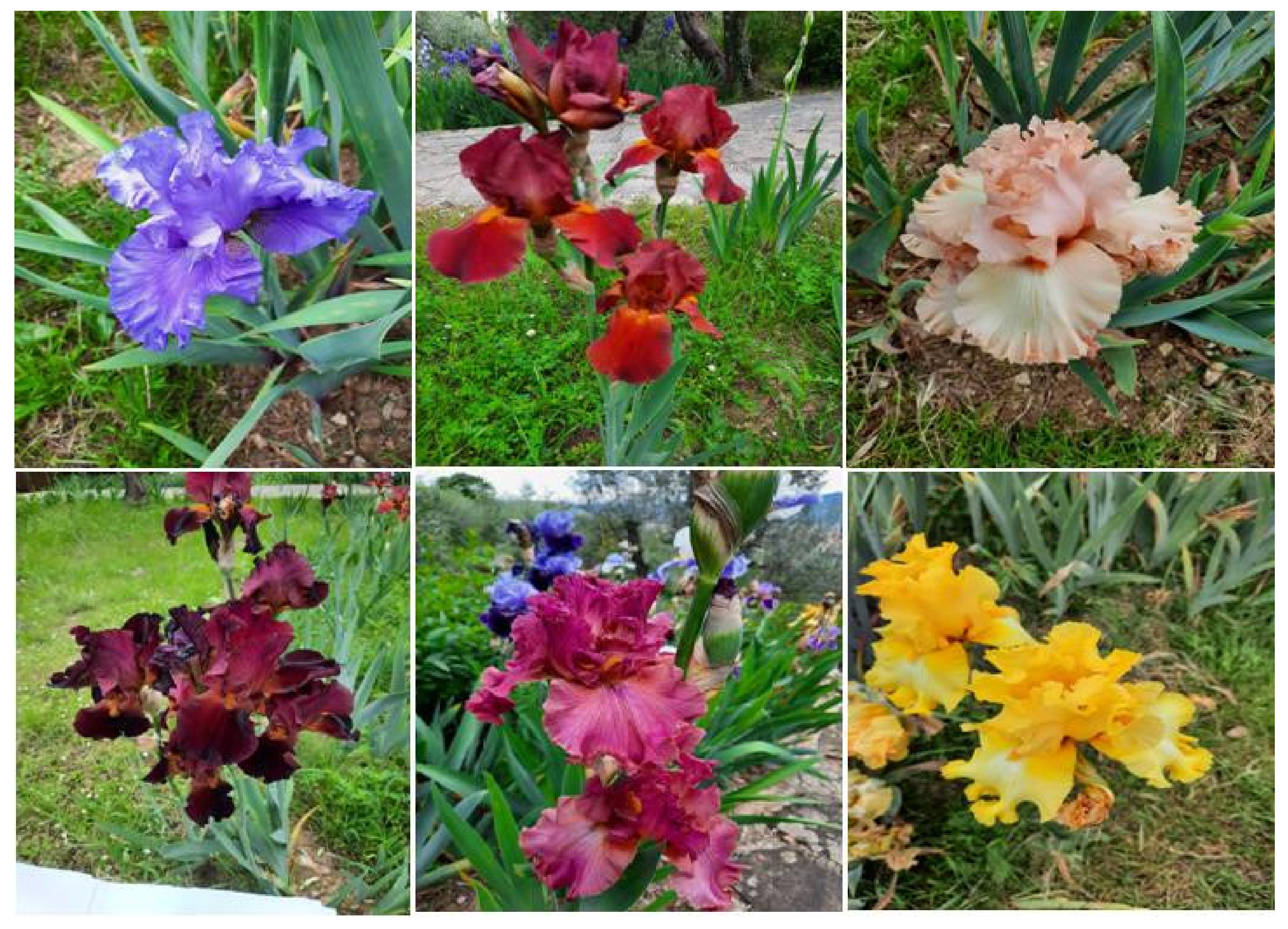 Figure 1. A collection of pictures of various Iris spp. taken at “Iris Garden”, Florence, Italy. ©2022.
Currently, Iris species are still finding application in numerous sectors, including cosmetics, pharmaceutics and the food industry. In Morocco, the rhizomes of Iris species, commonly known as Orris roots, are used as one of the many ingredients in Ras el hanout, a Moroccan spice blend [8]. Similarly, I. germanica L. rhizomes are peeled and used as a flavoring in ice cream, confectionery, baked products and alcoholic beverages [7][9]. In Southern Europe, Iris species are still grown for commercial purposes and are used in tooth powder, toothpaste and teething rings [10], while in the cosmetic field, some Iris spp., such as I. florentina L. and I. germanica L., are currently used in the manufacturing of high-priced luxury perfumes and lotions such as “Iris Ganach”©, Guerlain; “Extravagance d’Amarige”©, Givenchy; “Chanel 19”©; and “So pretty”©, Cartier [10][11][12][13].
Recently, phytochemical investigations of Iris species have resulted in the identification of various bioactive compounds belonging to different classes, including alkaloids [11], flavonoids and their derivatives [12][13][14], quinones, terpenes, steroids and simple phenolics [15]. Modern pharmacological studies have reported that these compounds exhibit significant effects on human health, such as cancer chemopreventive properties [16] and anticancer [17], antioxidant [18], antiplasmodial [19], immunomodulatory and anti-inflammatory activities [20].
2. Botany (Taxonomy, Geographic Distribution and Edaphic Conditions)
The genus Iris (Table 1) is a well-reputed rhizomatous plant belonging to the Iridaceae, a family of herbaceous, perennial and bulbous plants [5]. This plant comprises over 260 species widely distributed in temperate regions across the Northern Hemisphere, occurring particularly across North America and Eurasia, with approximately four species in northern Africa [21][22]. Although numerous Iris species have been found to be growing in mesic or wetland environments, the majority of Iris species thrives in montane, desert, semi-desert, or dry and rocky habitats [22]. Therefore, Iris species can withstand a wide variety of harsh environments, from cold areas where the hard grounds freeze to subtropical climates [10]. In terms of edaphic conditions, several Iris spp., such as I. aucheri (Baker) Sealy and I. persica L., prefer relatively acid soil, whilst the majority grows in slightly acid–alkaline soil, such as I. danfordiae (Baker) Boiss [5][10]. Some other species favor sunny borders with well-drained soil and full shade, whereas others thrive in dappled shade [10].
Table 1. Taxonomy of the genus Iris [23].
| Taxonomic Hierarchy | Classification |
|---|---|
| Kingdom | Plantae |
| Subkingdom | Viridiplantae |
| Infrakingdom | Streptophyta |
| Superdivision | Embryophyta |
| Division | Tracheophyta |
| Subdivision | Spermatophytina |
| Class | Magnoliopsida |
| Superorder | Lilianae |
| Order | Asparagales |
| Family | Iridaceae |
| Genus | Iris L.—Iris |
The genus Iris is identified by the basal fan of unifacial leaves, colorful perianth of three horizontal sepals and three upright petals that are basally fused into the tube and style branches that are fused at the base [24]. They are petaloid and distally expand beyond the tiny flap-like, transverse stigma as a bifid crest; they also have three stamens that are opposite to the sepals and are petaloid in style [22][24].
3. Pharmacological Properties of Iris spp.
3.1. Antioxidant Activity
Antioxidants are stable molecules that scavenge free radicals and maintain a lowered redox state inside cells to prevent or postpone cell damage [25]. The imbalance between free radicals and antioxidants leads to oxidative-stress-related diseases, such as diabetes, cancers, atherosclerosis, and inflammatory and neurodegenerative diseases [26]. Recently, several synthetic antioxidants, such as butylated hydroxytoluene and butylated hydroxyanisole, were discovered to be harmful to human health [26]. As such, the quest for effective, non-toxic, natural substances with potent antioxidative effects has recently intensified.
Studies have shown that there is a substantial relationship between chemical composition and antioxidant activity. In particular, the contents of polyphenols, flavonoids and saponins are responsible for the antioxidant properties. Polyphenolic compounds act as antiradical activity, reducing agents, and complexes of pro-oxidant metals and quenchers of singlet oxygen, promoting the natural antioxidative defense mechanisms and protecting enzyme activity [27]. The genus Iris has been proven to contain substantial amounts of phenolic compounds, particularly flavonoids and their derivatives. Therefore, various extracts of this plant have been evaluated for their antioxidant potency.
Mahdinezhad et al. [28] investigated the in vivo protective effects of I. germanica L. hydroalcoholic extract at doses of 100 and 200 mg/kg on the liver and pancreas of a streptozotocin-induced diabetic rat model for 4 weeks. Accordingly, the repeated oral administration of the extract lowered the high level of aspartate aminotransferase (AST), alanine aminotransferase (ALT) and alkaline phosphatase (ALP) compared with diabetic control rats. The extract also improved the liver antioxidant capacity (increase in thiol groups). The protective effect was ascribed to the significant amounts of flavonoids and anthocyanins in the hydroalcoholic extract. The authors supported the use of the plant as a natural antioxidant source to preserve the human body from free-radical-related disorders, especially diabetes mellitus and hepatic injury [28].
The in vitro antioxidant activity of Iris has been shown to be significantly correlated with the total content of phenolic compounds. The antioxidant activity of petroleum ether, chloroform and methanol crude extracts of fresh I. suaveolens Boiss & Reut rhizomes was tested using the β-carotene–linoleic acid and CUPRAC techniques; quercetin and butylated hydroxytoluene (BHT) served as positive controls [29]. The results disclosed that both petroleum ether and chloroform extracts exhibited pronounced antioxidant potency. Thirteen phenolic and flavonoid compounds were isolated from the petroleum ether and chloroform extracts and were screened in vitro for their antioxidant effects. Coniferaldehyde, a phenolic compound obtained from the chloroform extract, displayed the greatest activity among all the investigated compounds at 25 and 50 mg/mL in both β-carotene-bleaching and CUPRAC systems [29].
Moreover, the aqueous and ethanol extracts of I. germanica L. were evaluated for their in vitro antioxidant activity using several testing systems, namely, free radical scavenging, reducing power, superoxide anion radical scavenging, metal chelating activities and hydrogen peroxide scavenging [30]. The results indicated that at concentrations of 15, 30 and 50 µg/mL both aqueous and ethanol fractions exhibited excellent antioxidant properties, displaying 95.9, 88.4 and 79.9% and 90.5, 78.0 and 65.3% inhibition of peroxidation of linoleic acid emulsion, respectively. At concentrations of 20, 40 and 60 µg/mL, both extracts showed remarkable reducing power, free radical scavenging, hydrogen peroxide scavenging, metal chelating and superoxide anion radical scavenging activities [30].
Similarly, the antioxidant activity of the ethanolic extracts I. germanica L. areal parts and rhizomes was assessed using free radical DPPH scavenging and β-carotene–linoleic acid assays [31]. The results showed that, in the DPPH system, the aerial part and rhizome extracts exhibited significant IC50 values of 5.38 and 12.3 mg/mL, respectively, while at the concentration of 3.15 mg/mL, the total antioxidant activity of the extracts was 98.7% and 97.4%, respectively [31].
In a recent study, the antioxidant activity of the petroleum ether, ethyl acetate and methanol extracts of I. ensata leaves was analyzed using various antioxidant assays such as the DPPH radical scavenging assay and FRAP (ferric ion reducing assay) [32]. Accordingly, all the extracts exhibited pronounced antioxidant potential. In addition, the research reported that the IC50 values decreased with the increase in polarity. In the ferric reducing assay, the IC50 values of the three extracts were found to be 226.66, 188.94 and 124.63 µg/mL, respectively [32].
The genus Iris contains substantial amounts of glycosylated flavonoids and phenolic acids, which are, generally, water-soluble products and can be detected in great quantities in the bloodstream, thus exhibiting high oral bioavailability. Due to all these properties, polyphenols are involved in a wide range of biological effects, such as antibacterial, anti-inflammatory, antiallergic, hepatoprotective, antiviral, antithrombotic, anticarcinogenic, cardioprotective and vasodilatory effects.
3.2. Anticancer Activity
Recently, the use of anticancer drugs has been hampered by the emergence of several impediments, with these mostly being the cellular resistance to chemotherapy drugs and toxicities [33]. Therefore, the global trend is being shifted toward medicinal plants and plant-based compounds owing to their accessibility, affordability and effectiveness [33]. Several Iris-based compounds have been isolated from various extracts and tested in vitro (Table 2) for their cytotoxicity and chemopreventive activities (Figure 2).
 Figure 2. General approach applying to assess the anticancer effect of Iris spp. in vitro.
Irilone, iriflogenin, genistein and iris kashmirianin are only a few of the flavonoids isolated from I. germanica L. that have been shown to exert chemopreventive benefits by reducing cytochrome P450 1A activity and enhancing NAD(P)H: quinone reductase (QR)activity [16].
Alam et al. [34] evaluated the cytotoxicity potential of glycosides and isoflavonoids newly isolated from the rhizomes of I. kashmiriana Baker against several cancer cell lines, namely, MCF-7 and MDA-MB-231 (breast cancer), HeLa (cervical cancer), PC-3 (prostate cancer) and A-549 (lung cancer), using the MTT cellular viability assay. Accordingly, the compounds 5,7,8-trihydroxy-3-(4-methoxyphenyl)-4H-chromen-4-one,5,7,8-trihydroxy-3-(4-hydroxyphenyl)-4H-chromen-4-one,5,7,8-triacetoxyoxy-3-(4-methoxyphenyl)-4H-chromen-4-one and 6,7-diacetoxyoxy-3-(4-methoxyphenyl)-4H-chromen-4-one showed prominent anticancer activity against all cell lines, with IC50 values ranging from 3.8 to 5.6 mg/mL. These compounds were also found to induce cell-cycle block at the G2/M phase [34].
Similarly, Tantry et al. [35] studied the in vitro cytotoxicity activity of a new alkylated 1,4-benzoquinone derivative obtained from the chloroform extract of I. nepalensis rhizomes against various cancer cell lines using the MTT colorimetric assay. The compound revealed remarkable cytotoxicity against HCT116 (colon carcinoma), HL-60 (blood cancer) and ZR-75 (breast cancer), with IC50 values of 10 ± 1.1002, 34 ± 1.1205 and 31 ± 1.1001, respectively. Likewise, the cytotoxicity potential of two flavonoids, 7-O-methylaromadendrin and tectorigenin, as well as four iridal-type triterpenes, iritectols A and B, isoiridogermanal and iridobelamal A, isolated from the rhizomes of I. tectorum Maxim were assessed against four cancer cell lines using the SRB method (sulphorhodamine B) [36]. The results indicated that iritectol B, isoiridogermanal and iridobelamal A displayed identical cytotoxicity against both MCF-7 and C32 cell lines, with IC50 values for a range of 11 µM and 23 µM. Moreover, they found that iritectol B exhibited a dose-dependent apoptotic effect against COR-L23, while both 7-O-methylaromadendrin and tectorigenin flavonoids were discovered to be capable of triggering cell-cycle arrest at the S and G2/M phases, respectively (Table 2). In vivo experiments based on animal models and molecular targets involved in the anticancer effects studies are mandatory to confirm the anticancer potential of Iris spp.
Table 2. In vitro anticancer and cytotoxic activities of Iris spp. extracts against various cell lines.
| Species | Parts | Extract | Cancer Type |
Cell Line | Method | IC50 | Results | References | |
|---|---|---|---|---|---|---|---|---|---|
| I.nertschinskia Lodd. | Rhizomes | EtOH | Breast | MCF-7 | TBE | - | Induced apoptosis; triggered cell cycle block at G1 phase; ↑ p53 phosphorylation in a dose-dependent fashion; ↑ Bax expression; induced caspase-7 cleavage. | [17] | |
| I.nertschinskia Lodd. | Whole plant | EtOH | Breast | Hs578T | TBE | - | Triggered apoptosis hallmarked by cells accumulation in the sub-G 1 phase. | [37] | |
| MDA-MB-231 | |||||||||
| I. pseudopumila Tineo | Rhizomes | PET | Breast | MCF-7 | SRB | 48 h | 96.79 µg/mL | Induced potent cytotoxic effects against the three cell lines. | [38] |
| Skin | C32 | 57 ± 1.04 µg/mL | |||||||
| Kidney | ACHN | 99 ± 1.95 µg/mL | |||||||
| I. variegata L. | Rhizomes | H2O | Skin | IGR39 | MTT | 0.53 mg/mL | Reduced significantly cell viability; the ethanolic extract was shown to be more efficient against both cell lines. | [39] | |
| Breast | MDA-MB-231 | 0.33 mg/mL | |||||||
| I. hungarica Waldst. & Kit. | H2O | Skin | IGR39 | 1.15 mg/mL | |||||
| Breast | MDA-MB-231 | 0.57 mg/mL | |||||||
| 70% EtOH | Skin | IGR39 | 0.53 mg/mL | ||||||
| Breast | MDA-MB-231 | 0.33 mg/mL | |||||||
| I. pseudopumila Tineo | Rhizomes | MeOH | lung | CORL-23 | MTT | 31.5 ± 2.6 µg/mL | Both extracts revealed strong antiproliferative effects towards both cell lines. | [40] | |
| Skin | C32 | 48.7 ± 2.6 µg/mL | |||||||
| Flowers | lung | CORL-23 | 25.4 ± 2.6 µg/mL | ||||||
| Skin | C32 | 50.9 ± 2.6 µg/mL | |||||||
| I. Spuria L. | Rhizomes | MeOH | Lung | A549 | MTT | 123.04 µg/mL | All extracts displayed a dose dependent inhibitory potential against both cell lines A549, and Caco-2. | [41] | |
| Colon | Caco-2 | 302.94 µg/mL | |||||||
| I. kashmiriana Baker | Lung | A549 | 128.7µg/mL | ||||||
| Colon | Caco-2 | 237.76 µg/mL | |||||||
| I. germanica L. | Lung | A549 | 134.72 µg/mL | ||||||
| Colon | Caco-2 | 230.82 µg/mL | |||||||
| I. crocea Jacquem. ex R.C.Foster | Lung | A549 | 149.80 µg/mL | ||||||
| Colon | Caco-2 | 368.88µg/mL | |||||||
| I. ensata Thunb. | Lung | A549 | 137.98 µg/mL | ||||||
| Colon | Caco-2 | 358.81 µg/mL | |||||||
| I. kashmiriana Baker | Whole plant | MeOH | Lung | A549 | MTT | 128.7 µg/mL | The ethanol extract exhibited a dose-dependent selective antiproliferative effect on epithelial cancers. | [42] | |
| Colon | Caco-2 | 237.76 µg/mL | |||||||
| I. hungarica | Rhizomes | H2O | Colon | HCT116 | MTT | 42.3 µg/mL | Cell lines HCT116, HeLa, HL-60 were sensitive to the plant aqueous extract. The highest cytotoxicity was noticed against HL-60. | [43] | |
| Cervical | HeLa | 78.7 µg/mL | |||||||
| Leukemia | HL-60 | 3.6 µg/mL | |||||||
Abbreviations, H2O: aqueous extract; EtOH: ethanol extract; PET: Petroleum ether extract; SRB: Sulforodamine B; TBE: Tris-Borate-EDTA; MTT: 3-(4,5-dimethylthiazol-2-yl)-2,5-diphenyltetrazolium bromide, a tetrazole) assay; Bax: Bcl-2-associated X protein.
3.3. Neuroprotective Activity
The neuroprotective activity of Iris spp. has been shown to be related to the presence of flavonoid compounds, which, interestingly, prevent brain-related diseases due to their powerful antioxidant effect. The neuroprotective effect of the total content of flavonoids extracted from I. tenuifolia Pall was assessed on cultured cortical neurons under oxidative stress induced via H2O2 exposure [44]. Pre-treatment with I. tenuifolia Pall flavonoids prevented H2O2-induced cell death in cortical neuronal cultures. The research reported that the mechanism underlying the neuroprotective effect was related to the activation of both ERK1/2 and was enacted by flavonoid-triggered Shp-2 pathways.
Similarly, the in vivo neuroprotective potential of I. tenuifolia Pall ethanolic extract was evaluated for the first time in a middle cerebral artery occlusion model (MCAO) using C57BL/6J mice [45]. Accordingly, the applications of I. tenuifolia Pall ethanolic extract one hour before or immediately after the surgery outstandingly decreased the infarct size. However, treatment with the same extract less than one hour after surgery did not show any protective effect. The reduction in infarct volume is likely attributable to the richness of I. tenuifolia Pall in flavonoid compounds, which acted as protective agents in the MCAO model due to their significant antioxidant potential. The other factor that might be involved in the protective effect is the activation of both ERK1/2 stimulated by I. tenuifolia Pall flavonoids. The research likewise reported an increase in interleukin-6 concentration in blood plasma. However, the mechanism via which interleukin-6 exerted its protective effects was not determined.
In a similar approach, the in vitro neuroprotective activity of three iridals, namely, Spirioiridotectal A, Spirioiridotectal Band and Spirioiridotectal F, isolated from the ethanolic extract of the rhizomes of I. tectorum Maxim was evaluated at the concentration of 10 μM against serum-deprivation-induced PC12 cell damage using the MTT method [46]. The results revealed that all the tested compounds exhibited moderate neuroprotective effects against serum-deprivation-induced PC12 cell damage. Despite some promising results in terms of neurological disease prevention, the neuroprotective activities of Iris species are still poorly investigated. In vitro and in vivo studies are still mandatory, especially against neurodegenerative diseases such as Alzheimer’s disease.
3.4. Hepatoprotective Activity
The in vivo hepatoprotective activity of the methanolic extract of I. spuria rhizomes was evaluated against paracetamol-induced hepatotoxicity in Wistar rats at the two doses of 100 and 200 mg/kg [47]. The results revealed an increase in serum enzymes and bilirubin level as a sign of hepatic injury in intoxicated rats. Interestingly, the administration of paracetamol along with I. spuria L. methanolic extract was shown to exert a dose-dependent protective effect, bringing the levels of ALT, AST, ALP and total bilirubin to normal ranges as a consequence. Furthermore, the research reported that the methanolic extract restored the serum levels of albumin and glutathione (GSH) and prevented both elevated triglyceride and lipid peroxidation [47].
Likewise, the in vitro hepatoprotective potential of three iridal metabolites, iridojaponal A, B and C, isolated from the ethanolic extract of I. japonica whole plant was assessed against N-acetyl-p-aminophenol (APAP)-induced toxicity in HepG2 cells [48]. Accordingly, iridojaponal A and B exhibited moderate hepatoprotective effects, with cell survival rates of 55.27 and 56.45%, respectively, while the positive control displayed a cell survival rate of 59.28%.
3.5. Anthelmintic Activity
Standard anthelmintic drugs are widely utilized against internal parasites and encompass several classes, such as benzimidazoles and avermectins. They are classified based on their chemical structure and mode of action [49]. Although synthetic anthelmintics have effectively been applied to control helminth infections, their usage has lately been hampered by nematode resistance; they may also affect the host itself and remain as residues in edible tissue [49]. These drawbacks have prompted researchers to look for alternate control strategies, such as using traditional medicinal herbs.
Data have shown that I. hookeriana Linn and I. kashmiriana Linn exhibit significant in vitro and in vivo anthelmintic activities. To corroborate the ethnoveterinary use of I. kashmiriana Linn, Khan et al. [50] evaluated the in vitro anthelmintic activity of I. kashmiriana Linn aqueous and methanolic extracts against Haemonchus contortus nematodes using the motility inhibition test. The positive control was the standard treatment Levamisole 0.5 mg/mL, while the negative control was 0.95% (PBS solution). The worms were exposed to 50, 25 and 12.5 mg/mL crude extracts and their motility was examined 0, 1, 2, 5 and 8 h post-exposure. After 6 h of treatment, the authors observed that the aqueous extract of I. kashmiriana inhibited worm motility by 85.0% at 50 mg/mL, whereas the methanolic extract exhibited better anthelmintic activity, displaying a mean worm-motility inhibition of 100.0%. The anthelmintic effect was attributed to the presence of alcohol-soluble and water-soluble active molecules in the extracts.
Using the same method, Tariq et al. [51] tested the crude aqueous extract and crude ethanolic extract of I. hookeriana Linn rhizomes against Trichuris ovis worms to validate the ethnoveterinary uses of I. hookeriana Linn. They proved that both extracts had significant anthelmintic activity and the highest worm-motility inhibition was exhibited by the ethanolic extract (84.6%) at 25 mg/mL.
Likewise, I. kashmiriana aqueous extract at 2 g/kg body weight exhibited a maximum (70.27%) egg-count reduction in sheep naturally infected with mixed gastrointestinal nematodes after 15 days of treatment [51]. In the same way, I. hookeriana ethanolic extract at 2 g/kg displayed a maximum (45.62%) egg-count reduction in sheep naturally infected with mixed gastrointestinal nematodes after 10 days of treatment. The authors of both studies supported the application of I. hookeriana and I. kashmiriana as natural veterinary agents to control sheep gastrointestinal nematode parasites [50][51].
3.6. Antibacterial Activity
The ethanol/water extracts (70/30, v/v) of I. haphylla L. rhizomes at the concentration of 1% were tested in vitro against standard Gram-positive and Gram-negative bacterium strains. The optimal activity was noticed against the Gram-positive strains, Basillus subtilis ATCC 6633 and Staphyloccocus aureus ATCC 25923, with diameters of growth inhibition of 16.00 and 15.60 nm, respectively. Meanwhile, Gram-negative strains were relatively resistant to the plant extracts [52].
The ethyl acetate fractions derived from 70% of ethanolic extract of I. unguicularis Poir rhizomes at concentrations of 25, 50 and 100 µg/mL were investigated for their antibacterial activity against two Gram-positive and five Gram-negative bacterium strains using the disk diffusion method [18]. The best antibacterial activity was observed against S. aureus (11–23 mm zone of inhibition) followed by B. subtilis (8–13 mm zone of inhibition). The lowest activity was noticed against M. Morganii [18]. The antibacterial activity of the methanolic extract of I. pseudopumila Tineo rhizomes was assessed against four Gram-negative and nine Gram-positive strains using the broth dilution method [53]. The extract exhibited prominent inhibition against all the bacterial strains with minimum inhibitory concentrations (MIC) ranging between 7.8 and 250 μg/mL. It is worth mentioning that the Gram-negative strains, especially E. coli and E. aerogenes, were more sensitive to the Iris species extract.
3.7. Antifungal Activity
The in vitro antifungal activity of I. unguicularis Poir methanolic extract was tested against the Aspergillus Niger 2CA936, Aspergillus flavus NRRL3357 and Candida albicans ATCC1024 fungal strains [54]. The results revealed that the methanolic extract exhibited potent antifungal properties, mainly against Aspergillus Niger 2CA936. I. unguicularis Poir antifungal activity was attributed to the lipophilic properties of the phenolic compounds. The essential oils of I. persica L. extracted from flowers, leaves and rhizomes were evaluated against three human pathogenic fungal strains, Candida albicans, Trichophyton mentagrophytes and Microsporum canis, using the broth microdilution assay. All the extracts exhibited moderate antifungal properties.The research also reported that the highest antifungal activity was detected for essential oils extracted from leaves and flowers.
Moreover, the antifungal activity of iridal, a triterpenoid compound isolated from the rhizomes of I. germanica L., was performed against Plasmodium falciparum chloroquine-resistant and -sensitive strains. Iridal was less effective against both fungal strains, with minimal inhibitory concentration values exceeding 50 mg/mL from 24 to 48 h of incubation [19]. Furthermore, the ethanolic extract of I. hungarica rhizomes was evaluated in vitro against Candida albicans ATCC 653/885 at the concentration of 1%. The fungal strain was interestingly sensitive to the ethanolic extract, with 16.30 nm as a diameter of growth inhibition [52].
3.8. Antiviral Activity
The aqueous and ethanolic extracts of I. sibirica L. were evaluated against herpes simplex virus type 1. Accordingly, the rhizome ethanolic extract was the most effective on the herpes simplex virus when compared with the aqueous extract [55].
3.9. Antidiabetic Activity
Standard antidiabetic drugs, especially α-amylase and α-glucosidase inhibitors, have recently been linked to a number of serious side effects in humans, including diarrhea, bloating and abdominal pain [56]. Thus, researchers have switched their attention to a plethora of medicinal plants that have been exploited by indigenous people worldwide, which has led to a rich know-how related to diabetes treatment. Researchers have lent credence to their ethnomedicinal uses and identified many bioactive compounds endowed with substantial antidiabetic activity, primarily flavonoids and phenolic acids [57].
Although there are more than 260 accepted species of the genus Iris worldwide, data have shown that the only Iris spp. that have been evaluated for their antidiabetic activity are I. germanica L. and I. ensata Thunb. In this sense, Mahdinezhad et al. [28] studied the hypoglycemic effect of the hydroalcoholic extract of I. germanica L. rhizomes on streptozotocin-induced diabetic rats. The repeated oral administration of the doses of 100 and 200 mg/kg for 4 weeks significantly decreased the levels of glucose, triglycerides and oxidative stress markers levels such as ALT (alanine aminotransferase), AST (aspartate aminotransferase) and ALP (alkaline phosphatase). The authors stated that the antihyperglycemic and antihypertriglyceridemic effects of I. germanica L. could be attributed to the abundance of phenolic constituents in the hydroalcoholic extract, especially anthocyanins.
Furthermore, Suresh et al. [58] used normal, glucose-loaded and streptozotocin-induced diabetic rats to evaluate the hyperglycemic effect of I. Ensata Thunb dried root extract for 21 days. The authors reported that the oral administration of the extract reduced blood glucose in both normal and streptozotocin-diabetic rats. They associated the observed effect with the capacity of the extract to lower the intestinal uptake of glucose (digestive-enzyme inhibition), increase the glucose absorption at the tissue level (sensitize the cells) and enhance the activity of the β-cells of the pancreas.
On the other hand, the increase in blood glucose levels is mainly ascribed to the degradation of carbohydrates in the intestine, which is under the control of α-amylase, β-amylase and α-glucosidase [59]. Inhibiting or slowing down the activity of these key enzymes might be an effective therapeutic approach for preventing glucose from entering the bloodstream [56].
Therefore, Ibrahim et al. [60] identified eight known isoflavonoids, as well as two novel isoflavonoids, 8-hydroxyirilone 5-methyl ether and 8-hydroxyirilone, from the methanolic extract of I.germanica L. powdered rhizomes. Using acarbose as a reference, they assessed the in vitro α-amylase inhibitory potency of these compounds. They reported that, among all the tested components, 8-hydroxyirilone 5-methyl ether, 8-hydroxyirilone, irilone and irisolidone exhibited prominent α-amylase inhibitory capacity at the concentration of 250 μg/mL with inhibition rates of 66.1, 78.3, 67.3 and 70.1%, respectively. They indicated that the α-amylase inhibitory potency increased with the presence of C-7 hydroxyl and C-5 hydroxyl or with the methylation of the hydroxyl groups in the A and B rings of isoflavonoids.
References
- Kicel, A. An Overview of the Genus Cotoneaster (Rosaceae): Phytochemistry, biological activity, and toxicology. Antioxidants 2020, 9, 1002.
- Sahoo, N.; Manchikanti, P.; Dey, S. Herbal drugs: Standards and regulation. Fitoterapia 2010, 81, 462–471.
- Van Wyk, B.-E.; Wink, M. Phytomedicines, Herbal Drugs, and Poisons; The University of Chicago Press: Chicago, IL, USA, 2015; pp. 1–304.
- Zougagh, S.; Belghiti, A.; Rochd, T.; Zerdani, I.; Mouslim, J. Medicinal and Aromatic Plants Used in Traditional Treatment of the Oral Pathology: The Ethnobotanical Survey in the Economic Capital Casablanca, Morocco (North Africa). Nat. Prod. Bioprospect. 2019, 9, 35–48.
- Fan, L.; Gao, Y.; Hasenstein, K.H.; Wang, L. ‘Flower Angel’: A New Iris sanguinea Cultivar. HortScience 2021, 56, 617–618.
- Roguz, K.; Gallagher, M.K.; Senden, E.; Bar-Lev, Y.; Lebel, M.; Heliczer, R.; Sapir, Y. All the Colors of the Rainbow: Diversification of Flower Color and Intraspecific Color Variation in the Genus Iris. Front. Plant Sci. 2020, 11, 1519.
- Crişan, I.; Cantor, M. New perspectives on medicinal properties and uses of Iris sp. Hop. Med. Plants 2016, 24, 24–36.
- Lim, T.K. Edible Medicinal and Non-Medicinal Plants: Modified Stems, Roots, Bulbs; Springer International Publishing: Cham, Switzerland, 2016; Volume 11, pp. 1–392.
- Crișan, I.; Vidican, R.; Olar, L.; Stoian, V.; Morea, A.; Ştefan, R. Screening for changes on Iris germanica L. rhizomes following inoculation with arbuscular mycorrhiza using Fourier transform infrared spectroscopy. Agronomy 2019, 9, 815.
- Austin, C. Irises. A Gardener’s Encyclopedia; Timber Press: Portland, OR, USA, 2005.
- Xie, G.; Qin, X.; Chen, Y.; Wen, R.; Wu, S.; Qin, M. Alkaloids from the Rhizomes of Iris germanica. Chem. Nat. Compd. 2017, 53, 196–198.
- Amin, H.I.M.; Hussain, F.H.S.; Najmaldin, S.K.; Thu, Z.M.; Ibrahim, M.F.; Gilardoni, G.; Vidari, G. Phytochemistry and Biological Activities of Iris Species Growing in Iraqi Kurdistan and Phenolic Constituents of the Traditional Plant Iris postii. Molecules 2021, 26, 264.
- Mykhailenko, O. Composition of volatile oil of Iris pallida Lam. from Ukraine. Turk. J. Pharm. Sci. 2018, 15, 85–90.
- Wang, H.; Cui, Y.; Zhao, C. Flavonoids of the genus Iris (Iridaceae). Mini Rev. Med. Chem. 2010, 10, 643–661.
- Kukula-Koch, W.; Sieniawska, E.; Widelski, J.; Urjin, O.; Głowniak, P.; Skalicka-Woz’niak, K. Major secondary metabolites of Iris spp. Phytochem. Rev. 2015, 14, 51–80.
- Wollenweber, E.; Stevens, J.F.; Klimo, K.; Knauft, J.; Frank, N.; Gerhäuser, C. Cancer chemopreventive in vitro activities of isoflavones isolated from Iris germanica. Planta Med. 2003, 69, 15–20.
- Shin, J.S.; Hong, S.W.; Lee, J.G.; Lee, Y.M.; Kim, D.W.; Kim, J.E.; Jung, D.J.; An, S.K.; Hong, N.J.; Kim, D.; et al. An ethanol extract of Iris nertschinskia induces p53-dependent apoptosis in the MCF7 human breast cancer cell line. Int. J. Mol. Med. 2011, 27, 401–405.
- Bensari, S.; Ouelbani, R.; Yimaz, M.A.; Bensouici, C.; Gokalp, E.; Khelifi, D. Phytochemical profiles of Iris unguicularis Poir. with antioxidant, antibacterial, and anti-Alzheimer activities. Acta Nat. Sci. 2020, 7, 74–87.
- Benoit-Vical, F.; Imbert, C.; Bonfils, J.P.; Sauvaire, Y. Antiplasmodial and antifungal activities of iridal, a plant triterpenoid. Phytochemistry 2003, 62, 747–751.
- Nazir, N. Immunomodulatory activity of isoflavones isolated from Iris kashmiriana: Effect on T-lymphocyte proliferation and cytokine production in Balb/c mice. Biomed. Prev. Nutr. 2013, 3, 151–157.
- Qi, X.Y.; Fan, L.J.; Gao, Y.; Shang, Y.; Liu, H.Y.; Wang, L. ‘NEFU-1′: A new Iris sanguine cultivar. HortScience 2020, 55, 109–111.
- Wilson, C.A. Subgeneric classification in Iris re-examined using chloroplast sequence data. Taxon 2011, 60, 27–35.
- Hussain, H.; Al-Harrasi, A.; Green, I.R.; Rehman, U. Iris (Iris germanica) Oils. In Essential Oils in Food Preservation, Flavor and Safety; Preedy, V.R., Ed.; Elsevier: London, UK, 2016; pp. 481–486.
- Kaššák, P. Secondary metabolites of the choosen genus Iris species. Acta Univ. Agric. Silvic. Mendel. Brun. 2013, 60, 269–280.
- Henriksen, E.J. Role of oxidative stress in the pathogenesis of insulin resistance and type 2 diabetes. In Bioactive Food as Dietary Interventions for Diabetes; Academic Press: London, UK; Oxford, UK; San Diego, CA, USA; Cambridge, MA, USA, 2019; pp. 3–17.
- Lobo, V.; Patil, A.; Phatak, A.; Chandra, N. Free radicals, antioxidants and functional foods: Impact on human health. Pharmacogn. Rev. 2010, 4, 118.
- Huwaitat, S.; Al-Khateeb, E.; Finjan, S.; Maraqa, A. Antioxidant and antimicrobial activities of Iris nigricans methanolic extracts containing phenolic compounds. Eur. Sci. J. 2018, 9, 83–91.
- Mahdinezhad, M.R.; Hooshmand, S.; Soukhtanloo, M.; Jamshidi, S.T.; Ehtiati, S.; Ghorbani, A. Protective effects of a standardized extract of Iris germanica on pancreas and liver in streptozotocin-induced diabetic rats. Int. J. Pharm. Sci. Res. 2021, 16, 71.
- Hacıbekiroğlu, I.; Kolak, U. Antioxidant and anticholinesterase constituents from the petroleum ether and chloroform extracts of Iris suaveolens. Phytother. Res. 2011, 25, 522–529.
- Nadaroğlu, H.; Demir, Y.; Demir, N. Antioxidant and radical scavenging properties of Iris germanica. Pharm. Chem. J. 2007, 41, 409–415.
- Machalska, A.; Skalicka-Woźniak, K.; Widelski, J.; Głowniak, K.; Purevsuren, G.; Oyun, Z.; Khishgéé, D.; Urjin, B. Screening for phenolic acids in five species of iris collected in Mongolia. Acta Chromatogr. 2008, 20, 259–267.
- Ganaie, A.A.; Mishra, R.P.; Allaie, A.H. Antioxidant activity of some extracts of Iris ensata. J. Pharmacogn. Phytochem. 2018, 7, 230–235.
- Deyno, S.; Eneyew, K.; Seyfe, S.; Wondim, E. Efficacy, safety and phytochemistry of medicinal plants used for the management of diabetes mellitus in Ethiopia: A systematic review. Clin. Phytoscience 2021, 7, 16.
- Alam, A.; Jaiswal, V.; Akhtar, S.; Jayashree, B.S. Isolation of isoflavones from Iris kashmiriana Baker as potential anti proliferative agents targeting NF-κB. Phytochemistry 2017, 136, 70–80.
- Tantry, M.A.; Ghazanfar, K.; Zargar, U.R. New alkylated benzoquinone from Iris nepalensis. Nat. Prod. Res. 2013, 27, 1832–1836.
- Fang, R.; Houghton, P.J.; Hylands, P.J. Cytotoxic effects of compounds from Iris tectorum on human cancer cell lines. J. Ethnopharmacol. 2008, 118, 257–263.
- Shin, J.S.; Maeng, H.G.; Hong, S.W.; Moon, J.H.; Kim, J.S.; Suh, Y.A.; Kim, E.S.; Choi, E.K.; Kim, I.; Lee, S.K.; et al. Iris Nertschinskia ethanol extract differentially induces cytotoxicity in human breast cancer cells depending on AKT1/2 activity. Asian Pac. J. Cancer Prev. 2012, 13, 6511–6516.
- Rigano, D.; Conforti, F.; Formisano, C.; Menichini, F.; Senatoter, F. Comparative free radical scavenging potential and cytotoxicity of different extracts from Iris pseudopumila Tineo flowers and rhizomes. Nat. Prod. Res. 2009, 23, 17–25.
- Mykhailenko, O.; Korinek, M.; Ivanauskas, L.; Bezruk, I.; Myhal, A.; Petrikaitė, V.; El-Shazly, M.; Lin, G.H.; Lin, C.H.; Yen, C.H.; et al. Qualitative and Quantitative Analysis of Ukrainian Iris Species: A Fresh Look on Their Antioxidant Content and Biological Activities. Molecules 2020, 25, 4588.
- Conforti, F.; Menichini, F.; Rigano, D.; Senatore, F. Antiproliferative activity on human cancer cell lines after treatment with polyphenolic compounds isolated from Iris pseudopumila flowers and rhizomes. Z. Nat. C 2009, 64, 490–494.
- Wani, S.H.; Padder, B.A.; Mokhdomi, T.; Mir, J.I.; Bhat, H.A.; Hassan, Q.P.; Qadri, R.A. Antiproliferative activity of methanolic extracts of different Iris plant species against A549 and Caco-2 cell lines. J. Pharmacogn. Phytochem. 2017, 6, 1034–1037.
- Amin, A.; Wani, S.H.; Mokhdomi, T.A.; Bukhari, S.; Wafai, A.H.; Mir, J.I.; Hassan, Q.P.; Qadri, R.A. Investigating the pharmacological potential of Iris kashmiriana in limiting growth of epithelial tumors. Pharmacogn J. 2013, 5, 170–175.
- Mykhailenko, O.; Lesyk, R.; Finiuk, N.; Stoika, R.; Yushchenko, T.; Ocheretniuk, A.; Vaschuk, V.; Mishchenko, V.; Georgiyants, V. In vitro anticancer activity screening of Iridaceae plant extracts. J. Appl. Pharm. Sci. 2020, 10, 59–63.
- Jalsrai, A.; Numakawa, T.; Numakawa, Y.; Adachi, N.; Kunugi, H. Phosphatase-mediated intracellular signaling contributes to neuroprotection by flavonoids of Iris tenuifolia. Am. J. Chin. Med. 2014, 42, 119–130.
- Jalsrai, A.; Reinhold, A.; Becker, A. Ethanol Iris tenuifolia extract reduces brain damage in a mouse model of cerebral ischaemia. Phytother. Res. 2018, 32, 333–339.
- Zhang, C.L.; Wang, Y.; Liu, Y.F.; Ni, G.; Liang, D.; Luo, H.; Song, X.Y.; Zhang, W.Q.; Chen, R.Y.; Chen, N.H.; et al. Iridal-type triterpenoids with neuroprotective activities from Iris tectorum. J. Nat. Prod. 2014, 77, 411–415.
- Akther, N.; Andrabi, K.; Nissar, A.; Ganaie, S.; Chandan, B.K.; Gupta, A.P.; Khuswant, M.; Sultana, S.; Shawl, A.S. Hepatoprotective activity of LC–ESI-MS standardized Iris spuria rhizome extract on its main bioactive constituents. Phytomedicine 2014, 21, 1202–1207.
- Shi, G.R.; Wang, X.; Liu, Y.F.; Zhang, C.L.; Ni, G.; Chen, R.Y.; Chen, D.Q. Novel iridal metabolites with hepatoprotective activities from the whole plants of Iris japonica. Tetrahedron Lett. 2016, 57, 5761–5763.
- Romero-González, R.; Garrido Frenich, A.; Martínez Vidal, J.L. Veterinary Drugs Residues: Anthelmintics. Encycl. Food Saf. 2014, 45–54.
- Khan, A.; Tak, H.; Nazir, R.; Lone, B.A. In vitro and in vivo anthelmintic activities of Iris kashmiriana Linn. J. Saudi Soc. Agric. Sci. 2018, 17, 235–240.
- Tariq, K.A.; Chishti, M.Z.; Ahmad, F.; Shawl, A.S.; Tantray, M.A. Evaluation of anthelmintic activity of Iris hookeriana against gastrointestinal nematodes of sheep. J. Helminthol. 2008, 82, 135–141.
- Mykhailenk, O.; Kovalyov, V.; Kovalyov, S.; Krechun, A. Isoflavonoids from the rhizomes of Iris hungarica and antibacterial activity of the dry rhizomes extract. Ars Pharm. 2017, 58, 39–45.
- Rigano, D.; Grassia, A.; Formisano, C.; Basile, A.; Sorbo, S.; Sorbo, F. Antibacterial and allelopathic activity of methanolic extract from Iris pseudopumila rhizomes. Fitoterapia 2006, 77, 460–462.
- Sofiane, G.; Wafa, N.; Loubna, A. Evaluation of antioxidant and antifungal activities of methanolic aerial part extract of Iris unguicularis Poiret. Asian J. Plant Sci. Res. 2016, 6, 18–23.
- Tikhomirova, L.I.; Ilyicheva, T.N. Preparation of biotechnological raw materials of Iris sibirica L. with a given content of mangiferin and antiviral activity. IOP Conf. Ser. Earth Environ. 2020, 421, 022049.
- Gong, L.; Feng, D.; Wang, T.; Ren, Y.; Liu, Y.; Wang, J. Inhibitors of α-amylase and α-glucosidase: Potential linkage for whole cereal foods on prevention of hyperglycemia. Food Sci. Nutr. 2020, 8, 6320–6337.
- Bouyahya, A.; El Omari, N.; Elmenyiy, N.; Guaouguaou, F.; Balahbib, A.; Belmehdi, O.; Salhi, N.; Imtara, H.; Mrabti, H.N.; El-Shazly, M.; et al. Moroccan antidiabetic medicinal plants: Ethnobotanical studies, phytochemical bioactive compounds, preclinical investigations, toxicological validations and clinical evidences; challenges, guidance and perspectives for future management of diabetes worldwide. Trends Food Sci. 2021, 115, 147–254.
- Suresh, D.K.; Ahemad, W.; Khalid, M.S.; Aasim, S.M. Anti-hyperglycemic activity of iris ensata Thunb root extracts in normal, glucose fed and streptozotocin induced diabetic rats. Adv. Pharmacol. Toxicol. 2010, 11, 93.
- Lin, A.H.M.; Nichols, B.L.; Quezada-Calvillo, R.; Avery, S.E.; Sim, L.; Rose, D.R.; Naim, H.Y.; Hamaker, B.R. Unexpected high digestion rate of cooked starch by the Ct-maltase-glucoamylase small intestine mucosal α-glucosidase subunit. PLoS ONE 2012, 7, e35473.
- Ibrahim, S.R.; Mohamed, G.A.; Zayed, M.F.; Ross, S.A. 8-Hydroxyirilone 5-methyl ether and 8-hydroxyirilone, new antioxidant and α-amylase inhibitors isoflavonoids from Iris germanica rhizomes. Bioorg. Chem. 2017, 70, 192–198.
More
Information
Subjects:
Plant Sciences
Contributor
MDPI registered users' name will be linked to their SciProfiles pages. To register with us, please refer to https://encyclopedia.pub/register
:
View Times:
1.8K
Revisions:
3 times
(View History)
Update Date:
29 Mar 2022
Table of Contents


Notice
You are not a member of the advisory board for this topic. If you want to update advisory board member profile, please contact office@encyclopedia.pub.
OK
Confirm
Only members of the Encyclopedia advisory board for this topic are allowed to note entries. Would you like to become an advisory board member of the Encyclopedia?
Yes
No
${ textCharacter }/${ maxCharacter }
Submit
Cancel
Back
Comments
${ item }
|
${ item.createdUser.fullName }
${ item.createdAt }
${ item.vote }
${ item.reply }
Delete
${ reply.createdUser.fullName }
${ reply.createdAt }
${ reply.vote }
Delete
There is no reply to this comment~
${ item.replyTextCharacter }/${ item.replyMaxCharacter }
Submit
Cancel
More
No more~
There is no comment~
${ textCharacter }/${ maxCharacter }
Submit
Cancel
${ selectedItem.replyTextCharacter }/${ selectedItem.replyMaxCharacter }
Submit
Cancel
Confirm
Are you sure to Delete?
Yes
No