Submitted Successfully!
To reward your contribution, here is a gift for you: A free trial for our video production service.
Thank you for your contribution! You can also upload a video entry or images related to this topic.
Version Summary Created by Modification Content Size Created at Operation
1 + 3577 word(s) 3577 2022-03-14 03:27:43 |
2 format Meta information modification 3577 2022-03-28 03:51:09 | |
3 Remove from the EC Meta information modification 3577 2022-03-28 04:52:37 |

Video Upload Options

Do you have a full video?

Confirm

Are you sure to Delete?
Cite
If you have any further questions, please contact Encyclopedia Editorial Office.
Suzuki, H. Research on Neuroelectric Disruption after Aneurysmal Subarachnoid Hemorrhage. Encyclopedia. Available online: https://encyclopedia.pub/entry/21056 (accessed on 27 July 2024).
Suzuki H. Research on Neuroelectric Disruption after Aneurysmal Subarachnoid Hemorrhage. Encyclopedia. Available at: https://encyclopedia.pub/entry/21056. Accessed July 27, 2024.
Suzuki, Hidenori. "Research on Neuroelectric Disruption after Aneurysmal Subarachnoid Hemorrhage" Encyclopedia, https://encyclopedia.pub/entry/21056 (accessed July 27, 2024).
Suzuki, H. (2022, March 25). Research on Neuroelectric Disruption after Aneurysmal Subarachnoid Hemorrhage. In Encyclopedia. https://encyclopedia.pub/entry/21056
Suzuki, Hidenori. "Research on Neuroelectric Disruption after Aneurysmal Subarachnoid Hemorrhage." Encyclopedia. Web. 25 March, 2022.
Research on Neuroelectric Disruption after Aneurysmal Subarachnoid Hemorrhage
Edit

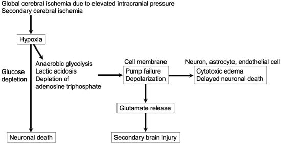
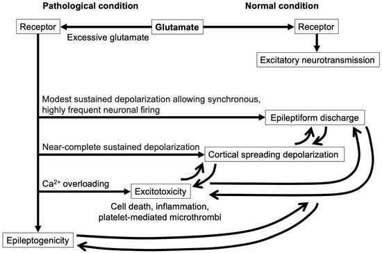
Delayed cerebral ischemia (DCI) remains a challenging but very important condition, because DCI is preventable and treatable for improving functional outcomes after aneurysmal subarachnoid hemorrhage (SAH). The pathologies underlying DCI are multifactorial. Classical approaches to DCI focus exclusively on preventing and treating the reduction of blood flow supply. However, recently, glutamate-mediated neuroelectric disruptions, such as excitotoxicity, cortical spreading depolarization and seizures, and epileptiform discharges, have been reported to occur in high frequencies in association with DCI development after SAH. Each of the neuroelectric disruptions can trigger the other, which augments metabolic demand. If increased metabolic demand exceeds the impaired blood supply, the mismatch leads to relative ischemia, resulting in DCI. The neuroelectric disruption also induces inverted vasoconstrictive neurovascular coupling in compromised brain tissues after SAH, causing DCI. Although glutamates and the receptors may play central roles in the development of excitotoxicity, cortical spreading ischemia and epileptic activity-related events, more studies are needed to clarify the pathophysiology and to develop novel therapeutic strategies for preventing or treating neuroelectric disruption-related DCI after SAH.

cortical spreading depolarization delayed cerebral ischemia early brain injury excitotoxicity glutamate inflammation microcirculation receptor seizure subarachnoid hemorrhage

1. Introduction

A rupture of an intracranial aneurysm causes subarachnoid hemorrhage (SAH), for which the prognosis remains poor [1]. Aneurysmal rupture-induced elevation of intracranial pressure (ICP) and extravasated intracranial blood components trigger early brain injury (EBI) and systemic complications such as cardiopulmonary dysfunction and systemic inflammatory response syndrome, which are sometimes fatal [2][3]. Delayed cerebral ischemia (DCI) is an important modifiable prognostic factor and develops at day four or later post-SAH in patients surviving the initial aneurysmal rupture [4]. Cerebral vasospasm has classically been considered to be only cause of DCI, but now multiple concurrent and synergistic mechanisms have been suggested as a cause of DCI [5]. EBI may be a precursor or a contributor to DCI, and therefore some pathophysiologies may be shared or interrelated between EBI and DCI [6]. These shared or interrelated pathophysiologies may include cortical spreading depolarization (CSD) [7], which is intimately related to epileptic discharge and excitotoxicity, leading to metabolic derangement, that is, mismatch of metabolic supply and demand, with the resultant relative cerebral ischemia and neuronal death [8][9]. In fact, recent clinical studies reported that CSD and epileptic discharge were in frequently observed in association with the development of DCI after aneurysmal SAH [10][11]. A common inducer of CSD and epileptic discharge, glutamate, was also reported to increase in brain parenchyma after SAH, followed by the development of DCI [12].

2. Glutamate

Brain tissues contain high concentrations of free glutamates, in the 5–15 mmol/kg range intracellularly, which are involved in endogenous neural signaling in multiple pathways as a major excitatory amino acid neurotransmitter throughout the central nervous system [13]. Glutamates are released from the presynaptic vesicles, while excess glutamates are removed from the extracellular space by astrocytes and endothelial cells with uptake and metabolizing functions, and are exhausted into the blood through diffusion when endothelial glutamate concentrations become higher than in the blood [14]. Thus, pathologically excessive glutamates are caused by a derangement between glutamate release and reuptake.

2.1. Signaling via Glutamates

In normal brain, glutamate is the most abundant neurotransmitter used by excitatory synapses [15]. Once released into the synaptic cleft from presynaptic vesicles in neurons and astrocytes, glutamates activate postsynaptic glutamate receptors, which consist of ionotropic glutamate receptors and metabotropic glutamate receptors (mGluRs). Ionotropic glutamate receptors are ligand-gated ion channels regulating fast synaptic transmission, including α-amino-3-hydroxy-5-methyl-4-isoxazolepropionic acid (AMPA; GluA1–4), N-methyl-D-aspartate (NMDA; GluN1, GluN2A–D, and GluN3A, B), kainate (GluK1–5), and δ (GluD1 and GluD2) receptors, and mGluRs (mGluR1–8) G-protein coupled receptors, leading to activation of intracellular metabolic pathways to modulate postsynaptic responses, synapse activities, and glutamate releases and to regulate different cell functions ranging from cell cycle to gene expression [16][17][18][19]. AMPA receptors are the first ones to be activated by released glutamates and then to depolarize the postsynaptic membrane, defining the strength of the postsynaptic responses to activate downstream signaling pathways, resulting in the propagation of the excitatory signal [15]. That is, synaptic activity initially stimulates the influx of sodium ions (Na+) through AMPA receptor channels, which increases the concentrations of positively charged ions in the cytoplasm, causing cell depolarization [20]. As a result, NMDA receptor channels become permeable to calcium ions (Ca2+), and Ca2+ influx activates Ca2+/calmodulin-dependent protein kinase II, protein kinase C, protein kinase A, and tyrosine kinase, which phosphorylate AMPA and NMDA receptors [20]. The affinity of AMPA receptors for glutamates is relatively low, and the number of glutamate molecules bound to AMPA receptors determines the open probability; in contrast, NMDA receptors have a higher affinity for glutamates and desensitize slower than AMPA receptors, but the slow binding rate puts a considerable limit on the opening probability of NMDA receptors during the short-lived glutamate peak [21]. This neurotransmitter system not only drives physiological excitatory signal transmission as well as abnormal hyperexcitable circuitry during a seizure but also may initiate neurodegenerative processes by excessive calcium uptake and pushing cells toward apoptotic cell death [22].

2.2. Major Glutamate Receptors

AMPA receptors are highly dynamic receptors formed by tetrameric assembly of four subunits, GluA1–4, and their functional properties largely depend on the composition of these subunits [23]. AMPA receptors are primarily of the GluA1/GluA2 and GluA2/GluA3 configuration [17]. GluA1–4 are widely expressed in both neurons and glia, but the predominantly expressed subunits are GluA1 and GluA2 [24]. GluA1–3 are expressed in the majority of neurons in the nervous system of mature animals, while GluA4 is primarily expressed early in development and in cerebellar granule neurons and some populations of interneurons in the mature brain [25]. The protein levels of GluA1 subunits were higher than that of GluA2 subunits in mature neurons [26]. AMPA receptors are not normally Ca2+ permeable, by virtue of their GluA2 subunits [27]. AMPA receptors lacking GluA2 subunits or containing unedited (Q form) GluA2 subunits are rendered permeable to Ca2+ [17]. The subunit switch of GluA1 and GluA2 may lead to the excessive intracellular Ca2+ influx, resulting in neuronal injury after SAH [28].
NMDA receptors are composed of three subunits (GluN1–3) and are involved in various processes from learning and memory to neurodegeneration [19]. At the synaptic level, GluN2A is activated to mediate the prosurvival signaling, while abruptly elevated extracellular glutamates stimulate the extrasynaptic GluN2B to trigger excitotoxic neuronal death [18]. As well, under nonexcitotoxic conditions, mGluR1α couples to the neuroprotective phosphoinositide 3-kinase (PI3K)-Akt signaling cascades [29]; however, under excitotoxicity, excess stimulation of NMDA receptors activates Ca2+-dependent protease calpain, which causes calpain-mediated truncation of mGluR1α at Ser936 [30]. The truncated mGluR1α disrupts the link between the PI3K-Akt signaling and mGluR1α, while the truncated mGluR1α-mediated intracellular Ca2+ release from the endoplasmic reticulum is maintained and contributes to Ca2+ overloading through the enhancement of NMDA receptor-mediated Ca2+ influx [30]. The truncated mGluR1α translocates to axons and enhances glutamate releases and thereby excitotoxicity, suggesting that there is a positive feedback loop between the truncation of mGluR1α and excitotoxicity [30]. Endogenous releases of glutamates and NMDA receptors were also reported to be involved in both CSDs and epileptiform activities [31].
There are eight major mGluRs and several splice variants, subclassified into three groups (I, II, and III) based on the structure, G-protein coupling or function, and ligand selectivity: Group I (mGluR1 and mGluR5) is linked to activation of phospholipase C (PLC); and Groups II (mGluR2 and mGluR3) and III (mGluR4, mGluR6, mGluR7, and mGluR8) are linked to inhibition of adenylate cyclase to change levels of cyclic adenosine monophosphate [32][33]. mGluR1 is one of the most abundantly expressed mGluRs in the mammalian brain [16]. mGluR modulates synaptic transmission and plasticity and probably plays no primary roles in mediating excitotoxic brain injuries; however, mGluR influences excitotoxic injuries and can be the secondary therapeutic target in strokes [34]. Group I mGluRs-activated PLC generates diacylglycerol and inositol 1,4,5-trisphosphate triggering Ca2+ release from endoplasmic reticulum, which together through protein kinase C can enhance excitotoxic Ca2+ entry through NMDA receptors, reverse operation of the electrogenic Na+/Ca2+ exchangers and membrane Na+/hydrogen ions (H+) exchangers, and activate phospholipase A2, promoting reactive oxygen species (ROS) formation and lipid peroxidation [13]. In contrast, Group II and III mGluRs usually exert inhibitory effects on neural circuits and thereby anti-excitotoxic effects [35], although neuronal mGluR2 activation may enhance excitotoxicity, possibly by limiting the release of γ-aminobutyric acid (GABA) [36].

2.3. Glutamate in Blood Vessels

AMPA, NMDA, kainate receptors, and mGluRs are all expressed on cerebral microvascular cells and perivascular astrocytic processes [37][38][39]. Glutamates contribute to the dilatory tone of cerebral microvessels under physiological conditions via these receptors [40]. In normal brain tissues with preserved neurovascular coupling, an increase in neuronal activities is associated with arteriolar dilation to increase local blood flow (functional hyperemia) via glutamate release [5]. That is, the activated neuron releases glutamates, which bind to at least mGluRs and NMDA receptors on perivascular astrocytes and trigger intracellular Ca2+ increases to activate large-conductance Ca2+-activated potassium ions (K+) channels for outflow of K+ at astrocytic end-feet [5][40]. As a result, overall mild increases in perivascular K+ concentrations (<20 mM) hyperpolarize arteriolar smooth muscle cells and induce vasorelaxation [41]. Glutamates have been also demonstrated to increase vascular permeability through activation of NMDA receptors in rat cerebral cortex [42] and induce an increase in cultured brain endothelial cell permeability via the action on endothelial AMPA, NMDA, kainate receptors, and group I and III mGluRs [43][44]. Glutamate-mediated activation of group I or III mGluRs disturbs the barrier function of endothelial cells by promoting dephosphorylation of vasodilator-stimulated phosphoproteins (VASPs), which increases actin filament formation as well as cell retraction, thus impairing cell-cell junctions [43][45]. On binding of glutamates, mGluR1 and mGluR5 activate multiple intracellular signaling pathways, such as PLC, protein kinase C, and mitogen-activated protein kinase pathways [16]. mGluR1 and mGluR5 expressed on glial and Purkinje cells are activated by glutamates and are reported to promote the secretion of thromboxane A2, endothelin-1, and 20-hydroxyeicosatetraenoic acid by these cells, leading to constriction of microvessels [46][47].

2.4. Glutamate in SAH

Aneurysmal rupture-induced elevated ICP and the subsequent global cerebral ischemia induce energy storage loss, metabolic failure, and disturbed ionic hemostasis, resulting in plasma membrane depolarization, which causes an excessive and uncontrolled release of neurotransmitters such as glutamates (Figure 1) [48][49]. Neurons release glutamates under decreased cerebral blood flow of less than 20 mL/100 g brain/min [50]. SAH increases the permeability of the paravascular space, through which blood components and the degradation products are perfused into the brain parenchyma, inducing intraparenchymal microvascular constriction, inflammation, and microthrombus formation [51]. Post-SAH blood-brain barrier (BBB) disruption is also associated with abluminal and intraparenchymal platelet aggregates [41]. Although massive SAH and secondary tissue ischemia induce ROS and proinflammatory cytokines, which damage arterial, capillary and venous endothelial cells to activate inflammatory cells and platelets, leading to microthrombus formation throughout the entire cerebral hemisphere even distant from the aneurysm rupture site [5], platelet-mediated microthrombi were reported to release glutamates [52]. Furthermore, excessive glutamates were synthesized and released by activated astrocytes, microglia, and neutrophils in experimental SAH [4]. On the other hand, excitatory amino acid transporters on astrocytes, which uptake glutamates, were downregulated in experimental SAH, at least partly explaining the excessive glutamates and thereby the excitotoxicity in EBI [53].
Figure 1. Glutamate release at the rupture of intracranial aneurysm. Released glutamate causes secondary brain injury.
In a clinical setting, cerebral glutamate levels are considered to increase within minutes after aneurysmal SAH [12]. Studies using cerebral microdialysis showed that glutamate levels in an acute phase were already high in poor-grade SAH patients with neurological impairments and cerebral edema; a trend toward normalization of the values was associated with clinical improvement, whereas further deterioration led to permanent neurological deficits [12]. In addition, elevated intraparenchymal concentrations of glutamates at 1 to 7 days post-SAH were an independent predictor of DCI and 12-month poor outcomes in clinical settings [12]. Cerebrospinal fluid levels of glutamates were significantly correlated with angiographic vasospasm and DCI [54][55]. In a rat model of SAH, an increase in glutamate concentrations was accompanied by vasospasm of the basilar artery [56].
Excessive glutamate overactivates the receptors, which mediate intracellular Ca2+ overload by Ca2+ influx from the extracellular space through ionotropic glutamate receptors as well as intracellular Ca2+ release from the endoplasmic reticulum via mGluRs [5][57]. Then, excitotoxicity or CSD follows and causes mitochondrial dysfunction and compromised energy metabolism, inducing ionic imbalance; as a result, neurons, astrocytes, pericytes, and vascular endothelial cells constituting the neurovascular unit develop apoptotic or necrotic cell death [5][49]. Persistent CSDs are known to be followed by spreading depression of electrocorticographic activities and increased glutamate releases, the latter of which leads to excitotoxicity [5]. A positive feedback loop was also reported between the stimulation of glutamate receptors such as the NMDA receptor or mGluR1 and glutamate releases [57].

2.5. Glutamate and Inverse Neurovascular Coupling after SAH

CSD and seizure or epilepsy are distinct entities, but both develop related to glutamate releases after aneurysmal SAH and have similar toxic effects, such as increased metabolic demand, inverse vasoconstrictive neurovascular coupling, disruption of the BBB, and cellular death [4]. CSD may lower the threshold of seizure (inappropriate neuronal firing) [58], while seizure may induce CSD (Figure 2) [59].
Figure 2. Glutamate action via the receptors. In normal conditions, glutamate is a major excitatory neurotransmitter via the receptor. In pathological conditions, excessive glutamates activate the receptors excessively and cause epileptiform discharges, cortical spreading depolarization, and excitotoxicity, depending on the extent of the cell membrane depolarization. Each of the neuroelectric disruptions triggers the other. Surviving cells irrespective of these events achieve epileptogenicity.
In SAH, hemolysis causes an increase in basal perivascular K+ concentrations and a decrease in basal nitric oxide (NO) [5]. In addition, infiltration of blood degradation products into intraparenchymal perivascular space induces higher amplitude in spontaneous Ca2+ oscillations in hypertrophic astrocyte end-feet surrounding parenchymal arterioles, causing a surge of extracellular and perivascular K+ [5]. Thus, a physiological mechanism of neurovascular coupling, that is, activated neuron-induced glutamate release, causes an excessive concentration (>20 mM) and impaired clearance of perivascular K+, which result in depolarization of parenchymal arteriolar smooth muscle cells, inducing vasoconstriction or pathological inversion of neurovascular coupling [41]. The phenomenon may form the basis of spreading ischemia and DCI development associated with CSDs after aneurysmal SAH [5]. In experimental SAH models in rats or mice, impaired neurovascular coupling developed time-dependently up to 96 h, and any neuronal or metabolic activation such as sensory stimulation, an increase in carbon dioxide, and a decrease in pH resulted in parenchymal arteriolar constriction, mismatch of cerebral metabolism and blood flow, and relative cerebral ischemia, causing further brain damage after SAH [60].

3. Excitotoxity in Post-SAH Ischemic Brain

Excitotoxity contributes to both EBI and DCI after aneurysmal SAH. In EBI, massive aneurysmal rupture causes severe elevation of ICP, followed by transient cerebral circulation arrest, which leads to cessation of neuronal electrical activity within seconds, mitochondrial dysfunction associated with decreased production of adenosine triphosphate to deteriorate the energy state and to disrupt the Na+-K+ pump, and ion homeostasis, resulting in disturbed membrane ion gradients (depolarization), Ca2+ influx, and extracellular release of a large amount of glutamates from depolarized nerve terminals and astrocytes within minutes [61]. In DCI, secondary cerebral ischemia also triggers excessive glutamate releases. Massive releases of glutamates over-activate AMPA, NMDA, and kainate receptors on neurons, as well as other cellular components of the neurovascular unit, causing excessive intracellular Ca2+ entry as the primary mediator of excitotoxicity through the receptors, which is augmented by Ca2+ releases from endoplasmic reticulum via activation of mGluRs [13][61]. Intracellular Ca2+ overload induces further release of glutamates and overactivation of multiple Ca2+-dependent enzymes such as calpains, other proteases, protein kinases, calcineurins, endonucleases, phospholipases A2, and xanthine oxidases [13]. The activation of theses enzymes impairs mitochondrial energy production and causes increased production of ROS to promote lipid peroxidation, membrane failure, and cell damage, as well as alterations in the organization of the cytoskeleton, activation of genetic signals leading to cell death, and an increase in expressions of immediate early genes [13][33][62]. Cellular damage also causes further glutamate releases [13]. In addition, Ca2+ influx activates neuronal NO synthases to produce NO, which may be involved in both normal neuronal signaling and free-radical-mediated glutamate excitotoxicity harnessed by macrophages [13][63]. Ca2+ overload and oxidative stress, including NO, cause apoptosis and necrosis, which can occur caspase-dependently or -independently [13]. Glutamates also increase deoxyribonucleic acid binding of the redox-regulated transcription factors, nuclear factor-κB, and activating protein 1, as well as upregulate the immediate early gene, c-fos, leading to glutamate-induced apoptosis or necrosis [33]. Glutamate levels in cerebrospinal fluid and peripheral blood have been reported to be positively correlated with infarct size, infarct growth, and functional outcomes in clinical settings [64]. In contrast, the neuroprotective neurotransmitter GABA rapidly and transiently increases in the extracellular space like most neurotransmitters, but the expressions of both GABA-A and GABA-B receptors decrease after cerebral ischemia to cause impaired GABA-mediated neurotransmission, which contributes to ongoing neuronal excitability and possibly to neuronal death [61]. Excitotoxic neuronal death is not a uniform event but, rather, a continuum of necrotic, apoptotic, and autophagic morphologies [33].
Excitotoxicity is composed of two components: the first one is an acute, intracellular influx of Na+ and chloride ions followed by water influx, resulting in cell swelling, tissue edema and, consequently, impaired perfusion of the surrounding brain tissues, even in the absence of extracellular Ca2+; the second one is Ca2+-dependent delayed cellular degeneration [62]. In contrast to neuronal swelling, which developed immediately after excessive glutamate exposure, delayed neuronal death was observed 24 h post-glutamate exposure and was abolished by the removal of Ca2+ while potentiated by the addition of Ca2+ [18]. The removal of Na+ from the culture medium prior to glutamate exposure prevented neuronal swelling but had no effects on delayed neuronal death [18].

3.1. Glutamate Receptors and Ions in Excitotoxicity

Although the NMDA receptor was initially considered to be a critical mediator in focal cerebral ischemia, subsequent studies support a more central role for AMPA receptors in hippocampal injuries associated with global cerebral ischemia [65]. AMPA receptors mediate Na+ influx and therefore can contribute to excitotoxic Ca2+ overload and neuronal death [13]. Although AMPA receptors lack direct linkage to NO synthases and nicotinamide adenine dinucleotide phosphate (NADPH) oxidases, AMPA receptors participate substantially in brain damage after focal and global ischemia; in global ischemia, AMPA receptors typically contribute more than NMDA receptors to delayed death of selectively vulnerable neurons, possibly due to upregulation of Ca2+-permeable AMPA receptors [13]. Ischemia upregulates Ca2+-permeable AMPA receptors in vulnerable neuronal populations, which constitute a dominant route for toxic Ca2+/zinc ion (Zn2+) entry [66].
In addition to Na+ and Ca2+, H+ participates in excitotoxicity. In ischemic brain tissues, extracellular pH typically drops within minutes toward 6.5 or lower due to anaerobic glycolysis for resynthesis of adenosine triphosphate, and an increase in extracellular H+ attenuates NMDA receptor channel openings and NADPH oxidase 2 activity, resulting in reduced NMDA receptor-mediated excitotoxicity [67]. However, ischemic acidosis is itself cytotoxic to both neurons and glia by enhancing neurotoxic Ca2+ overload via the gating of acid-sensing ion channels and by being accompanied by an increase in intracellular Zn2+ [13]. Relatively low concentrations (≈20 μM) of Zn2+ elicit apoptotic neuronal death, while higher concentrations (50–100 μM) of Zn2+ cause neuronal death, with the characteristics of necrosis [68]. In addition to promoting neuronal death, intracellular release of excitotoxic Zn2+ contributes to the death of adjacent non-neuronal cells such as astrocytes, oligodendroglia, and capillary endothelial cells in ischemic brain tissues [13]. An intracellular increase in Zn2+ is considered to mediate peroxynitrite-induced death through activation of extracellular signal-regulated kinases 1/2 and arachidonate 12-lipoxygenases and thereby further ROS generation [69], and to upregulate intercellular adhesion molecule-1 expression in vascular endothelial cells, promoting leukocyte attraction and microvascular leakage [70][71]. Several other membrane channels may be activated in part as a result of overstimulation of glutamate receptors and can contribute to toxic Ca2+/Zn2+ overload and other ionic derangements in ischemic brain tissues [13].
Although excitotoxicity was originally described as specific to neurons, oligodendrocytes and astrocytes also suffer excitotoxic injury and death [13]. Astrocytes are less insensitive to excitotoxicity compared with oligodendrocytes: this is because most astrocytes express AMPA receptors and mGluRs, but NMDA receptors and Ca2+-permeable AMPA receptors are generally not abundant [72][73][74]. However, as astrocytes are vulnerable to Zn2+ or H+-induced damages, astrocytic death may increase secondary to excitotoxicity occurring in nearby neurons or oligodendrocytes [13].

3.2. Relationships among Inflammation, Microthrombus, and Excitotoxity

Excitotoxicity per se triggers and augments inflammatory reactions to continue destroying brain tissues. The neurovascular unit-constituent cells release cytokines and chemokines, recruiting leukocytes to the evolving ischemic region over hours to days, and advance microvascular damage and oxidative stress [75][76]. Inducible NO synthases are induced in infiltrating neutrophils and endothelial cells in ischemic brain tissues to produce NO and to synergize oxidatively with superoxide emanating from neutrophil NADPH oxidase 2 and endothelial NADPH oxidase 4, aggravating brain damage [13]. Activated microglia are a significant source of redundant extracellular glutamates that induce excitotoxic neuronal death [33]. Although microglia-mediated neuroinflammation in EBI may have impacts on neuronal excitotoxicity, BBB disruption, and the further changes of immune responses, all of which may lower the seizure threshold, activated microglia themselves may promote epilepsy development independent of the inflammatory responses [77].
In DCI after SAH, platelet aggregates are also observed to be associated with or without focal microvascular constriction and to be extravasated into the brain parenchyma by platelet-mediated release of collagenase and subsequent depletion of collagen IV in vessel walls [78]. The extravasated platelet aggregates or platelet-mediated microthrombi are reported not only to propagate pro-inflammatory signaling [79] but also to release glutamates, causing excitotoxic brain injuries [52]. Although glutamate does not cross the BBB, BBB disruption at sites of microthrombi or extravasated platelets that release glutamates during their lysis or aggregation may allow neurons to be exposed to excessive glutamates [52]. Platelets have dense granules, carrying a considerable amount of glutamates, and also express glutamate receptors on their surface [80]. Excessive glutamate is reported to induce platelet activation and synthesis of thrombogenic peptides, plasminogen activator inhibitor-1, and hypoxia-inducible factor-2α from pre-existing messenger ribonucleic acids in anucleate platelets, which are mediated mostly through AMPA receptors [80].

References

  1. Kanamaru, H.; Kawakita, F.; Asada, R.; Miura, Y.; Shiba, M.; Toma, N.; Suzuki, H.; pSEED group. Prognostic factors varying with age in patients with aneurysmal subarachnoid hemorrhage. J. Clin. Neurosci. 2020, 76, 118–125.
  2. Suzuki, H.; Fujimoto, M.; Kawakita, F.; Liu, L.; Nakatsuka, Y.; Nakano, F.; Nishikawa, H.; Okada, T.; Kanamaru, H.; Imanaka-Yoshida, K.; et al. Tenascin-C in brain injuries and edema after subarachnoid hemorrhage: Findings from basic and clinical studies. J. Neurosci. Res. 2020, 98, 42–56.
  3. Suzuki, H. Inflammation: A good research target to improve outcomes of poor-grade subarachnoid hemorrhage. Transl. Stroke Res. 2019, 10, 597–600.
  4. Geraghty, J.R.; Testai, F.D. Delayed Cerebral Ischemia after Subarachnoid Hemorrhage: Beyond Vasospasm and Towards a Multifactorial Pathophysiology. Curr. Atheroscler. Rep. 2017, 19, 50.
  5. Suzuki, H.; Kanamaru, H.; Kawakita, F.; Asada, R.; Fujimoto, M.; Shiba, M. Cerebrovascular pathophysiology of delayed cerebral ischemia after aneurysmal subarachnoid hemorrhage. Histol. Histopathol. 2021, 36, 143–158.
  6. Suzuki, H. What is early brain injury? Transl. Stroke Res. 2015, 6, 1–3.
  7. Xiao, M.; Li, Q.; Feng, H.; Zhang, L.; Chen, Y. Neural vascular mechanism for the cerebral blood flow auto-regulation after hemorrhagic stroke. Neural Plast. 2017, 2017, 5819514.
  8. Dreier, J.P.; Major, S.; Pannek, H.W.; Woitzik, J.; Scheel, M.; Wiesenthal, D.; Martus, P.; Winkler, M.K.; Hartings, J.A.; Fabricius, M.; et al. Spreading convulsions, spreading depolarization and epileptogenesis in human cerebral cortex. Brain 2012, 135, 259–275.
  9. Nakano, F.; Liu, L.; Kawakita, F.; Kanamaru, H.; Nakatsuka, Y.; Nishikawa, H.; Okada, T.; Shiba, M.; Suzuki, H. Morphological characteristics of neuronal death after experimental subarachnoid hemorrhage in mice using double immunoenzymatic technique. J. Histochem. Cytochem. 2019, 67, 919–930.
  10. Chung, D.Y.; Oka, F.; Ayata, C. Spreading depolarizations: A therapeutic target against delayed cerebral ischemia after subarachnoid hemorrhage. J. Clin. Neurophysiol. 2016, 33, 196–202.
  11. Zafar, S.F.; Postma, E.N.; Biswal, S.; Boyle, E.J.; Bechek, S.; O’Connor, K.; Shenoy, A.; Kim, J.; Shafi, M.S.; Patel, A.B.; et al. Effect of epileptiform abnormality burden on neurologic outcome and antiepi-leptic drug management after subarachnoid hemorrhage. Clin. Neurophysiol. 2018, 129, 2219–2227.
  12. Helbok, R.; Kofler, M.; Schiefecker, A.J.; Gaasch, M.; Rass, V.; Pfausler, B.; Beer, R.; Schmutzhard, E. Clinical Use of Cerebral Microdialysis in Patients with Aneurysmal Subarachnoid Hemorrhage—State of the Art. Front. Neurol. 2017, 8, 565.
  13. Choi, D.W. Excitotoxicity: Still Hammering the Ischemic Brain in 2020. Front. Neurosci. 2020, 14, 579953.
  14. Castillo, J.; Loza, M.I.; Mirelman, D.; Brea, J.; Blanco, M.; Sobrino, T.; Campos, F. A novel mechanism of neuroprotection: Blood glutamate grabber. J. Cereb. Blood Flow Metab. 2016, 36, 292–301.
  15. Baranovic, J. AMPA receptors in the synapse: Very little space and even less time. Neuropharmacology 2021, 196, 108711.
  16. Niswender, C.M.; Conn, P.J. Metabotropic glutamate receptors: Physiology, pharmacology, and disease. Annu. Rev. Pharmacol. Toxicol. 2010, 50, 295–322.
  17. Casillas-Espinosa, P.M.; Powell, K.L.; O’Brien, T.J. Regulators of synaptic transmission: Roles in the pathogenesis and treatment of epilepsy. Epilepsia 2020, 53 (Suppl. S9), 41–58.
  18. Lai, T.W.; Zhang, S.; Wang, Y.T. Excitotoxicity and stroke: Identifying novel targets for neuroprotection. Prog. Neurobiol. 2014, 115, 157–188.
  19. Serwach, K.; Gruszczynska-Biegala, J. STIM Proteins and Glutamate Receptors in Neurons: Role in Neuronal Physiology and Neurodegenerative Diseases. Int. J. Mol. Sci. 2019, 20, 2289.
  20. Lau, A.; Tymianski, M. Glutamate receptors, neurotoxicity and neurodegeneration. Pflug. Arch. Eur. J. Physiol. 2010, 460, 525–542.
  21. Scheefhals, N.; MacGillavry, H.D. Functional organization of postsynaptic glutamate receptors. Mol. Cell. Neurosci. 2018, 91, 82–94.
  22. Casillas-Espinosa, P.M.; Ali, I.; O’Brien, T.J. Neurodegenerative pathways as targets for acquired epilepsy therapy development. Epilepsia Open 2020, 5, 138–154.
  23. Hollmann, M.; Heinemann, S. Cloned glutamate receptors. Annu. Rev. Neurosci. 1994, 17, 31–108.
  24. Isaac, J.T.; Ashby, M.C.; McBain, C.J. The role of the GluR2 subunit in AMPA receptor function and synaptic plasticity. Neuron 2007, 54, 859–871.
  25. Diering, G.H.; Huganir, R.L. The AMPA receptor code of synaptic plasticity. Neuron 2018, 100, 314–329.
  26. Chang, D.T.W.; Reynolds, I.J. Differences in mitochondrial movement and morphology in young and mature primary cortical neurons in culture. Neuroscience 2006, 141, 727–736.
  27. Doyle, K.P.; Simon, R.P.; Stenzel-Poore, M.P. Mechanisms of ischemic brain damage. Neuropharmacology 2008, 55, 310–318.
  28. Chen, T.; Zhu, J.; Wang, Y.H. RNF216 mediates neuronal injury following experimental subarachnoid hemorrhage through the Arc/Arg3.1-AMPAR pathway. FASEB J. 2020, 34, 15080–15092.
  29. Chong, Z.Z.; Li, F.; Maiese, K. Group I metabotropic receptor neuroprotection requires Akt and its substrates that govern FOXO3a, Bim, and beta-catenin during oxidative stress. Curr. Neurovasc. Res. 2006, 3, 107–117.
  30. Xu, W.; Wong, T.P.; Chery, N.; Gaertner, T.; Wang, Y.T.; Baudry, M. Calpain-mediated mGluR1alpha truncation: A key step in excitotoxicity. Neuron 2007, 53, 399–412.
  31. Marrannes, R.; Willems, R.; De Prins, E.; Wauquier, A. Evidence for a role of the N-methyl-D-aspartate (NMDA) receptor in cortical spreading depression in the rat. Brain Res. 1988, 457, 226–240.
  32. Pin, J.P.; Duvoisin, R. The metabotropic glutamate receptors: Structure and functions. Neuropharmacology 1995, 34, 1–26.
  33. Wang, Y.; Qin, Z.H. Molecular and cellular mechanisms of excitotoxic neuronal death. Apoptosis 2010, 15, 1382–1402.
  34. Sun, Y.; Feng, X.; Ding, Y.; Li, M.; Yao, J.; Wang, L.; Gao, Z. Phased Treatment Strategies for Cerebral Ischemia Based on Glutamate Receptors. Front. Cell. Neurosci. 2019, 13, 168.
  35. Bruno, V.; Battaglia, G.; Copani, A.; Giffard, R.G.; Raciti, G.; Raffaele, R.; Shinozaki, H.; Nicoletti, F. Activation of class II or III metabotropic glutamate receptors protects cultured cortical neurons against excitotoxic degeneration. Eur. J. Neurosci. 1995, 7, 1906–1913.
  36. Corti, C.; Battaglia, G.; Molinaro, G.; Riozzi, B.; Pittaluga, A.; Corsi, M.; Mugnaini, M.; Nicoletti, F.; Bruno, V. The use of knock-out mice unravels distinct roles for mGlu2 and mGlu3 metabotropic glutamate receptors in mechanisms of neurodegeneration/neuroprotection. J. Neurosci. 2007, 27, 8297–8308.
  37. Matsui, K.; Jahr, C.E. Differential control of synaptic and ectopic vesicular release of glutamate. J. Neurosci. 2004, 24, 8932–8939.
  38. Parfenova, H.; Fedinec, A.; Leffler, C.W. Ionotropic glutamate receptors in cerebral microvascular endothe-lium are functionally linked to heme oxygenase. J. Cereb. Blood Flow Metab. 2003, 23, 190–197.
  39. Brand-Schieber, E.; Lowery, S.L.; Werner, P. Select ionotropic glutamate AMPA/kainate receptors are expressed at the astrocyte-vessel interface. Brain Res. 2004, 1007, 178–182.
  40. Fergus, A.; Lee, K.S. Regulation of cerebral microvessels by glutamatergic mechanisms. Brain Res. 1997, 754, 35–45.
  41. Tso, M.K.; Macdonald, R.L. Subarachnoid hemorrhage: A review of experimental studies on the microcirculation and the neurovascular unit. Transl. Stroke Res. 2014, 5, 174–189.
  42. Vazana, U.; Veksler, R.; Pell, G.S.; Prager, O.; Fassler, M.; Chassidim, Y.; Roth, Y.; Shahar, H.; Zangen, A.; Raccah, R.; et al. Glutamate-mediated blood-brain barrier opening: Implications for neuroprotection and drug delivery. J. Neurosci. 2016, 36, 7727–7739.
  43. Collard, C.D.; Park, K.A.; Montalto, M.C.; Alapati, S.; Buras, J.A.; Stahl, G.L.; Colgan, S.P. Neutrophil-derived glutamate regulates vascular endothelial barrier function. J. Biol. Chem. 2002, 277, 14801–14811.
  44. András, I.E.; Deli, M.A.; Veszelka, S.; Hayashi, K.; Hennig, B.; Toborek, M. The NMDA and AMPA/KA receptors are involved in glutamate-induced alterations of occludin expression and phosphorylation in brain endothelial cells. J. Cereb. Blood Flow Metab. 2007, 27, 1431–1443.
  45. Pula, G.; Krause, M. Role of Ena/VASP proteins in homeostasis and disease. Handb. Exp. Pharmacol. 2008, 186, 39–65.
  46. Mulligan, S.J.; MacVicar, B.A. Calcium transients in astrocyte endfeet cause cerebrovascular constrictions. Nature 2004, 431, 195–199.
  47. Rancillac, A.; Rossier, J.; Guille, M.; Tong, X.K.; Geoffroy, H.; Amatore, C.; Arbault, S.; Hamel, E.; Cauli, B. Glutamatergic control of microvascular tone by distinct GABA neurons in the cerebellum. J. Neurosci. 2006, 26, 6997–7006.
  48. Kanamaru, H.; Suzuki, H. Potential therapeutic molecular targets for blood-brain barrier disruption after subarachnoid hemorrhage. Neural Regen. Res. 2019, 14, 1138–1143.
  49. Van Lieshout, J.H.; Dibué-Adjei, M.; Cornelius, J.F.; Slotty, P.J.; Schneider, T.; Restin, T.; Boogaarts, H.D.; Steiger, H.J.; Petridis, A.K.; Kamp, M.A. An introduction to the pathophysiology of aneurysmal subarachnoid hemorrhage. Neurosurg. Rev. 2018, 41, 917–930.
  50. Mindt, S.; Tokhi, U.; Hedtke, M.; Groß, H.J.; Hänggi, D. Mass spectrometry-based method for quantification of nimodipine and glutamate in cerebrospinal fluid. Pilot study with patients after aneurysmal subarachnoid haemorrhage. J. Clin. Pharm. Ther. 2020, 45, 81–87.
  51. Luo, C.; Yao, X.; Li, J.; He, B.; Liu, Q.; Ren, H.; Liang, F.; Li, M.; Lin, H.; Peng, J.; et al. Paravascular pathways contribute to vasculitis and neuroinflammation after subarachnoid hemorrhage independently of glymphatic control. Cell Death Dis. 2016, 7, e2160.
  52. Bell, J.D.; Thomas, T.C.; Lass, E.; Ai, J.; Wan, H.; Lifshitz, J.; Baker, A.J.; Macdonald, R.L. Platelet-mediated changes to neuronal glutamate receptor expression at sites of microthrombosis following experimental subarachnoid hemorrhage. J. Neurosurg. 2014, 121, 1424–1431.
  53. Feng, D.; Wang, W.; Dong, Y.; Wu, L.; Huang, J.; Ma, Y.; Zhang, Z.; Wu, S.; Gao, G.; Qin, H. Ceftriaxone allevi-ates early brain injury after subarachnoid hemorrhage by increasing excitatory amino acid transporter 2 expression via the PI3K/Akt/NF-κB signaling pathway. Neuroscience 2014, 268, 21–32.
  54. Jung, C.S.; Lange, B.; Zimmermann, M.; Seifert, V. CSF and Serum Biomarkers Focusing on Cerebral Vasospasm and Ischemia after Subarachnoid Hemorrhage. Stroke Res. Treat. 2013, 2013, 560305.
  55. Wang, H.B.; Wu, Q.J.; Zhao, S.J.; Hou, Y.J.; Li, H.X.; Yang, M.F.; Wang, B.J.; Sun, B.L.; Zhang, Z.Y. Early high cerebrospinal fluid glutamate: A potential predictor for delayed cerebral ischemia after aneurysmal subarachnoid hemorrhage. ACS Omega 2020, 5, 15385–15389.
  56. Wu, C.T.; Wen, L.L.; Wong, C.S.; Tsai, S.Y.; Chan, S.M.; Yeh, C.C.; Borel, C.O.; Cherng, C.H. Temporal changes in glutamate, glutamate transporters, basilar arteries wall thickness, and neuronal variability in an experimental rat model of subarachnoid hemorrhage. Anesth. Analg. 2011, 112, 666–673.
  57. Zhang, Z.; Liu, J.; Fan, C.; Mao, L.; Xie, R.; Wang, S.; Yang, M.; Yuan, H.; Yang, X.; Sun, J.; et al. The GluN1/GluN2B NMDA receptor and metabotropic glutamate receptor 1 negative allosteric modulator has enhanced neuroprotection in a rat subarachnoid hemorrhage model. Exp. Neurol. 2018, 301, 13–25.
  58. Ahmadabad, R.A.; Ghadiri, M.K.; Gorji, A. The role of Toll-like receptor signaling pathways in cerebrovascular disorders: The impact of spreading depolarization. J. Neuroinflamm. 2020, 17, 108.
  59. Kramer, D.R.; Fujii, T.; Ohiorhenuan, I.; Liu, C.Y. Cortical spreading depolarization: Pathophysiology, implications, and future directions. J. Clin. Neurosci. 2016, 24, 22–27.
  60. Balbi, M.; Koide, M.; Wellman, G.C.; Plesnila, N. Inversion of neurovascular coupling after subarachnoid hemorrhage in vivo. J. Cereb. Blood Flow Metab. 2017, 37, 3625–3634.
  61. Amantea, D.; Bagetta, G. Excitatory and inhibitory amino acid neurotransmitters in stroke: From neurotoxicity to ischemic tolerance. Curr. Opin. Pharmacol. 2017, 35, 111–119.
  62. Arundine, M.; Tymianski, M. Molecular mechanisms of glutamate-dependent neurodegeneration in ischemia and traumatic brain injury. Cell. Mol. Life Sci. 2004, 61, 657–668.
  63. Bredt, D.S.; Hwang, P.M.; Snyder, S.H. Localization of nitric oxide synthase indicating a neural role for nitric oxide. Nature 1990, 347, 768–770.
  64. Nicolo, J.-P.; Chen, Z.; Moffat, B.; Wright, D.K.; Sinclair, B.; Glarin, R.; Neal, A.; Thijs, V.; Seneviratne, U.; Yan, B.; et al. Study protocol for a phase II randomised, double-blind, placebo-controlled trial of perampanel as an antiepileptogenic treatment following acute stroke. BMJ Open 2021, 11, e043488.
  65. Pulsinelli, W.; Sarokin, A.; Buchan, A. Antagonism of the NMDA and non-NMDA receptors in global versus focal brain ischemia. Prog. Brain Res. 1993, 96, 125–135.
  66. Pellegrini-Giampietro, D.E.; Zukin, R.S.; Bennett, M.V.; Cho, S.; Pulsinelli, W.A. Switch in glutamate receptor subunit gene expression in CA1 subfield of hippocampus following global ischemia in rats. Proc. Natl. Acad. Sci. USA 1992, 89, 10499–10503.
  67. Giffard, R.G.; Monyer, H.; Christine, C.W.; Choi, D.W. Acidosis reduces NMDA receptor activation, glutamate neurotoxicity, and oxygen-glucose deprivation neuronal injury in cortical cultures. Brain Res. 1990, 506, 339–342.
  68. Kim, Y.H.; Kim, E.Y.; Gwag, B.J.; Sohn, S.; Koh, J.Y. Zinc-induced cortical neuronal death with features of apoptosis and necrosis: Mediation by free radicals. Neuroscience 1999, 89, 175–182.
  69. Zhang, Y.; Wang, H.; Li, J.; Dong, L.; Xu, P.; Chen, W.; Neve, R.L.; Volpe, J.J.; Rosenberg, P.A. Intracellular zinc release and ERK phosphorylation are required upstream of 12-lipoxygenase activation in peroxynitrite toxicity to mature rat oligodendrocytes. J. Biol. Chem. 2006, 281, 9460–9470.
  70. Yeh, S.C.; Tsai, F.Y.; Chao, H.R.; Tsou, T.C. Zinc ions induce inflammatory responses in vascular endothe-lial cells. Bull. Environ. Contam. Toxicol. 2011, 87, 113–116.
  71. Sumagin, R.; Lomakina, E.; Sarelius, I.H. Leukocyte-endothelial cell interactions are linked to vascular permeability via ICAM-1-mediated signaling. Am. J. Physiol. Heart Circ. Physiol. 2008, 295, H969–H977.
  72. Skowrońska, K.; Obara-Michlewska, M.; Zielińska, M.; Albrecht, J. NMDA Receptors in Astrocytes: In Search for Roles in Neurotransmission and Astrocytic Homeostasis. Int. J. Mol. Sci. 2019, 20, 309.
  73. Ceprian, M.; Fulton, D. Glial Cell AMPA Receptors in Nervous System Health, Injury and Disease. Int. J. Mol. Sci. 2019, 20, 2450.
  74. Bradley, S.J.; Challiss, R.A.J. G protein-coupled receptor signalling in astrocytes in health and disease: A focus on metabotropic glutamate receptors. Biochem. Pharmacol. 2012, 84, 249–259.
  75. Anrather, J.; Iadecola, C. Inflammation and stroke: An overview. Neurotherapeutics 2016, 13, 661–670.
  76. Jayaraj, R.L.; Azimullah, S.; Beiram, R.; Jalal, F.Y.; Rosenberg, G.A. Neuroinflammation: Friend and foe for ischemic stroke. J. Neuroinflamm. 2019, 16, 142.
  77. Wang, J.; Liang, J.; Deng, J.; Liang, X.; Wang, K.; Wang, H.; Qian, D.; Long, H.; Yang, K.; Qi, S. Emerging role of microglia-mediated neuroinflammation in epilepsy after subarachnoid hemorrhage. Mol. Neurobiol. 2021, 58, 2780–2791.
  78. Friedrich, V.; Flores, R.; Muller, A.; Sehba, F.A. Luminal platelet aggregates in functional deficits in parenchymal vessels after subarachnoid hemorrhage. Brain Res. 2010, 1354, 179–187.
  79. McBride, D.W.; Blackburn, S.L.; Peeyush, K.T.; Matsumura, K.; Zhang, J.H. The Role of Thromboinflammation in Delayed Cerebral Ischemia after Subarachnoid Hemorrhage. Front. Neurol. 2017, 8, 555.
  80. Gautam, D.; Tiwari, A.; Chaurasia, R.N.; Dash, D. Glutamate induces synthesis of thrombogenic peptides and extracellular vesicle release from human platelets. Sci. Rep. 2019, 9, 8346.
More
Information
Subjects: Clinical Neurology
Contributor MDPI registered users' name will be linked to their SciProfiles pages. To register with us, please refer to https://encyclopedia.pub/register :
View Times: 473
Revisions: 3 times (View History)
Update Date: 28 Mar 2022
1000/1000
Video Production Service