Submitted Successfully!
To reward your contribution, here is a gift for you: A free trial for our video production service.
Thank you for your contribution! You can also upload a video entry or images related to this topic.
Version Summary Created by Modification Content Size Created at Operation
1 + 2680 word(s) 2680 2022-03-10 14:03:27 |
2 format corrected. + 17 word(s) 2697 2022-03-23 10:14:15 | |
3 typos corrected. -1 word(s) 2696 2022-03-25 06:58:59 |

Video Upload Options

Do you have a full video?

Confirm

Are you sure to Delete?
Cite
If you have any further questions, please contact Encyclopedia Editorial Office.
Katzav, S. Vav1 Promotes B-Cell Lymphoma Development. Encyclopedia. Available online: https://encyclopedia.pub/entry/20913 (accessed on 29 June 2024).
Katzav S. Vav1 Promotes B-Cell Lymphoma Development. Encyclopedia. Available at: https://encyclopedia.pub/entry/20913. Accessed June 29, 2024.
Katzav, Shulamit. "Vav1 Promotes B-Cell Lymphoma Development" Encyclopedia, https://encyclopedia.pub/entry/20913 (accessed June 29, 2024).
Katzav, S. (2022, March 23). Vav1 Promotes B-Cell Lymphoma Development. In Encyclopedia. https://encyclopedia.pub/entry/20913
Katzav, Shulamit. "Vav1 Promotes B-Cell Lymphoma Development." Encyclopedia. Web. 23 March, 2022.
Vav1 Promotes B-Cell Lymphoma Development
Edit

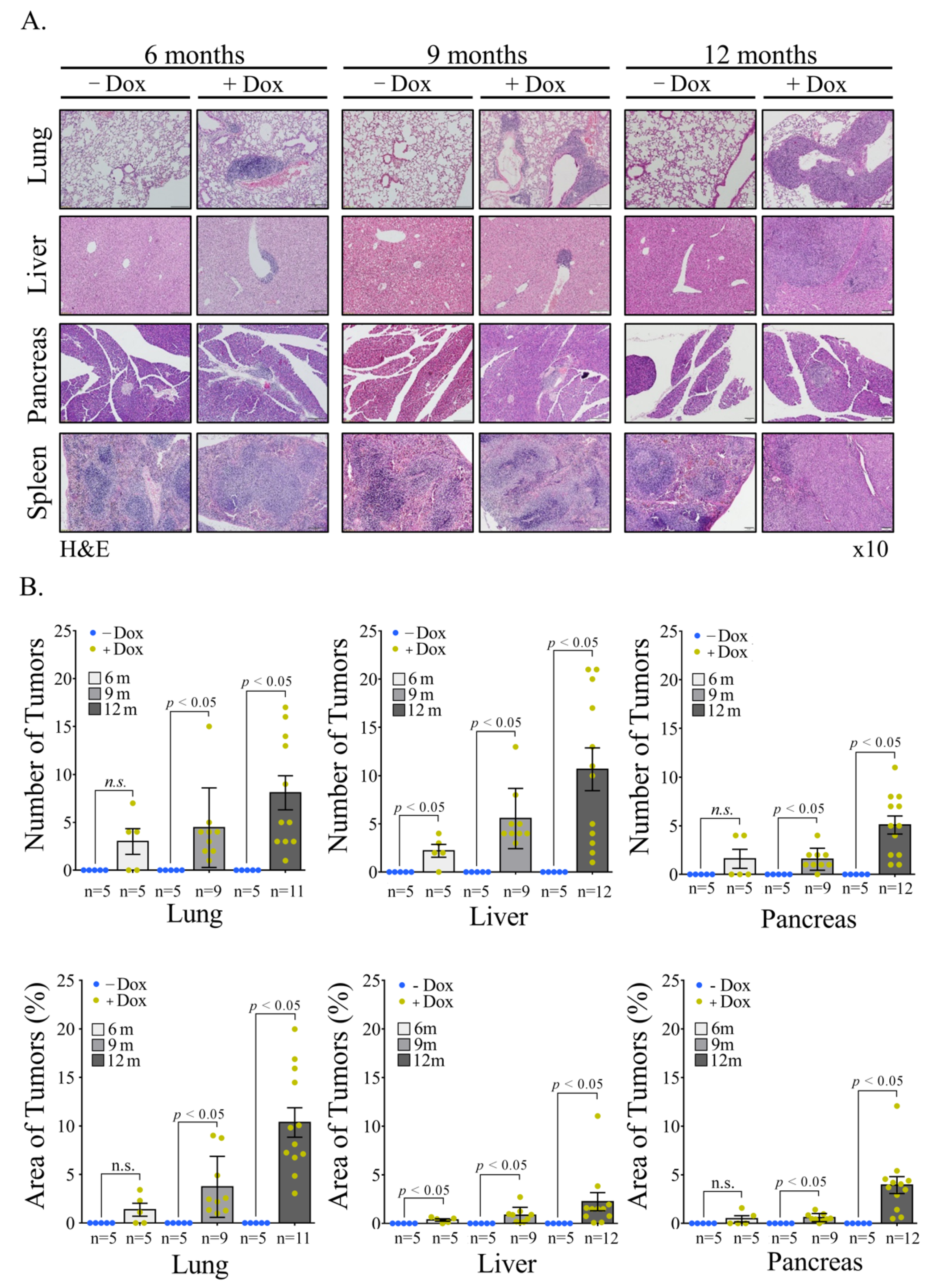
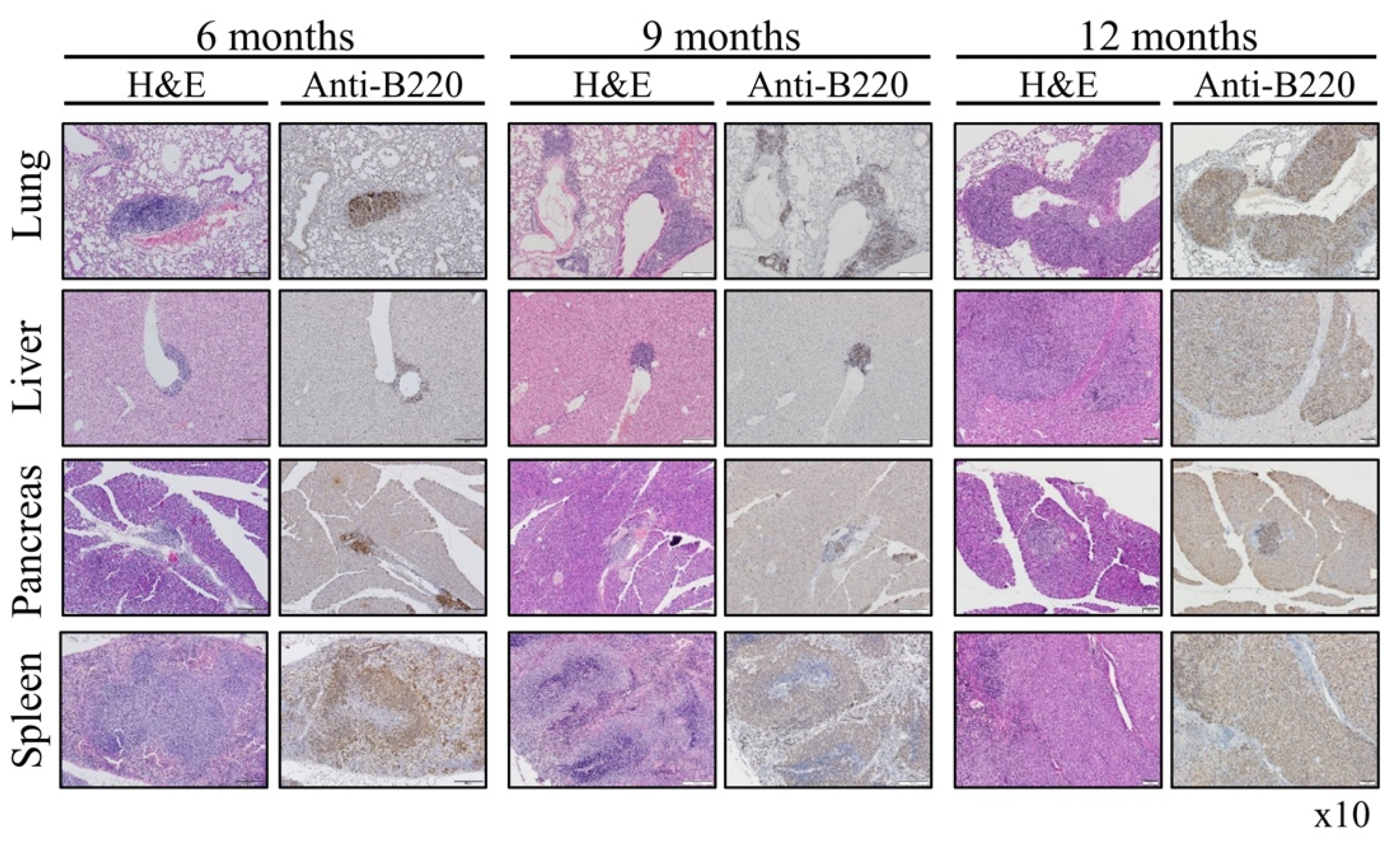
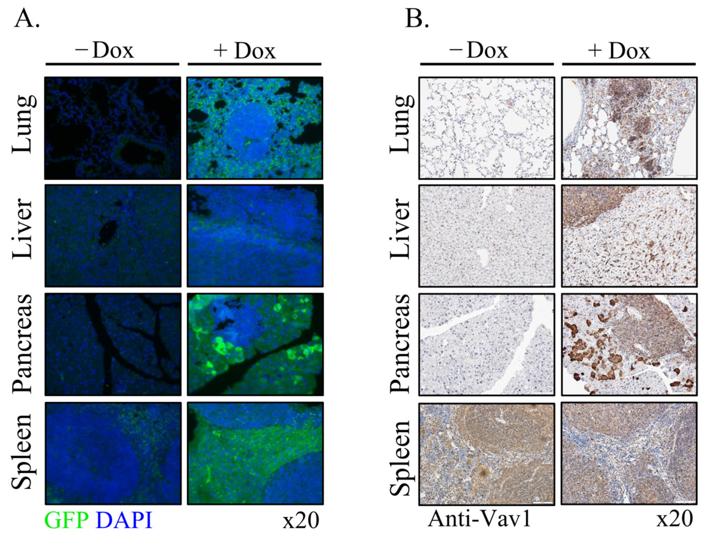
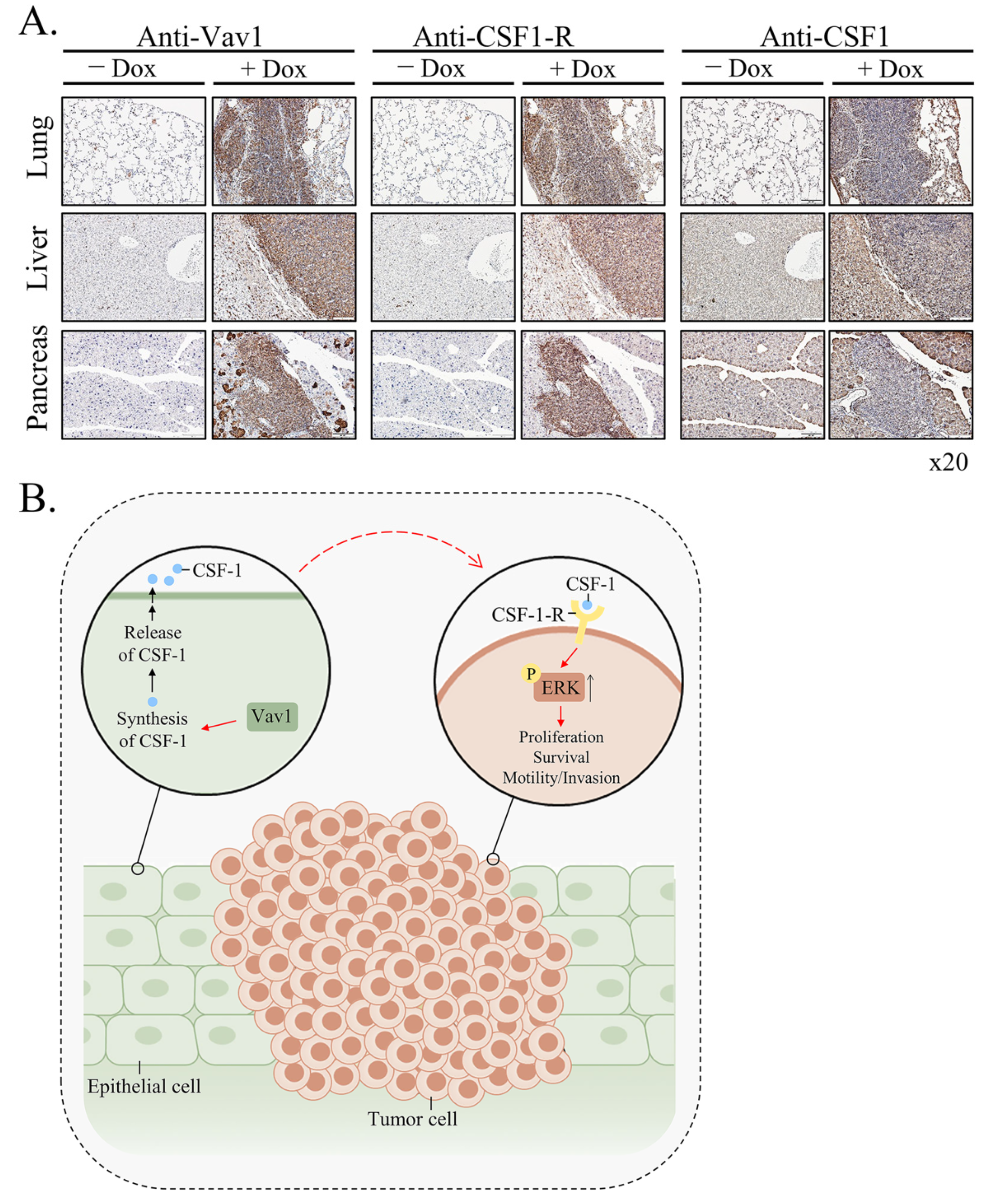
Vav1 is normally and exclusively expressed in the hematopoietic system where it functions as a specific GDP/GTP nucleotide exchange factor (GEF), firmly regulated by tyrosine phosphorylation. Mutations and overexpression of Vav1 in hematopoietic malignancies, and in human cancers of various histologic origins, are well documented. The research results suggest that overexpressing Vav1 in epithelial tissues induced chronic inflammatory reactions eventually leading to B-cell lymphomas development. The development of the lymphomas was accompanied by an increase in ERK phosphorylation, elevation of CSF- in the epithelial tissue, and an increase in CSF1-R expression in the lymphomas. These findings provide a novel mechanism by which Vav1 contributes to tumor propagation.

Vav1 Rac-GTP B-cell lymphoma Rosa26

1. Introduction

Many deregulated signal transducer proteins that are involved in cancer contribute to the ability of cells to over proliferate and escape mechanisms that normally control their survival and migration. These alterations might also lead to cancer progression through changes in the tumor microenvironment, angiogenesis, and inflammation. Deciphering the involvement of such signaling proteins is of utmost importance to the understanding of cancer development. The research is focused on the role of the signal transducer Vav1 in cancer of various histological origins.
Vav1 was identified as a transforming gene, due to the loss of its amino-terminus in a nude mice tumorigenicity assay [1]. Wild-type Vav1 (herein Vav1) was subsequently recognized as a cardinal signal transducer protein in the hematopoietic system, where it is exclusively expressed [2][3][4]. Tyrosine phosphorylation regulates the activity of Vav1 functions as a specific GDP/GTP nucleotide exchange factor (GEF) [2][3][4][5]. Mutations in various domains of the Vav1 protein are present in human cancers such as adult T-cell leukemia/lymphoma [6], peripheral T-cell lymphomas [7], lung adenocarcinoma and squamous cell carcinomas [7], as well other cancers (https://cancer.sanger.ac.uk/cosmic/ (accessed on 15 December 2021)). In addition, numerous studies have reported the unexpected expression of Vav1 in a variety of human cancers of various histological origins including the hematopoietic system [7][8][9], as well as solid tumors such as neuroblastoma [10], lung [11], pancreas [12], breast [13][14], ovarian [15], prostate [16], esophageal [17], and brain tumors [18]. Overexpression of signal transducer proteins, such as EGFR, HER2, Ras, and others in human cancers was reported [19]. Thus, the expression of a signal transducer protein in a non-physiological context might overwhelm the normal regulatory control of cellular proliferation. Vav1 can be activated by various receptors in human cancers when it is overexpressed in non-hematopoietic cells, leading to signaling cascades similar to those it regulates in hematopoietic cells, including cytoskeletal reorganization and transcription [20][21].
Despite the reports on the involvement of Vav1 in human cancer, either as a mutated gene or an abnormally expressed gene, little work has been conducted to decipher its involvement in the development of cancer by using genetically engineered mouse models (GEMMs). The researchers' laboratory was the first to analyze whether wild-type Vav1 leads to tumor generation in vivo [22]. The researchers generated a novel transgenic mouse model that inducibly expresses Vav1 and mutant K-RasG12D in the pancreas. The researchers showed that co-expression of Vav1 and mutant K-Ras dramatically increases the prevalence and decreases the time course required for malignant pancreatic lesions to appear compared to results obtained in mice that express only K-RasG12D. The results undoubtedly indicated that Vav1 and mutant K-Ras proteins synergize to augment the development of pancreatic tumors [22]. Fukumoto K, et al., analyzed the effects of activating mutations in Vav1 in the development of T cell neoplasms in transgenic mice and established the association between Vav1 mutations and malignant transformation of T cells [23].
The question is whether the mere overexpression of Vav1 in certain cells of non-hematopoietic origin suffices to lead to tumor development. The researchers approached this issue by using animal models. The expression of Vav1 alone in the pancreas did not generate any malignant lesions. To find out whether the sole expression of Vav1 in other tissues could lead to the development of malignant lesions, the researchers sought to express Vav1 ubiquitously in transgenic mice by using the Reverse Orientation Splice Acceptor (ROSA)βgeo26 (ROSA26) promoter (RosaVav1) [24]. The resulting RosaVav1 mice overexpress transgenic Vav1 in multiple epithelial tissues, but the researchers could not detect its expression in lymphoid tissues. Surprisingly, overexpressing Vav1 in epithelial tissues induced chronic inflammatory reactions eventually leading to B-cell lymphomas development. The analysis indicated that ERK phosphorylation increased in the lymphomas, suggesting that signaling pathways are evoked. Finally, while the growth factor CSF-1 is highly expressed in the epithelial compartment, the expression of its specific receptor is noticeable in the lymphomas. Taken together, the results suggest that abnormal expression of wild-type Vav1 in certain histological compartment leads to changes in signal transduction pathways as well as in growth factor/cytokine expression, which might contribute to development of cancer in adjacent histological compartments.

2. Current Insights

The overexpression of Vav1 in hematopoietic malignancies [8][9], and in human cancers of varied histologic origins is well documented [10][11][12][13][14][15][16][17][18]. Prieto-Sanchez et al. [8], reported overexpression of Vav1 in 10 of 14 cases of B-CLL with 13q deletion. Hollmann et al. [25] demonstrated a link between the expression of Vav1 and CD40-mediated apoptosis in diffuse large B-cell lymphoma cell lines. Vav1 expression was linked to sensitivity/resistance to CD40 stimulation between DLBCL cell lines. In a malignant T-cell line, Yin, et al. [26] have established an association between increased Vav1 expression and increased Bcl2 expression which is associated with decreased sensitivity to fas-mediated apoptosis, but to the researchers' knowledge, this has not been described in malignant B-cell. Vav1 was shown to be required ATRA-induced differentiation in acute myeloid leukemia (AML) cell lines to neutrophils and to maturation of these same cell lines to monocytes/macrophages following PMA treatment [27][28]. Vav1’s overexpression in epithelial tissues where it leads to tumor generation was thus far attributed to its promoter methylation status. In human lymphocytes, Vav1’s promoter is unmethylated, but it is methylated to various degrees in cancers of non-hematopoietic tissues, that do not normally express Vav1 [12][18][29]. Another mechanism pointed to the involvement of changes in putative transcription factor binding sites at the Vav1 promoter that affect its transcription in cells of various histological origins [30].
The potential contribution of Vav1 to oncogenesis was mainly investigated by IHC analysis of human specimens [11][12][13][15][16][17][20] and by the use of cell lines [1][31][32]. These studies provided invaluable insights into the association between Vav1 expression and prognosis in multiple tumor types. The researchers experiments with transgenic mice expressing Vav1 in the pancreas demonstrated that the mere expression of Vav1 in this organ does not lead to tumor formation. To find out whether this is a general phenomenon, the researchers expressed Vav1 under a ubiquitous promoter, Rosa26, and followed tumor generation. Although the ROSA26 locus is widely used as a locus for expressing transgene sequences, the results indicate that it mainly drives the Vav1 transgene’s expression in the epithelial tissue, and not in the lymphoid tissues. It is possible that such an event is the result of transcriptional interference with the endogenous ROSA26 promoter, as was shown to take place by Strathdee et al. [33][34]. The same phenomenon was demonstrated for the β-globin locus control region that can silence as well as activate gene expression [34]. The researchers show here, for the first time, that expression of Vav1 in the epithelial tissue compartment leads to the generation of B-cell lymphomas in various organs such as lungs, liver, pancreas, and spleen (Figure 1, Figure 2, and Figure 3).
Figure 1. Appearance of Malignant lesions in Rosa Vav1 mice. (A) Representative images of hematoxylin and eosin (H&E) staining of lung, liver, pancreas, and spleen sections from Rosa Vav1 mice either treated (+Dox) or non-treated (−Dox), at indicated time points after transgene induction. Magnification at ×10. (B) Quantitative analysis of tumor numbers (upper panel) and percentage area (lower panel) in H&E-stained sections. The quantitation of area of tumors is detailed in the Material section. Number of mice used: non-treated (−Dox; 6, 9, 12 months n = 5); treated (+Dox; 6 months n = 5; 9 months n = 9; 12 months n = 12). SEM and significance between the treated (+Dox) and the non-treated (−Dox) at each time point analyzed by t-test are indicated.
Figure 2. Presence of B cells in the Lymphomas of Rosa Vav1 mice. Sections of lung, liver, pancreas, and spleen from Rosa Vav1 mice either treated (+Dox) or non-treated (−Dox) at indicated time points after transgene induction, were stained with H&E and anti-B220 antibodies. Representative pictures are shown. Magnification at ×10.
Figure 3. Expression of transgenic human Vav1(indicated by GFP) in the epithelial tissue at various organs of Rosa Vav1 mice. Sections of lung, liver, pancreas, and spleen from Rosa Vav1 mice either treated (+Dox) or non-treated (−Dox), Dox 12 months post transgene induction were stained with anti-GFP antibodies that identify the human Vav1 transgene (immunofluorescence; green) (A) or anti-Vav1 antibodies that identify murine and human Vav1 (B). Representative pictures are shown. Magnification at ×20.
Host defense, inflammation, organogenesis, tissue repair, cancer growth, and immunity are all regulated by a complex network of epithelial cells and leukocytes. Bidirectional interactions, rather than the functions of individual cell types, contribute to tissue integrity and immunological homeostasis under steady-state conditions, while they can produce a complex pathologic tissue microenvironment leading to disease development. Indeed, the question then arises as to is how can the expression of Vav1 in one histologic compartment, the epithelium, lead to the development of tumors of a different histologic origin, B-lymphomas. One possible example is that of Mucosa-associated lymphoid tissue (MALT) lymphomas that originate in sites of chronic epithelial inflammation in various sites of the body, where it plays a role in regulating mucosal immunity [35]. MALT lymphomas are present in the gastrointestinal tract [36], nasopharynx [37], thyroid [38], breast [39], lung [40], salivary glands [41], eye [42], and skin [43] and exhibit characteristics with B cells located in the marginal zone of lymph node follicles. MALT lymphomas originate in sites of chronic epithelial inflammation. One example are MALT lymphomas in the stomach which are commonly caused by Helicobacter pylori infection [36]. The association between epithelial cells and lymphoma generated is best explored in patients with primary Sjögren’s syndrome (pSS), which is characterized by chronic hyperactivation of B lymphocytes, that exhibit an increased risk of development of non-Hodgkin lymphoma [36]. Salivary gland epithelial cells (SGECs) were shown to play a role in promoting B cell activation, differentiation and survival through direct interaction and cytokine production [36][41][44]. These cytokines include IL-6, B-cell activating factor (BAFF), and type I interferon leading to B cell activation, homeostasis, and survival [45][46][47][48][49][50]. SGECs were shown to express immune-competent molecules that regulate lymphocyte recruitment, homing, activation, differentiation, and survival [51]. Thus, the crosstalk between SGECs and B cells suggests that salivary gland epithelial cells play a critical role in SS pathogenesis. The activation of Vav1 by various pathogens, such as Helicobacter pylori [52] and mycoplasma [53], that may drive MALT carcinogenesis was also suggested, yet it is not clear whether the B-cell lymphomas developed in Rosa Vav1 mice fit the same pathological classification.
One of the likeliest possibilities that B-cell lymphoma develop in Rosa Vav1-transgenic mice is that Vav1-epithelial expressing cells secrete ligands that affect B-cell proliferation. Indeed, the results clearly demonstrate the increased expression of CSF-1 in epithelial cells, while the expression of its receptor is found on the lymphoma cells (Figure 4). Vav1 was shown to be involved in increased secretion of ligands which function in an autocrine or paracrine fashion. Thus, the human mammary epithelial cell line MCF-10A, which ectopically expresses an oncogenic form of Vav1, exhibits increased migration and morphological changes, accompanied by secretion of an autocrine EGF receptor ligand [54]. Also, diminished Vav1′s expression in lung cancer cell lines reduced the expression of the growth factors, TGFα and EGF [54][55], and CSF-1 [56], which led to reduced tumorigenicity. Depletion of CSF1 in lung cancer cells led to decreased proliferation and focus-formation in vitro, as well as diminished tumor growth in immune-compromised mice, suggesting that CSF-1 secretion is cardinal for tumorigenicity [56]. Immunohistochemical analysis of Vav1 and CSF-1 expression of primary human lung tumors pointed to a strong link between these proteins, associated with tumor grade. The researchers also demonstrated that conditioned media from lung cancer cells contain a growth factor, potentially CSF-1, that activates U937 monocytic cells, and that conditioned media from U937 cells instigates signaling in lung cancer cells, thus suggesting a cross-talk mechanism between immune cells and lung cancer cells mediated by CSF-1. Thus, Vav1 could influence tumor growth and the tumor’s microenvironment via CSF-1 in an autocrine/paracrine mechanism. Such a potential mechanism was also shown for breast and ovarian carcinomas. Thus, the rate of tumor progression is reduced substantially in CSF-1-knockout mice [57]. Macrophages expressing EGF promote migration and invasiveness of breast carcinoma cells as well as CSF-1 expression by the latter, and cancer cell-derived CSF-1 is able to induce EGF production in macrophages [58]. Thus, cytokines/growth factors such as CSF-1 can mediate the interaction between epithelial tumor cells and inflammatory cells.
CSF-1 contributes to the survival, proliferation, and differentiation of mononuclear phagocytes and the female’s reproductive tract [59]. Under physiological conditions, CSF-1 is produced by fibroblasts, endothelial cells, monocytes, macrophages, osteoblasts, microglia, keratinocytes, bone marrow stromal cells, natural killer cells, B-cells and T-cells, and epithelial cells [60][61]. It is an essential regulator of development and homeostasis of the mononuclear phagocyte system and, by extension, a key factor of CSF1-dependent macrophage control of development and homeostasis [59][62][63]. CSF-1 appears to play an autocrine and/or paracrine role in cancers of the ovary, endometrium, breast, lung, nervous system, and myeloid and lymphoid tissues, within which overexpression of CSF-1 receptor is considered as a prognostic factor for survival in cancer [63].
These results demonstrating the presence of CSF-1 in the epithelia of the various organs of Rosa Vav1 transgenic mice that develop B-cell lymphoma, points to the possibility that it can be produced in cells other than the hematopoietic system, once signaling driven by proteins such as Vav1 are abnormally expressed (Figure 4). In the hematopoietic system, CSF-1 exerts its pleiotropic effects by binding to a single class of high-affinity receptors (CSF-1R) expressed predominantly on monocytes, macrophages, and their committed BM precursors [62]. Based on this knowledge, the researchers expected the CSF-1 receptor to be expressed on monocytes within the B-Cell lymphomas identified in the Rosa Vav1 transgenic mice. However, no staining was detected when the researchers used F4/80 antibodies that usually bind to macrophages from different sites including the peritoneal cavity, lung, spleen, and thymus, to blood monocytes and to macrophages derived from bone marrow precursors in culture (data not shown) [64], thus suggesting that the marked expression of CSF-1 receptor noted by the researchers is expressed on other cells (Figure 4). Although CSF-1R expression is generally thought to be restricted to myeloid lineage cells, recent studies convincingly demonstrated its aberrant expression on non-myeloid lineage cells, including malignant B cells and classic Hodgkin lymphoma [65][66][67][68][69].
Figure 4. Mechanism of generation of B-cell Lymphomas in Rosa Vav1 mice. (A) Sections of lung, liver, and pancreas from Rosa Vav1 mice either treated (+Dox) or non-treated (−Dox), 12 months post transgene induction stained with anti-Vav1, anti-CSF-1R, and anti-CSF1 antibodies are depicted. Representative pictures are shown. Magnification at ×20. (B) The model proposed for the generation of B-cell lymphomas due to the aberrant expression of Rosa Vav1 in epithelial cells suggests that CSF-1 secretion from the epithelial compartment activates the CSF-1R on B-cells, leading eventually to the development of B-cell lymphoma.
The emerging model from the researchers' studies depicted in Figure 4B is that aberrant expression of Rosa Vav1 in epithelial cells leads to CSF-1 secretion (Figure 4A). CSF-1, in turn, activates the CSF-1R on B-cells and leads to the enhancement of ERK phosphorylation and cell propagation, leading eventually to the development of B-cell lymphoma. The fact that the mere expression of Vav1 in epithelial cells does not lead to the development of carcinomas further substantiates the researchers' previous studies with Vav1-pancreatic transgenic mice [22]. One would have expected, based on the researchers' current results, that Vav1 will lead to the generation of B-Cell lymphomas when expressed in the pancreas, but it’s expression in both studies was under different promoters, rosa26 in this research versus Ptf1a promoter in the pancreas, which might affect its expression and influence. Yet, it is obvious that Vav1’s ectopic expression impacts tumor development either in the pancreas when it is co-expressed with mutant K-Ras or when it is expressed in various tissues under the Rosa promoter.

References

  1. Katzav, S.; Martin-Zanca, D.; Barbacid, M. Vav, a novel human oncogene derived from a locus ubiquitously expressed in hematopoietic cells. EMBO J. 1989, 8, 2283–2290.
  2. Katzav, S. Flesh and blood: The story of Vav1, a gene that signals in hematopoietic cells but can be transforming in human malignancies. Cancer Lett. 2007, 255, 241–254.
  3. Katzav, S. Vav1: A hematopoietic signal transduction molecule involved in human malignancies. Int. J. Biochem. Cell Biol. 2009, 41, 1245–1248.
  4. Tybulewicz, V.L.J. Vav-family proteins in T-cell signalling. Curr. Opin. Immunol. 2005, 17, 267–274.
  5. Crespo, P.; Schuebel, K.E.; Ostrom, A.A.; Gutkind, J.S.; Bustelo, X.R. Phosphotyrosine-dependent activation of Rac-1 GDP/GTP exchange by the vav proto-oncogene product. Nature 1997, 385, 169–172.
  6. Kataoka, K.; Nagata, Y.; Kitanaka, A.; Shiraishi, Y.; Shimamura, T.; Yasunaga, J.-I.; Totoki, Y.; Chiba, K.; Sato-Otsubo, A.; Nagae, G.; et al. Integrated molecular analysis of adult T cell leukemia/lymphoma. Nat. Genet. 2015, 47, 1304–1315.
  7. Abate, F.; da Silva-Almeida, A.C.; Zairis, S.; Robles-Valero, J.; Couronne, L.; Khiabanian, H.; Quinn, S.A.; Kim, M.-Y.; Laginestra, M.A.; Kim, C.; et al. Activating mutations and translocations in the guanine exchange factor VAV1 in peripheral T-cell lymphomas. Proc. Natl. Acad. Sci. USA 2017, 114, 764–769.
  8. Prieto-Sánchez, R.M.; Hernández, J.A.; García, J.L.; Gutiérrez, N.C.; San Miguel, J.; Bustelo, X.R.; Hernández, J.M. Overexpression of the VAV proto-oncogene product is associated with B-cell chronic lymphocytic leukaemia displaying loss on 13q. Br. J. Haematol. 2006, 133, 642–645.
  9. Oberley, M.J.; Wang, D.-S.; Yang, D.T. Vav1 in hematologic neoplasms, a mini review. Am. J. Blood Res. 2012, 2, 1–8.
  10. Hornstein, I.; Pikarsky, E.; Groysman, M.; Amir, G.; Peylan-Ramu, N.; Katzav, S. The haematopoietic specific signal transducer Vav1 is expressed in a subset of human neuroblastomas. J. Pathol. 2003, 199, 526–533.
  11. Lazer, G.; Idelchuk, Y.; Schapira, V.; Pikarsky, E.; Katzav, S. The haematopoietic specific signal transducer Vav1 is aberrantly expressed in lung cancer and plays a role in tumourigenesis. J. Pathol. 2009, 219, 25–34.
  12. Fernandez-Zapico, M.E.; Gonzalez-Paz, N.C.; Weiss, E.; Savoy, D.N.; Molina, J.R.; Fonseca, R.; Smyrk, T.C.; Chari, S.T.; Urrutia, R.; Billadeau, D.D. Ectopic expression of VAV1 reveals an unexpected role in pancreatic cancer tumorigenesis. Cancer Cell 2005, 7, 39–49.
  13. Sebban, S.; Farago, M.; Gashai, D.; Ilan, L.; Pikarsky, E.; Ben-Porath, I.; Katzav, S. Vav1 fine tunes p53 control of apoptosis versus proliferation in breast cancer. PLoS ONE 2013, 8, e54321.
  14. Grassilli, S.; Brugnoli, F.; Lattanzio, R.; Rossi, C.; Perracchio, L.; Mottolese, M.; Marchisio, M.; Palomba, M.; Nika, E.; Natali, P.G.; et al. High nuclear level of Vav1 is a positive prognostic factor in early invasive breast tumors: A role in modulating genes related to the efficiency of metastatic process. Oncotarget 2014, 5, 4320–4336.
  15. Wakahashi, S.; Sudo, T.; Oka, N.; Ueno, S.; Yamaguchi, S.; Fujiwara, K.; Ohbayashi, C.; Nishimura, R. VAV1 represses E-cadherin expression through the transactivation of Snail and Slug: A potential mechanism for aberrant epithelial to mesenchymal transition in human epithelial ovarian cancer. Transl. Res. 2013, 162, 181–190.
  16. Kniazev, I.P.; Cheburkin, I.V.; Spikermann, K.; Peter, S.; Jenster, G.; Bangma, K.H.; Karelin, M.I.; Shkol’nik, M.I.; Urbanskiĭ, A.I.; Evtushenko, V.I.; et al. Gene expression profiles of protein kinases and phosphatases obtained by hybridization with cDNA arrays: Molecular portrait of human prostate carcinoma. Mol. Biol. 2003, 37, 97–111.
  17. Zhu, X.; Jin, H.; Xia, Z.; Wu, X.; Yang, M.; Zhang, H.; Shang, X.; Cheng, R.; Zhan, Z.; Yu, Z. Vav1 expression is increased in esophageal squamous cell carcinoma and indicates poor prognosis. Biochem. Biophys. Res. Commun. 2017, 486, 571–576.
  18. Lindsey, J.C.; Kawauchi, D.; Schwalbe, E.C.; Solecki, D.J.; Selby, M.P.; McKinnon, P.J.; Olson, J.M.; Hayden, J.T.; Grundy, R.G.; Ellison, D.W.; et al. Cross-species epigenetics identifies a critical role for VAV1 in SHH subgroup medulloblastoma maintenance. Oncogene 2015, 34, 4746–4757.
  19. Barnes, C.J.; Vadlamudi, R.K.; Kumar, R. Novel estrogen receptor coregulators and signaling molecules in human diseases. Cell. Mol. Life Sci. 2004, 61, 281–291.
  20. Hornstein, I.; Alcover, A.; Katzav, S. Vav proteins, masters of the world of cytoskeleton organization. Cell. Signal. 2004, 16, 1–11.
  21. Lazer, G.; Katzav, S. Guanine nucleotide exchange factors for RhoGTPases: Good therapeutic targets for cancer therapy? Cell. Signal. 2011, 23, 969–979.
  22. Salaymeh, Y.; Farago, M.; Sebban, S.; Shalom, B.; Pikarsky, E.; Katzav, S. Vav1 and mutant K-Ras synergize in the early development of pancreatic ductal adenocarcinoma in mice. Life Sci. Alliance 2020, 3, e202000661.
  23. Fukumoto, K.; Sakata-Yanagimoto, M.; Fujisawa, M.; Sakamoto, T.; Miyoshi, H.; Suehara, Y.; Nguyen, T.B.; Suma, S.; Yanagimoto, S.; Shiraishi, Y.; et al. VAV1 mutations contribute to development of T-cell neoplasms in mice. Blood 2020, 136, 3018–3032.
  24. Soriano, P.; Friedrich, G.; Lawinger, P. Promoter interactions in retrovirus vectors introduced into fibroblasts and embryonic stem cells. J. Virol. 1991, 65, 2314–2319.
  25. Hollmann, A.; Aloyz, R.; Baker, K.; Dirnhofer, S.; Owens, T.; Sladek, R.; Tzankov, A. Vav-1 expression correlates with NFκB activation and CD40-mediated cell death in diffuse large B-cell lymphoma cell lines. Hematol. Oncol. 2010, 28, 142–150.
  26. Yin, J.; Wan, Y.; Li, S.; Du, M.; Zhang, C.; Zhou, X.; Cao, Y. The distinct role of guanine nucleotide exchange factor Vav1 in Bcl-2 transcription and apoptosis inhibition in Jurkat leukemia T cells. Acta Pharmacol. Sin. 2011, 32, 99–107.
  27. Bertagnolo, V.; Grassilli, S.; Bavelloni, A.; Brugnoli, F.; Piazzi, M.; Candiano, G.; Petretto, A.; Benedusi, M.; Capitani, S. Vav1 modulates protein expression during ATRA-induced maturation of APL-derived promyelocytes: A proteomic-based analysis. J. Proteome Res. 2008, 7, 3729–3736.
  28. Bertagnolo, V.; Brugnoli, F.; Grassilli, S.; Nika, E.; Capitani, S. Vav1 in differentiation of tumoral promyelocytes. Cell. Signal. 2012, 24, 612–620.
  29. Huang, P.H.; Lu, P.J.; Ding, L.Y.; Chu, P.C.; Hsu, W.Y.; Chen, C.S.; Tsao, C.C.; Chen, B.H.; Lee, C.T.; Shan, Y.S.; et al. TGFβ promotes mesenchymal phenotype of pancreatic cancer cells, in part, through epigenetic activation of VAV1. Oncogene 2017, 36, 2202–2214.
  30. Ilan, L.; Katzav, S. Human Vav1 expression in hematopoietic and cancer cell lines is regulated by c-Myb and by CpG methylation. PLoS ONE 2012, 7, e29939.
  31. Zugaza, J.L.; López-Lago, M.A.; Caloca, M.J.; Dosil, M.; Movilla, N.; Bustelo, X.R. Structural determinants for the biological activity of Vav proteins. J. Biol. Chem. 2002, 277, 45377–45392.
  32. Shalom, B.; Farago, M.; Pikarsky, E.; Katzav, S. Vav1 mutations identified in human cancers give rise to different oncogenic phenotypes. Oncogenesis 2018, 7, 80.
  33. Strathdee, D.; Ibbotson, H.; Grant, S.G.N. Expression of transgenes targeted to the Gt(ROSA)26Sor locus is orientation dependent. PLoS ONE 2006, 1, e4.
  34. Feng, Y.-Q.; Warin, R.; Li, T.; Olivier, E.; Besse, A.; Lobell, A.; Fu, H.; Lin, C.M.; Aladjem, M.I.; Bouhassira, E.E. The human beta-globin locus control region can silence as well as activate gene expression. Mol. Cell. Biol. 2005, 25, 3864–3874.
  35. Mullangi, S.; Lekkala, M.R. Mucosa-associated lymphoma tissue. In StatPearls; StatPearls Publishing: Treasure Island, FL, USA, 2022.
  36. Alvarez-Lesmes, J.; Chapman, J.R.; Poveda, J.C. Pitfalls in gastrointestinal tract haematopoietic lesions. Pathology 2021, 54, 177–183.
  37. Mnatsakanian, A.; Heil, J.R.; Sharma, S. Anatomy, head and neck, adenoids. In StatPearls; StatPearls Publishing: Treasure Island, FL, USA, 2022.
  38. Farooq, U.; Chakinala, R.C. MALToma. In StatPearls; StatPearls Publishing: Treasure Island, FL, USA, 2022.
  39. Genco, I.S.; Gur, H.; Hajiyeva, S. Lymphoma of the breast: A clinicopathologic analysis of 51 cases with a specific emphasis on patients with a history of breast carcinoma. Breast J. 2021, 27, 456–460.
  40. Borie, R.; Wislez, M.; Antoine, M.; Copie-Bergman, C.; Thieblemont, C.; Cadranel, J. Pulmonary mucosa-associated lymphoid tissue lymphoma revisited. Eur. Respir. J. 2016, 48, 1252.
  41. Rivière, E.; Pascaud, J.; Tchitchek, N.; Boudaoud, S.; Paoletti, A.; Ly, B.; Dupré, A.; Chen, H.; Thai, A.; Allaire, N.; et al. Salivary gland epithelial cells from patients with Sjögren’s syndrome induce B-lymphocyte survival and activation. Ann. Rheum. Dis. 2020, 79, 1468–1477.
  42. Ahmed, S.; Shahid, R.K.; Sison, C.P.; Fuchs, A.; Mehrotra, B. Orbital lymphomas: A clinicopathologic study of a rare disease. Am. J. Med. Sci. 2006, 331, 79–83.
  43. Kogame, T.; Kabashima, K.; Egawa, G. Putative Immunological Functions of Inducible Skin-Associated Lymphoid Tissue in the Context of Mucosa-Associated Lymphoid Tissue. Front. Immunol. 2021, 12, 733484.
  44. Verstappen, G.M.; Pringle, S.; Bootsma, H.; Kroese, F.G.M. Epithelial-immune cell interplay in primary Sjögren syndrome salivary gland pathogenesis. Nat. Rev. Rheumatol. 2021, 17, 333–348.
  45. Jones, B.E.; Maerz, M.D.; Buckner, J.H. IL-6: A cytokine at the crossroads of autoimmunity. Curr. Opin. Immunol. 2018, 55, 9–14.
  46. Batten, M.; Groom, J.; Cachero, T.G.; Qian, F.; Schneider, P.; Tschopp, J.; Browning, J.L.; Mackay, F. BAFF mediates survival of peripheral immature B lymphocytes. J. Exp. Med. 2000, 192, 1453–1466.
  47. Mackay, F.; Schneider, P. Cracking the BAFF code. Nat. Rev. Immunol. 2009, 9, 491–502.
  48. Kyriakidis, N.C.; Kapsogeorgou, E.K.; Gourzi, V.C.; Konsta, O.D.; Baltatzis, G.E.; Tzioufas, A.G. Toll-like receptor 3 stimulation promotes Ro52/TRIM21 synthesis and nuclear redistribution in salivary gland epithelial cells, partially via type I interferon pathway. Clin. Exp. Immunol. 2014, 178, 548–560.
  49. Ittah, M.; Miceli-Richard, C.; Gottenberg, E.; Lavie, F.; Lazure, T.; Ba, N.; Sellam, J.; Lepajolec, C.; Mariette, X. B cell-activating factor of the tumor necrosis factor family (BAFF) is expressed under stimulation by interferon in salivary gland epithelial cells in primary Sjögren’s syndrome. Arthritis Res. Ther. 2006, 8, R51.
  50. Gottenberg, J.-E.; Cagnard, N.; Lucchesi, C.; Letourneur, F.; Mistou, S.; Lazure, T.; Jacques, S.; Ba, N.; Ittah, M.; Lepajolec, C.; et al. Activation of IFN pathways and plasmacytoid dendritic cell recruitment in target organs of primary Sjögren’s syndrome. Proc. Natl. Acad. Sci. USA 2006, 103, 2770–2775.
  51. Kapsogeorgou, E.K.; Manoussakis, M.N. Salivary gland epithelial cells (SGEC): Carriers of exquisite B7-2 (CD86) costimulatory molecules. J. Autoimmun. 2010, 35, 188–191.
  52. Kim, N. Chemoprevention of gastric cancer by Helicobacter pylori eradication and its underlying mechanism. J. Gastroenterol. Hepatol. 2019, 34, 1287–1295.
  53. Sinkovics, J.G. Molecular biology of oncogenic inflammatory processes. I. Non-oncogenic and oncogenic pathogens, intrinsic inflammatory reactions without pathogens, and microRNA/DNA interactions (Review). Int. J. Oncol. 2012, 40, 305–349.
  54. Wilsbacher, J.L.; Moores, S.L.; Brugge, J.S. An active form of Vav1 induces migration of mammary epithelial cells by stimulating secretion of an epidermal growth factor receptor ligand. Cell Commun. Signal. 2006, 4, 5.
  55. Schapira, V.; Lazer, G.; Katzav, S. Osteopontin is an oncogenic Vav1- but not wild-type Vav1-responsive gene: Implications for fibroblast transformation. Cancer Res. 2006, 66, 6183–6191.
  56. Sebban, S.; Farago, M.; Rabinovich, S.; Lazer, G.; Idelchuck, Y.; Ilan, L.; Pikarsky, E.; Katzav, S. Vav1 promotes lung cancer growth by instigating tumor-microenvironment cross-talk via growth factor secretion. Oncotarget 2014, 5, 9214–9226.
  57. Lin, E.Y.; Nguyen, A.V.; Russell, R.G.; Pollard, J.W. Colony-stimulating factor 1 promotes progression of mammary tumors to malignancy. J. Exp. Med. 2001, 193, 727–740.
  58. Goswami, S.; Sahai, E.; Wyckoff, J.B.; Cammer, M.; Cox, D.; Pixley, F.J.; Stanley, E.R.; Segall, J.E.; Condeelis, J.S. Macrophages promote the invasion of breast carcinoma cells via a colony-stimulating factor-1/epidermal growth factor paracrine loop. Cancer Res. 2005, 65, 5278–5283.
  59. Stanley, E.R.; Berg, K.L.; Einstein, D.B.; Lee, P.S.; Pixley, F.J.; Wang, Y.; Yeung, Y.G. Biology and action of colony--stimulating factor-1. Mol. Reprod. Dev. 1997, 46, 4–10.
  60. Achkova, D.; Maher, J. Role of the colony-stimulating factor (CSF)/CSF-1 receptor axis in cancer. Biochem. Soc. Trans. 2016, 44, 333–341.
  61. Sun, X.; Ingman, W.V. Cytokine networks that mediate epithelial cell-macrophage crosstalk in the mammary gland: Implications for development and cancer. J. Mammary Gland Biol. Neoplasia 2014, 19, 191–201.
  62. Guilbert, L.J.; Stanley, E.R. Specific interaction of murine colony-stimulating factor with mononuclear phagocytic cells. J. Cell Biol. 1980, 85, 153–159.
  63. Mo, H.; Hao, Y.; Lv, Y.; Chen, Z.; Shen, J.; Zhou, S.; Yin, M. Overexpression of macrophage-colony stimulating factor-1 receptor as a prognostic factor for survival in cancer: A systematic review and meta-analysis. Medicine 2021, 100, e25218.
  64. Dos Anjos Cassado, A. F4/80 as a major macrophage marker: The case of the peritoneum and spleen. Results Probl. Cell Differ. 2017, 62, 161–179.
  65. Martín-Moreno, A.M.; Roncador, G.; Maestre, L.; Mata, E.; Jiménez, S.; Martínez-Torrecuadrada, J.L.; Reyes-García, A.I.; Rubio, C.; Tomás, J.F.; Estévez, M.; et al. CSF1R protein expression in reactive lymphoid tissues and lymphoma: Its relevance in classical hodgkin lymphoma. PLoS ONE 2015, 10, e0125203.
  66. Ullrich, K.; Wurster, K.D.; Lamprecht, B.; Köchert, K.; Engert, A.; Dörken, B.; Janz, M.; Mathas, S. BAY 43-9006/Sorafenib blocks CSF1R activity and induces apoptosis in various classical Hodgkin lymphoma cell lines. Br. J. Haematol. 2011, 155, 398–402.
  67. Lamprecht, B.; Walter, K.; Kreher, S.; Kumar, R.; Hummel, M.; Lenze, D.; Köchert, K.; Bouhlel, M.A.; Richter, J.; Soler, E.; et al. Derepression of an endogenous long terminal repeat activates the CSF1R proto-oncogene in human lymphoma. Nat. Med. 2010, 16, 571–579.
  68. Murga-Zamalloa, C.; Rolland, D.C.M.; Polk, A.; Wolfe, A.; Dewar, H.; Chowdhury, P.; Onder, O.; Dewar, R.; Brown, N.A.; Bailey, N.G.; et al. Colony-Stimulating Factor 1 Receptor (CSF1R) Activates AKT/mTOR Signaling and Promotes T-Cell Lymphoma Viability. Clin. Cancer Res. 2020, 26, 690–703.
  69. Krause, G.; Hassenrück, F.; Hallek, M. Relevant Cytokines in the B Cell Lymphoma Micro-Environment. Cancers 2020, 12, 2525.
More
Information
Subjects: Cell Biology
Contributor MDPI registered users' name will be linked to their SciProfiles pages. To register with us, please refer to https://encyclopedia.pub/register :
View Times: 590
Revisions: 3 times (View History)
Update Date: 25 Mar 2022
1000/1000
Video Production Service