Vav1 Promotes B-Cell Lymphoma Development: Comparison
Please note this is a comparison between Version 1 by Shulamit Katzav and Version 3 by Beatrix Zheng.

Vav1 is normally and exclusively expressed in the hematopoietic system where it functions as a specific GDP/GTP nucleotide exchange factor (GEF), firmly regulated by tyrosine phosphorylation. Mutations and overexpression of Vav1 in hematopoietic malignancies, and in human cancers of various histologic origins, are well documented. The research results suggest that overexpressing Vav1 in epithelial tissues induced chronic inflammatory reactions eventually leading to B-cell lymphomas development. The development of the lymphomas was accompanied by an increase in ERK phosphorylation, elevation of CSF- in the epithelial tissue, and an increase in CSF1-R expression in the lymphomas. TheseOur findings provide a novel mechanism by which Vav1 contributes to tumor propagation.

  • Vav1
  • Rac-GTP
  • B-cell lymphoma
  • Rosa26

1. Introduction

Many deregulated signal transducer proteins that are involved in cancer contribute to the ability of cells to over proliferate and escape mechanisms that normally control their survival and migration. These alterations might also lead to cancer progression through changes in the tumor microenvironment, angiogenesis, and inflammation. Deciphering the involvement of such signaling proteins is of utmost importance to the understanding of cancer development. TheOur researchstudy is focused on the role of the signal transducer Vav1 in cancer of various histological origins.
Vav1 was identified as a transforming gene, due to the loss of its amino-terminus in a nude mice tumorigenicity assay [1]. Wild-type Vav1 (herein Vav1) was subsequently recognized as a cardinal signal transducer protein in the hematopoietic system, where it is exclusively expressed [2][3][4][2,3,4]. Tyrosine phosphorylation regulates the activity of Vav1 functions as a specific GDP/GTP nucleotide exchange factor (GEF) [2][3][4][5][2,3,4,5]. Mutations in various domains of the Vav1 protein are present in human cancers such as adult T-cell leukemia/lymphoma [6], peripheral T-cell lymphomas [7], lung adenocarcinoma and squamous cell carcinomas [7], as well other cancers (https://cancer.sanger.ac.uk/cosmic/ (accessed on 15 December 2021)). In addition, numerous studies have reported the unexpected expression of Vav1 in a variety of human cancers of various histological origins including the hematopoietic system [7][8][9][7,8,9], as well as solid tumors such as neuroblastoma [10], lung [11], pancreas [12], breast [13][14][13,14], ovarian [15], prostate [16], esophageal [17], and brain tumors [18]. Overexpression of signal transducer proteins, such as EGFR, HER2, Ras, and others in human cancers was reported [19]. Thus, the expression of a signal transducer protein in a non-physiological context might overwhelm the normal regulatory control of cellular proliferation. Vav1 can be activated by various receptors in human cancers when it is overexpressed in non-hematopoietic cells, leading to signaling cascades similar to those it regulates in hematopoietic cells, including cytoskeletal reorganization and transcription [20][21][20,21].
Despite the reports on the involvement of Vav1 in human cancer, either as a mutated gene or an abnormally expressed gene, little work has been conducted to decipher its involvement in the development of cancer by using genetically engineered mouse models (GEMMs). The Ouresearchers' laboratory was the first to analyze whether wild-type Vav1 leads to tumor generation in vivo [22]. ThWe researchers generated a novel transgenic mouse model that inducibly expresses Vav1 and mutant K-RasG12D in the pancreas. ThWe researchers sshowed that co-expression of Vav1 and mutant K-Ras dramatically increases the prevalence and decreases the time course required for malignant pancreatic lesions to appear compared to results obtained in mice that express only K-RasG12D. TheOur results undoubtedly indicated that Vav1 and mutant K-Ras proteins synergize to augment the development of pancreatic tumors [22]. Fukumoto K, et al., analyzed the effects of activating mutations in Vav1 in the development of T cell neoplasms in transgenic mice and established the association between Vav1 mutations and malignant transformation of T cells [23].
The question is whether the mere overexpression of Vav1 in certain cells of non-hematopoietic origin suffices to lead to tumor development. ThWe researchers aapproached this issue by using animal models. The expression of Vav1 alone in the pancreas did not generate any malignant lesions. To find out whether the sole expression of Vav1 in other tissues could lead to the development of malignant lesions, the researchers swe sought to express Vav1 ubiquitously in transgenic mice by using the Reverse Orientation Splice Acceptor (ROSA)βgeo26 (ROSA26) promoter (RosaVav1) [24]. The resulting RosaVav1 mice overexpress transgenic Vav1 in multiple epithelial tissues, but thwe researchers ccould not detect its expression in lymphoid tissues. Surprisingly, overexpressing Vav1 in epithelial tissues induced chronic inflammatory reactions eventually leading to B-cell lymphomas development. TheOur analysis indicated that ERK phosphorylation increased in the lymphomas, suggesting that signaling pathways are evoked. Finally, while the growth factor CSF-1 is highly expressed in the epithelial compartment, the expression of its specific receptor is noticeable in the lymphomas. Taken together, theour results suggest that abnormal expression of wild-type Vav1 in certain histological compartment leads to changes in signal transduction pathways as well as in growth factor/cytokine expression, which might contribute to development of cancer in adjacent histological compartments.

2. Current Insights

The overexpression of Vav1 in hematopoietic malignancies [8][9][8,9], and in human cancers of varied histologic origins is well documented [10][11][12][13][14][15][16][17][18][10,11,12,13,14,15,16,17,18]. Prieto-Sanchez et al. [8], reported overexpression of Vav1 in 10 of 14 cases of B-CLL with 13q deletion. Hollmann et al. [25][31] demonstrated a link between the expression of Vav1 and CD40-mediated apoptosis in diffuse large B-cell lymphoma cell lines. Vav1 expression was linked to sensitivity/resistance to CD40 stimulation between DLBCL cell lines. In a malignant T-cell line, Yin, et al. [26][32] have established an association between increased Vav1 expression and increased Bcl2 expression which is associated with decreased sensitivity to fas-mediated apoptosis, but to the ouresearchers' knowledge, this has not been described in malignant B-cell. Vav1 was shown to be required ATRA-induced differentiation in acute myeloid leukemia (AML) cell lines to neutrophils and to maturation of these same cell lines to monocytes/macrophages following PMA treatment [27][28][33,34]. Vav1’s overexpression in epithelial tissues where it leads to tumor generation was thus far attributed to its promoter methylation status. In human lymphocytes, Vav1’s promoter is unmethylated, but it is methylated to various degrees in cancers of non-hematopoietic tissues, that do not normally express Vav1 [12][18][29][12,18,35]. Another mechanism pointed to the involvement of changes in putative transcription factor binding sites at the Vav1 promoter that affect its transcription in cells of various histological origins [30][36].
The potential contribution of Vav1 to oncogenesis was mainly investigated by IHC analysis of human specimens [11][12][13][15][16][17][20][11,12,13,15,16,17,20] and by the use of cell lines [1][31][32][1,37,38]. These studies provided invaluable insights into the association between Vav1 expression and prognosis in multiple tumor types. The Ouresearchers experiments with transgenic mice expressing Vav1 in the pancreas demonstrated that the mere expression of Vav1 in this organ does not lead to tumor formation. To find out whether this is a general phenomenon, the researchers ewe expressed Vav1 under a ubiquitous promoter, Rosa26, and followed tumor generation. Although the ROSA26 locus is widely used as a locus for expressing transgene sequences, theour results indicate that it mainly drives the Vav1 transgene’s expression in the epithelial tissue, and not in the lymphoid tissues. It is possible that such an event is the result of transcriptional interference with the endogenous ROSA26 promoter, as was shown to take place by Strathdee et al. [33][34][39,40]. The same phenomenon was demonstrated for the β-globin locus control region that can silence as well as activate gene expression [34][40]. ThWe researchers shshow here, for the first time, that expression of Vav1 in the epithelial tissue compartment leads to the generation of B-cell lymphomas in various organs such as lungs, liver, pancreas, and spleen (Figure 12, Figure 23, and Figure 34).
Figure 12. Appearance of Malignant lesions in Rosa Vav1 mice. (A) Representative images of hematoxylin and eosin (H&E) staining of lung, liver, pancreas, and spleen sections from Rosa Vav1 mice either treated (+Dox) or non-treated (−Dox), at indicated time points after transgene induction. Magnification at ×10. (B) Quantitative analysis of tumor numbers (upper panel) and percentage area (lower panel) in H&E-stained sections. The quantitation of area of tumors is detailed in the Material section. Number of mice used: non-treated (−Dox; 6, 9, 12 months  n = 5); treated (+Dox; 6 months n = 5; 9 months n = 9; 12 months n = 12). SEM and significance between the treated (+Dox) and the non-treated (−Dox) at each time point analyzed by t-test are indicated.
Figure 23. Presence of B cells in the Lymphomas of Rosa Vav1 mice. Sections of lung, liver, pancreas, and spleen from Rosa Vav1 mice either treated (+Dox) or non-treated (−Dox) at indicated time points after transgene induction, were stained with H&E and anti-B220 antibodies. Representative pictures are shown. Magnification at ×10.
Figure 34. Expression of transgenic human Vav1(indicated by GFP) in the epithelial tissue at various organs of Rosa Vav1 mice. Sections of lung, liver, pancreas, and spleen from Rosa Vav1 mice either treated (+Dox) or non-treated (−Dox), Dox 12 months post transgene induction were stained with anti-GFP antibodies that identify the human Vav1 transgene (immunofluorescence; green) (A) or anti-Vav1 antibodies that identify murine and human Vav1 (B). Representative pictures are shown. Magnification at ×20.
Host defense, inflammation, organogenesis, tissue repair, cancer growth, and immunity are all regulated by a complex network of epithelial cells and leukocytes. Bidirectional interactions, rather than the functions of individual cell types, contribute to tissue integrity and immunological homeostasis under steady-state conditions, while they can produce a complex pathologic tissue microenvironment leading to disease development. Indeed, the question then arises as to is how can the expression of Vav1 in one histologic compartment, the epithelium, lead to the development of tumors of a different histologic origin, B-lymphomas. One possible example is that of Mucosa-associated lymphoid tissue (MALT) lymphomas that originate in sites of chronic epithelial inflammation in various sites of the body, where it plays a role in regulating mucosal immunity [35][41]. MALT lymphomas are present in the gastrointestinal tract [36][42], nasopharynx [37][43], thyroid [38][44], breast [39][45], lung [40][46], salivary glands [41][47], eye [42][48], and skin [43][49] and exhibit characteristics with B cells located in the marginal zone of lymph node follicles. MALT lymphomas originate in sites of chronic epithelial inflammation. One example are MALT lymphomas in the stomach which are commonly caused by Helicobacter pylori infection [36][42]. The association between epithelial cells and lymphoma generated is best explored in patients with primary Sjögren’s syndrome (pSS), which is characterized by chronic hyperactivation of B lymphocytes, that exhibit an increased risk of development of non-Hodgkin lymphoma [36][42]. Salivary gland epithelial cells (SGECs) were shown to play a role in promoting B cell activation, differentiation and survival through direct interaction and cytokine production [36][41][44][42,47,50]. These cytokines include IL-6, B-cell activating factor (BAFF), and type I interferon leading to B cell activation, homeostasis, and survival [45][46][47][48][49][50][51,52,53,54,55,56]. SGECs were shown to express immune-competent molecules that regulate lymphocyte recruitment, homing, activation, differentiation, and survival [51][57]. Thus, the crosstalk between SGECs and B cells suggests that salivary gland epithelial cells play a critical role in SS pathogenesis. The activation of Vav1 by various pathogens, such as Helicobacter pylori [52][58] and mycoplasma [53][59], that may drive MALT carcinogenesis was also suggested, yet it is not clear whether the B-cell lymphomas developed in Rosa Vav1 mice fit the same pathological classification.
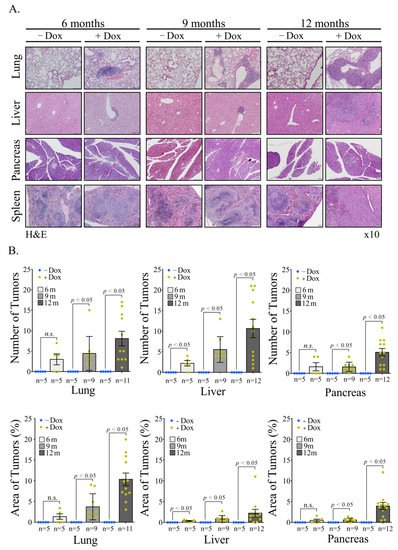
One of the likeliest possibilities that B-cell lymphoma develop in Rosa Vav1-transgenic mice is that Vav1-epithelial expressing cells secrete ligands that affect B-cell proliferation. Indeed, theour results clearly demonstrate the increased expression of CSF-1 in epithelial cells, while the expression of its receptor is found on the lymphoma cells (Figure 46). Vav1 was shown to be involved in increased secretion of ligands which function in an autocrine or paracrine fashion. Thus, the human mammary epithelial cell line MCF-10A, which ectopically expresses an oncogenic form of Vav1, exhibits increased migration and morphological changes, accompanied by secretion of an autocrine EGF receptor ligand [54][60]. Also, diminished Vav1′s expression in lung cancer cell lines reduced the expression of the growth factors, TGFα and EGF [54][55][60,61], and CSF-1 [56][30], which led to reduced tumorigenicity. Depletion of CSF1 in lung cancer cells led to decreased proliferation and focus-formation in vitro, as well as diminished tumor growth in immune-compromised mice, suggesting that CSF-1 secretion is cardinal for tumorigenicity [56][30]. Immunohistochemical analysis of Vav1 and CSF-1 expression of primary human lung tumors pointed to a strong link between these proteins, associated with tumor grade. ThWe researchers aalso demonstrated that conditioned media from lung cancer cells contain a growth factor, potentially CSF-1, that activates U937 monocytic cells, and that conditioned media from U937 cells instigates signaling in lung cancer cells, thus suggesting a cross-talk mechanism between immune cells and lung cancer cells mediated by CSF-1. Thus, Vav1 could influence tumor growth and the tumor’s microenvironment via CSF-1 in an autocrine/paracrine mechanism. Such a potential mechanism was also shown for breast and ovarian carcinomas. Thus, the rate of tumor progression is reduced substantially in CSF-1-knockout mice [57][62]. Macrophages expressing EGF promote migration and invasiveness of breast carcinoma cells as well as CSF-1 expression by the latter, and cancer cell-derived CSF-1 is able to induce EGF production in macrophages [58][63]. Thus, cytokines/growth factors such as CSF-1 can mediate the interaction between epithelial tumor cells and inflammatory cells.
CSF-1 contributes to the survival, proliferation, and differentiation of mononuclear phagocytes and the female’s reproductive tract [59][64]. Under physiological conditions, CSF-1 is produced by fibroblasts, endothelial cells, monocytes, macrophages, osteoblasts, microglia, keratinocytes, bone marrow stromal cells, natural killer cells, B-cells and T-cells, and epithelial cells [60][61][65,66]. It is an essential regulator of development and homeostasis of the mononuclear phagocyte system and, by extension, a key factor of CSF1-dependent macrophage control of development and homeostasis [59][62][63][64,67,68]. CSF-1 appears to play an autocrine and/or paracrine role in cancers of the ovary, endometrium, breast, lung, nervous system, and myeloid and lymphoid tissues, within which overexpression of CSF-1 receptor is considered as a prognostic factor for survival in cancer [63][68].
TheseOur results demonstrating the presence of CSF-1 in the epithelia of the various organs of Rosa Vav1 transgenic mice that develop B-cell lymphoma, points to the possibility that it can be produced in cells other than the hematopoietic system, once signaling driven by proteins such as Vav1 are abnormally expressed (Figure 46). In the hematopoietic system, CSF-1 exerts its pleiotropic effects by binding to a single class of high-affinity receptors (CSF-1R) expressed predominantly on monocytes, macrophages, and their committed BM precursors [62][67]. Based on this knowledge, thwe researchers eexpected the CSF-1 receptor to be expressed on monocytes within the B-Cell lymphomas identified in the Rosa Vav1 transgenic mice. However, no staining was detected when the researchers we used F4/80 antibodies that usually bind to macrophages from different sites including the peritoneal cavity, lung, spleen, and thymus, to blood monocytes and to macrophages derived from bone marrow precursors in culture (data not shown) [64][69], thus suggesting that the marked expression of CSF-1 receptor noted by the researcherus is expressed on other cells (Figure 46). Although CSF-1R expression is generally thought to be restricted to myeloid lineage cells, recent studies convincingly demonstrated its aberrant expression on non-myeloid lineage cells, including malignant B cells and classic Hodgkin lymphoma [65][66][67][68][69][70,71,72,73,74].
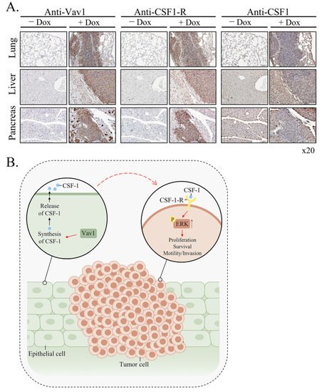
Figure 46. Mechanism of generation of B-cell Lymphomas in Rosa Vav1 mice. (A) Sections of lung, liver, and pancreas from Rosa Vav1 mice either treated (+Dox) or non-treated (−Dox), 12 months post transgene induction stained with anti-Vav1, anti-CSF-1R, and anti-CSF1 antibodies are depicted. Representative pictures are shown. Magnification at ×20. (B) The model proposed for the generation of B-cell lymphomas due to the aberrant expression of Rosa Vav1 in epithelial cells suggests that CSF-1 secretion from the epithelial compartment activates the CSF-1R on B-cells, leading eventually to the development of B-cell lymphoma.
The emerging model from the ouresearchers' studies depicted in Figure 46B is that aberrant expression of Rosa Vav1 in epithelial cells leads to CSF-1 secretion (Figure 46A). CSF-1, in turn, activates the CSF-1R on B-cells and leads to the enhancement of ERK phosphorylation and cell propagation, leading eventually to the development of B-cell lymphoma. The fact that the mere expression of Vav1 in epithelial cells does not lead to the development of carcinomas further substantiates the ouresearchers' previous studies with Vav1-pancreatic transgenic mice [22]. One would have expected, based on the reseaourchers' current results, that Vav1 will lead to the generation of B-Cell lymphomas when expressed in the pancreas, but it’s expression in both studies was under different promoters, rosa26 in this researchstudy versus Ptf1a promoter in the pancreas, which might affect its expression and influence. Yet, it is obvious that Vav1’s ectopic expression impacts tumor development either in the pancreas when it is co-expressed with mutant K-Ras or when it is expressed in various tissues under the Rosa promoter.
Video Production Service