Submitted Successfully!
To reward your contribution, here is a gift for you: A free trial for our video production service.
Thank you for your contribution! You can also upload a video entry or images related to this topic.
Version Summary Created by Modification Content Size Created at Operation
1 + 4679 word(s) 4679 2022-03-17 04:09:04 |
2 format correction Meta information modification 4679 2022-03-23 07:54:18 | |
3 format correction Meta information modification 4679 2022-03-23 07:56:04 |

Video Upload Options

Do you have a full video?

Confirm

Are you sure to Delete?
Cite
If you have any further questions, please contact Encyclopedia Editorial Office.
Koleva, L. Erythrocytes for Targeted Drug Delivery. Encyclopedia. Available online: https://encyclopedia.pub/entry/20899 (accessed on 26 April 2024).
Koleva L. Erythrocytes for Targeted Drug Delivery. Encyclopedia. Available at: https://encyclopedia.pub/entry/20899. Accessed April 26, 2024.
Koleva, Larisa. "Erythrocytes for Targeted Drug Delivery" Encyclopedia, https://encyclopedia.pub/entry/20899 (accessed April 26, 2024).
Koleva, L. (2022, March 23). Erythrocytes for Targeted Drug Delivery. In Encyclopedia. https://encyclopedia.pub/entry/20899
Koleva, Larisa. "Erythrocytes for Targeted Drug Delivery." Encyclopedia. Web. 23 March, 2022.
Erythrocytes for Targeted Drug Delivery
Edit

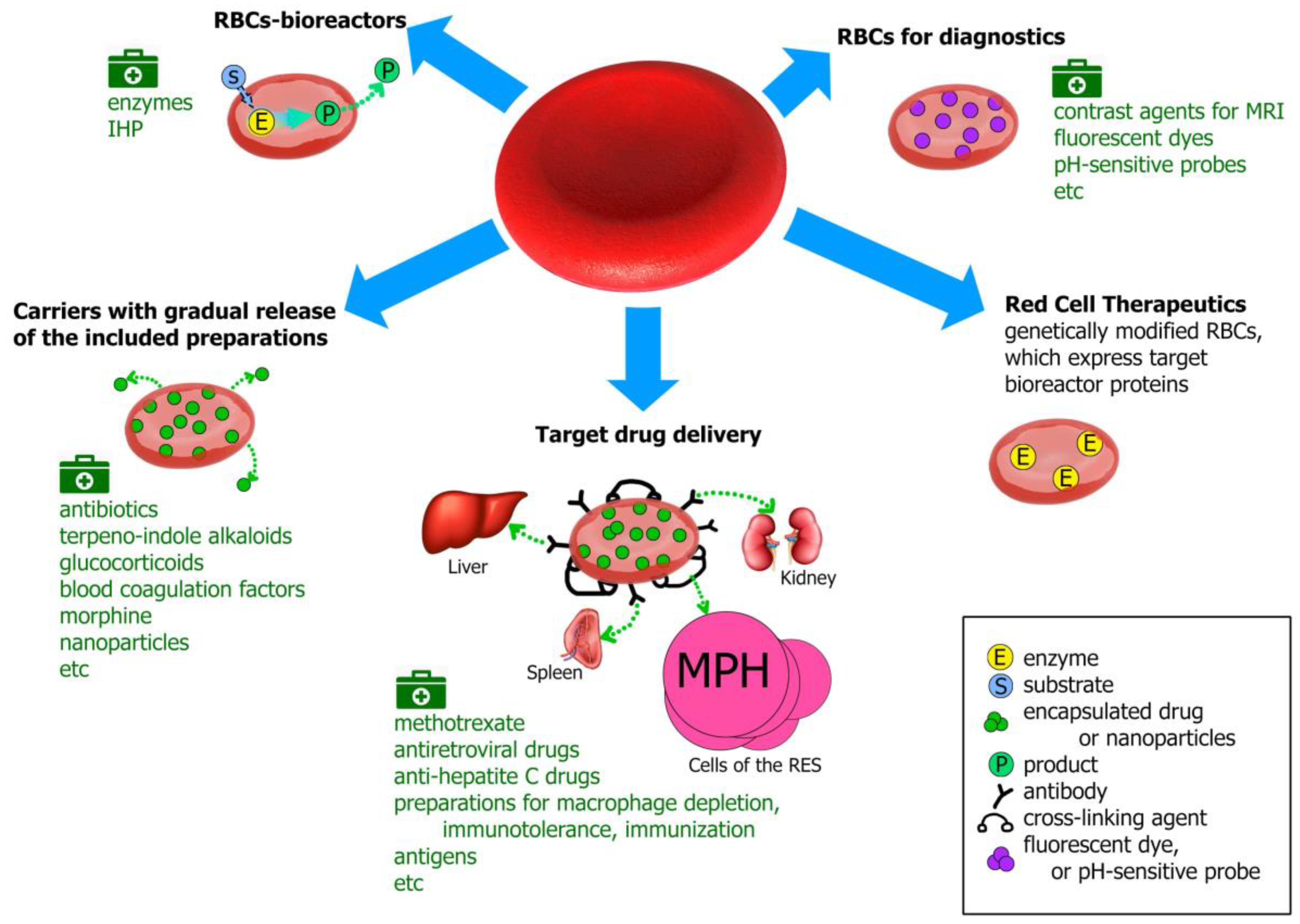
Erythrocytes (red blood cells, RBCs) are the largest population of blood cells in mammals. Their main function is oxygen transfer to cells and body tissues. The lifetime of erythrocytes in the bloodstream is 100–120 days, after which they are removed by the spleen. Due to the unique biophysical properties RBCs can be used as drug carriers in two different ways: by incorporating the drug into the cells or by binding it (using non-specific adsorption or a specific association, involving antibodies or various chemical cross-linking compounds) on the RBCs’ surface. Erythrocytes can act as carriers that prolong the drug’s action due to its gradual release from the carrier; as bioreactors with encapsulated enzymes performing the necessary reactions, while remaining inaccessible to the immune system and plasma proteases; or as a tool for targeted drug delivery to target organs, primarily to cells of the reticuloendothelial system, liver and spleen. To date, erythrocytes have been studied as carriers for a wide range of drugs, such as enzymes, antibiotics, anti-inflammatory, antiviral drugs, etc., and for diagnostic purposes. 

drug delivery erythrocyte carrier erythrocyte erythrocyte-bioreactor targeted drug delivery therapy

1. Erythrocytes as Drug Carriers

Drug delivery using natural biological carriers is a fast-developing field. Due to the unique biophysical properties, erythrocytes (red blood cells, RBCs) have great potential in this area. RBCs are the largest population of blood cells in mammals. Their main function is oxygen transfer to cells and body tissues [1]. Mature RBCs do not have a cell nucleus and most organelles, but they contain a large amount of a special protein, hemoglobin (Hb), which is able to bind to oxygen. The biconcave shape provides good flexibility and allows the erythrocyte to deform and pass through narrow capillaries. The lifetime of erythrocytes in the bloodstream is 100–120 days, after which they are removed by the spleen. Erythrocytes can be used as carriers in two different ways: by incorporating the drug into the cells or by binding it (using non-specific adsorption or a specific association, involving antibodies or various chemical cross-linking compounds) on the RBCs’ surface. The binding of drugs on the surface of RBCs has both advantages and disadvantages. A great contribution to the development of this direction was made by the team of Muzykantov et al. [2][3][4][5][6][7][8][9].
To incorporate the drug into the RBC, the cell must undergo some external influences so that pores can be reversibly formed in its membrane, through which the drug can penetrate. This unique property of RBCs allows to load them with biologically active substances of different molecular weights. For these reasons, erythrocytes are promising biocompatible cells for drug delivery.
The methods for incorporating various substances into red blood cells differ in the way that substances penetrate the cells. The cause of permeability may be the pores’ formation in the cell membrane due to a physical exposure (high voltage electric pulse [10][11] or ultrasound [12]). Drug molecules can also enter the RBCs by endocytosis in the presence of certain chemical compounds (for example, primaquine [13], vinblastine, chlorpromazine, hydrocortisone or tetracaine [14][15]), or using the cell-penetrating peptides bounded to the compound that should be encapsulated [16]. However, the most popular are different variants of osmotic methods.
In some cases, RBCs are first exposed to a hyperosmotic pulse of a low molecular weight substance that penetrates very well through the cell membrane (for example, dimethyl sulfoxide (DMSO) [17][18] or glucose [19][20]). After washing the cells, which decreases the external concentration of these compounds and creates a gradient of their concentration between both sides of the RBC membrane, the target drug is introduced into the external volume. Water with this drug begins to enter into the cells to decrease the osmotic pressure there. The process ends when the gradient of DMSO or glucose disappears. The pores close and part of the drug remains into RBCs. Other, the most popular of the osmotic methods are hypoosmotic. These methods are based on creating a hypotonic environment around RBCs, which causes swelling of the cells and opening pores in the cellular membrane, through which therapeutic compounds can penetrate RBCs. Then, a hypertonic solution is introduced into the cell suspension. The pores close, the cells restore their original size, trapping the drug molecules inside the cell. Osmotic methods are divided into several types. Simple reversible cell lysis in a hypotonic solution by dilutiing a cell suspension with a hypotonic medium causes the formation of erythrocyte ghosts [21][22]. The method of hypotonic pre-swelling is based on the initial controlled cells swelling in a hypotonic solution and their subsequent lysis by adding small portions of an aqueous solution of the drug for encapsulation [23][24][25]. Dialysis methods are based on a reduction of osmolality around the RBCs by a process of dialysis versus hypotonic solution in a dialysis bag [26][27] or in special dialyzers with increased area of contact of RBCs with a buffer solution in the case of flow dialysis [28][29][30]. As mentioned above, hypoosmotic methods are most preferable for incorporation of enzymes into RBCs in terms of efficacy and the properties of obtained cell carriers [31][32].
The history of carrier erythrocytes begins in 1973, when Ihler demonstrated in his article the possibility of incorporating enzymes such as β-glucosidase and β-galactosidase into these cells by reversible hypoosmotic lysis [21]. The analysis of the number of publications (relating only to medications inside the RBCs) shows that interest in this topic since 1973 has not declined, but, instead, has been constantly growing. The number of published articles on the subject of carrier erythrocytes increases every year, and currently, their total number is about 400 (Figure 1).
Figure 1. Change in the total number of articles published in the world on the subject of erythrocyte carriers of biologically active substances, since 1973.
RBCs for drug delivery have several advantages compared to the existing methods and systems for drug delivery. The erythrocyte is an ideal candidate for such delivery and meets all the requirements for such systems, namely:
- biocompatibility (human, both autologous and donor erythrocytes are used to treat patients);
- biodegradability (old or damaged erythrocytes are naturally removed by the reticuloendothelial system);
- long life in the bloodstream (the drug has an extended lifetime inside the cells because RBCs protect it from the immune system and plasma proteases and the cells survive in the body for a long time; thus, the pharmacokinetics and pharmacodynamics of the drug in RBCs can significantly increase the desired therapeutic effect);
- decreasing side effects of drugs (due to preventing allergic reactions, and the decrease in the peak concentrations of free drug in the blood to safer levels);
- ease of cell isolation in large quantities and the ability to scale production.
Carrier erythrocytes (CEs) can be used both in therapy and the diagnosis of some diseases, for example, as carriers of contrast agents for magnetic resonance imaging (MRI) or as biosensors that respond to changes in the concentration of metabolites or pH in the blood [33][34][35]. In therapy, depending on the drug that is loaded, the erythrocytes can be used as carriers with a gradual drug release, as bioreactors or a system for targeted drug delivery, primarily to the reticuloendothelial system (RES), liver and spleen [36]. In the first case, either a drug encapsulated into RBCs can slowly pass through the erythrocyte membrane into the bloodstream, or a membrane-nonpenetrating prodrug is loaded into RBCs, where it turns into a therapeutically effective compound that is able to exit the cell. This ensures prolonged drug circulation in the bloodstream with a decrease in the toxic effects on the body. In the second case, the enzyme encapsulated in erythrocytes works with substrates penetrating the cell membrane. Thus, the enzyme does not directly enter the bloodstream, which solves the problem of its immunogenicity, premature inactivation and increases its half-life (Figure 2).
Figure 2. Possible modes of use of RBCs loaded with biologically active compounds and nanoparticles. MRI—magnetic resonance imaging; IHP—inositol hexaphosphate; MPH—macrophages; RES—reticuloendothelial system.

2. Erythrocytes for Targeted Drug Delivery

The targeted delivery of drugs using RBCs can be carried out, firstly, to the cells of the RES (macrophages), as well as in the liver and spleen, i.e., in the body cells, that remove old and damaged RBCs. Thus, this approach may be successfully used to treat tumors of these tissues. To deliver the erythrocyte loaded with the drug into these target cells, it must be modified so that the target cells perceive it as being damaged. There are various methods of such modification. All of them lead to a modification of the erythrocyte membrane. This may be the opsonization of RBCs with antibodies to their membrane determinants (for example, by rhesus-antibodies [37]) or the binding of the complement component C3b to them, since there are receptors for the Fc fragment of IgG and for C3b on the cell surface. Treatment of RBCs with calcium ionophore leads to phosphatidylserine exposure on their surface [38], and treatment with glutaraldehyde cross-links the amino groups on the membrane surface, which makes the cell more rigid. Another method is treatment with reagents that cause clustering of the band 3 protein, for example, by a bifunctional amine–amine cross-linking agent, bisulfosuccinimidyl suberate (BS3) in ZnCl2 medium [39][40], which leads to the binding of Hb and proteins of the membrane and fixation of complement components on the cell surface [41]. Inactivation of intracellular hexokinase is also described, which leads to disruption of the cell metabolism and a decrease in the concentration of ATP necessary for cell survival [42].

2.1. Methotrexate

Methotrexate (MTX) is one of the cytostatic preparations (see above). In 1978, Zimmermann et al. were among the first to demonstrate, in mice, the advantage in the distribution of the erythrocytic form of methotrexate (MTX-RBC) in the body over the free form for intravenous administration. The authors encapsulated the drug by electroporation (i.e., created pores in the RBC membrane using an electrical impulse) through which methotrexate (MTX) penetrated the cell. When this form of the drug was administered to mice over 10 min, almost all the methotrexate that was administered in RBCs (0.75–1.0 doses) accumulated in the liver of animals, while in control experiments (with the introduction of the free form of methotrexate), only 0.25–0.3 of the administered dose accumulated [43].
DeLoach and Barton encapsulated methotrexate in erythrocytes by hypoosmotic methods and showed in dogs, in vivo, that in this case, the drug quickly leaves the RBCs. Thirty minutes after the injection of MTX-RBCs into the bloodstream, 50% of methotrexate appeared free in the plasma [44]. To slow the release of the drug from the cells, treatment of carrier erythrocytes with glutaraldehyde was proposed, which provides an additional advantage, since, as was shown in dogs, 50% of CEs treated with glutaraldehyde are rapidly detected in the liver, i.e., targeted delivery of methotrexate to RES occurs [44][45]. Another method for incorporating methotrexate into RBCs uses the pulse of a hyperosmotic glucose solution. In this case, the cells are incubated for 40 min in a 50% glucose solution. Then, they are gently washed and incubated for 30 min with a solution of methotrexate under normal tonicity. The half-life of such CEs with methotrexate was almost 3.5 times longer than for the free form of the drug (13.5 and 3.9 h, respectively) [46]. In addition, the peak plasma concentration of methotrexate after MTX-RBC administration was lower than with free MTX, but it decreased more slowly. A gradual release of the drug from RBCs was observed. In another study [47], N-hydroxysuccinimide biotin ester (NHS-biotin) was bound on the surface of CEs for targeted delivery of MTX-RBCs to the liver. In vivo experiments on rats showed that 1 h after administration of biotinylated MTX-RBCs to animals, 37.2% of biotin appears in the liver, which is almost three times more than after administration of free MTX (11.7%) and almost 1.8 times more than for non-biotinylated cells (20.4%). In an earlier work [48], the same authors modified MTX-RBCs with trypsin (Tt) or phenylhydrazine (PhT) to desialize the cell surface and induce hemichrome in cells, respectively. These two approaches were equally used for the recognition of erythrocytes by macrophages in order to deliver methotrexate for the treatment of RES tumors. Surface-modified erythrocytes loaded with MTX 1 h after administration to animals showed an increased level of methotrexate in the liver compared with the free form of the drug (approximately six times) and with unmodified cells (approximately two times). Phagocytosis by macrophages of surface-treated MTX-loaded erythrocytes was increased by three–five and five–six times for Tt- and PhT-treated CEs, respectively, compared with untreated CEs [48].
The presented examples demonstrate promising possibilities of using erythrocytes for targeted drug delivery to the liver and RES.

2.2. Erythrocytes-Carriers for Treatment of Retroviral Infection

Retroviruses are a family of RNA viruses that primarily infect vertebrates. The most famous and actively studied representative of retroviruses is the human immunodeficiency virus (HIV). Currently, nucleoside analogs, which are inhibitors of reverse transcriptase (after anabolic intracellular phosphorylation), are essential components of highly active antiretroviral therapy (HAART). The most famous of these are azidothymidine (and its analogs), dideoxycytidine and other 2′,3′-dideoxynucleosides [41][49]. Furthermore, the antiviral activity of reduced glutathione (GSH) against RNA and DNA viruses is well known. This activity is realized by interfering with protein-envelope folding and by blocking cell transcriptional factor (NF-kB) activation, which decreases the virus transcription and replication [50][51]. The nucleoside analogs protect lymphocytes, but cannot enter macrophages, while GSH inside specially modified RBCs can be captured by macrophages and protect them against viral infection.
Thus, to treat this immunodeficiency, both CEs containing antiretroviral drugs and CEs containing GSH or GSH + antiretroviral drugs can be used, since GSH-loaded RBCs has been shown to provide significant additional effects compared to monotherapy with antiretroviral drugs (nucleoside analogues) [50].
Since the 1990s, Magnani et al. has been actively developing CEs for the treatment of the human immunodeficiency virus. Since the targets and reservoirs of human immunodeficiency infection are cells of the monocyte/macrophage line, attempts have been made to deliver antiretroviral drugs directly to macrophages to prevent transmission of HIV from already infected macrophages to target lymphocytes [52]. The most popular nucleoside analogues, such as 3’-azido-2’,3’-dideoxythymidine and 2’,3’-dideoxycytidine (ddCTP), were encapsulated into RBCs.
It was shown [41] that for the manifestation of pharmacological activity, dideoxynucleosides must be phosphorylated to 5’-triphosphate by cell kinases. Different types of cells within the same species have different abilities to phosphorylate these compounds. To reduce the toxicity of nucleoside analogues, as well as to overcome the problem of the effectiveness of their phosphorylation, Magnani et al. incorporated ddCTP into RBCs in an active phosphorylated form (by the method of hypoosmotic dialysis). For targeted delivery of such RBCs to macrophages, the loaded cells were treated with a bifunctional amine–amine cross-linking agent BS3 in ZnCl2 medium. This makes the RBCs tougher and induces the binding of autologous immunoglobulin G (IgG) and complement component C3b on the cell surface. Such RBCs are recognized by macrophages and actively phagocitosed. In vitro and in vivo, it was shown that erythrocytes treated in this way loaded with phosphorylated ddCTP were able to significantly reduce typical symptoms of the disease within 3 months [41][52][53][54][55]. The ability to release 3’-azido-2′,3’-deoxythymidine (AZT) from erythrocytes loaded with the azidothymidine derivative di-(thymidine-3’-azido-2’,3’-dideoxy-d-β-riboside)-5’-5’-p1-p2-pyrophosphate (AZTp2AZT) has also been demonstrated in vitro. This prodrug is converted inside erythrocytes into the pharmacologically active AZT by sequential hydrolysis and dephosphorylation [56].
In [51][56][57], interesting results of combination therapy using oral AZT, AZT + DDI (2′,3′-dideoxyinosine) and the additional administration of erythrocytes encapsulated with GSH in each case were demonstrated. The experiments were performed on mice infected by the retrovirus complex (LP-BM5). Studies have shown a decrease in proviral DNA in the brain by about 50% with AZT + DDI treatment and 85% when GSH-loaded RBCs were added to AZT + DDI therapy. For bone marrow, this decrease was about 37% and 60%, respectively [57]. The addition of GSH-loaded RBCs to AZT monotherapy decreased proviral DNA in bone marrow by 60% [56].
RBCs encapsulated with fludarabine have become another possible approach for treating HIV-1 infection. As mentioned above, long-living macrophages in the infected body are the reservoir for the HIV-1 virus. It was shown that chronic infection of human macrophages with this virus increases the expression and phosphorylation of the protein STAT1, which is included in the regulation of many macrophage functions, including cell growth and proliferation [58]. The nucleoside analogue of 9-(β-d-arabinofuranosyl)-2-fluoroadenin-5’-monophosphate (FaraAMP, fludarabine) is active against STAT1-expressing cells and, in culture, is able to kill HIV-infected macrophages, but not uninfected cells. To direct fludarabine to macrophages, it was encapsulated into RBCs, which were then processed by the method described in [41], which causes clustering of the band 3 protein. The final concentration of fludarabine in macrophages after a single 18-h exposure with erythrocytes loaded with fludarabine was estimated at 10–20 μM. In that study, a powerful (>98%) and long-lasting (at least 4 weeks) effect of inhibiting the release of the virus from HIV-infected macrophages was obtained [59].

2.3. Drugs Loaded into RBCs for the Treatment of Hepatitis C

To enhance the effectiveness of the therapeutic effect of drugs used in the treatment of hepatitis C, and to minimize their side effects associated with an increase in the dose of drugs, Skorokhod et al. were searching for new ways to simultaneously deliver interferon (INF-α) and ribavirin (RIBA) to the liver [59]. Both drugs were loaded into human RBCs (RBCs-INF-α-RIBA) by the method of hypoosmotic reversible lysis. Cells were opsonized for targeted delivery to macrophages and liver. The entrapment efficiency was 40%. It was shown that RBCs-INF-α-RIBA were stored for up to 3 days at 4 °C without loss of antiviral activity. In vitro, monocyte activation by RBCs-INF-α-RIBA was also demonstrated, as well as the induction of surface receptors of the major histocompatibility complex type II (MHC class II) and Fc receptors that activate cell phagocytic activity. The authors argue that encapsulating INF-α and RIBA into RBCs and targeting the liver helps: (1) to release large amounts of INF-α and achieve higher therapeutically effective concentrations in the liver; (2) to induce autocrine stimulation of macrophages of the liver (and spleen) using INF-α to enhance cellular antiviral protection; (3) to control viral proliferation in macrophages. In this regard, it is advisable to further study a potentially therapeutically effective system in animals.
Forezesh and Zarrin proposed encapsulating a more modern hepatitis C drug, boceprevir, into RBCs in addition to interferon and ribavirin [60].

2.4. Macrophage Depletion

It is known that macrophages play an important role in the regulation of numerous biological processes in the body. In addition, it has been repeatedly shown that macrophages contribute to the development of pathologies such as autoimmune hemolytic anemia, immunothrombocytopenia, rheumatoid arthritis and sepsis, and play a key role in the spread of viruses in HIV infections [61]. Tumor-associated macrophages create favorable conditions for cancer progression, promoting angiogenesis and metastasis [62][63][64]. Rossi et al. studied the possibility of temporary depletion of macrophages by incorporating bisphosphonates (clodronate, zoledronate) into RBCs and the targeted delivery of such carriers to macrophages. They showed that RBCs loaded with zoledronate are able to deplete macrophages both in vitro and in vivo [65]. Balb/C mice were injected with 59 mg/mouse of zoledronate encapsulated into RBCs. For targeted delivery to macrophages, loaded erythrocytes were incubated in medium with BS3 and ZnCl2. After a single injection of encapsuled erythrocytes, macrophage depletion was 29% and 67% for liver and spleen macrophages, respectively.
Another study evaluated the effect of macrophage depletion to prevent Langerhans islet cell allograft rejection in diabetes mice [66]. Graft survival was 19–20 days for control groups of mice receiving unloaded erythrocytes or saline, 25 days for mice receiving free clodronate and 35 days for mice receiving clodronate in RBCs.

2.5. Antigens Loaded into Erythrocytes or Associated with Their Surface

2.5.1. Immunization

Binding antigens to the surface or encapsulating them inside the carrier erythrocytes opens up new possibilities for using such erythrocytes for immunization as an alternative to adjuvants (substances that adsorb antigen on their surface), namely, the possibility of delivering antigens directly to the immune system into antigen-presenting cells—macrophages or dendritic cells (DCs). Dendritic cells are believed to be most effective in initiating antigen-specific responses, but macrophages are also able to facilitate the presentation of peptides to T lymphocytes [67]. Magnani et al. has repeatedly shown that protein antigens (bovine serum albumin, porcine liver uricase, yeast hexokinase) and glycoproteins B of herpes simplex virus type 1 (HSV-I), which are associated with the surface of autologous RBCs via the biotin–avidin–biotin bridges, induce a higher immunological response (higher antibody levels) in mice than the response obtained using Freund’s adjuvant, which is often used in immunization [68][69]. Later, it was shown that the HIV-1 Tat protein, linked through the biotin–avidin–biotin bridges to the erythrocyte surface (RBC-Tat), has immunotherapeutic potential. This protein is important for virus replication and infectious activity (the presence of antibodies against Tat correlate with slower progression of the disease). Tat protein is immunogenic [70]. Erythrocytes associated with Tat (RBCs-Tat), in amounts 250 times less than the amount of soluble Tat in Freund’s adjuvant, are capable of eliciting specific responses of anti-Tat T killers. Moreover, the production of Tat neutralizing antibodies was observed in six out of six mice, in contrast to two out of six mice for Tat in Freund’s adjuvant.
In other works [71][72], using bacterial toxoids, proteins and enzymes as antigens, it was shown that immunization is also possible by encapsulating antigen in RBCs. In B6D2F1 and Balb/C mice, the total titers of specific antibodies (binding, lysing and neutralizing the antigens) and only neutralizing antibodies against introduced antigens were several times higher during immunization with antigens loaded into RBCs than after immunization with free forms of antigens [71].

2.5.2. Cancer Immunotherapy

Cancer immunotherapy is the use of the immune system to kill tumor cells that have specific tumor-associated antigens (TAA) [73]. Banz et al. proposed a strategy for using RBCs loaded with tumor-associated antigens in cancer immunotherapy. Immunization against TAA induces TAA-specific cytotoxic T lymphocytes (CTLs), which are capable of controlling tumor growth. Efficient and targeted delivery of TAA in vivo to DCs can be effective in tumor immunotherapy since it induces strong CTLs responses against the tumor [74]. It was shown in mice [75] that erythrocytes bearing an antigen (in this case, ovalbumin) in combination with polyinosine–polycytidylic acid (Poly (I:C)) introduced intravenously, can be effectively captured by antigen-presenting cells (APC). This causes antigen-specific responses of CD4+ and CD8+ T cells, which are able to induce in vivo ovalbumin-specific cell lysis even 30 days after CEs administration. Ovalbumin was loaded into RBCs by hypoosmotic dialysis (RBC-OVA). To enhance the phagocytosis of these erythrocytes with antigen-presenting cells, they were treated externally with antibodies (anti-TER119 mAb), and then were administered to C57BL/6 mice intravenously. RBC-OVA was mixed with Poly (I:C) before injection to enhance the induction of T-cell responses, as Poly (I:C) is a toll-like receptor III ligand that activates the CD4+ T cell response specific for alloantigen of RBCs [76][77]. Ninety minutes after injection of RBC-OVA + Poly (I:C) to mice, phagocytosis of the introduced RBCs by antigen-presenting macrophages and dendritic cells was observed.
The effectiveness of such a tumor-associated antigen delivery system was also demonstrated in two models of mice with melanoma [78]. The artificial ovalbumin antigen or tyrosinase 2 protein antigen (TRP-2) was encapsuled into red blood cells and tested on E.G7-OVA and B16F10 tumor models, respectively. The administration of a small amount of tumor-associated antigen (TRP-2) loaded into RBCs treated with antigen anti-TER119 in combination with Poly (I:C) caused an antigen-specific T-cell response and tumor growth control in mice, whereas the same amount of free TRP-2 did not cause a similar response.

2.5.3. Induction of Immune Tolerance

The opposite of immunization is the stimulation of immune tolerance, that is, the “training” of the immune system to create tolerance (resistance) to a particular antigen in order to prevent its attack. Such stimulation can be used in autoimmune diseases, when the immune system attacks its own antigens, during an allograft transplant, or in case of an allergy to a drug used in therapy. The induction of immune tolerance is often carried out using molecules that inhibit the immune system, such as rituximab (anti-CD20 monoclonal antibody), cyclophosphamide, and methotrexate, or by depleting B cells necessary for the immune response. In [79], the authors proposed the use of erythrocytes for the induction of immune tolerance. The drug, to which it was necessary to induce immune tolerance, was loaded into RBCs. That study showed that the drug inside the cell-carriers does not interact directly with antibodies, which may be present in plasma. The authors investigated the possibility of obtaining immune tolerance in mice for the enzyme alglucosidase α (AGA), a recombinant analogue of acidic α-glucosidase, which is currently used in enzyme replacement therapy for Pompe disease (glycogen storage disease caused by α-glucosidase deficiency). For targeted delivery of the drug to antigen-presenting cells of the liver and spleen, the erythrocytes loaded with the enzyme were treated with BS3/ZnCl2.
As mentioned above, therapy for Pompe disease is carried out by frequent intravenous administration of AGA, which ultimately causes a stable humoral response and leads to the need to discontinue treatment. This work showed that erythrocytes encapsulated with AGA and then BS3/ZnCl2-treated have tolerogenic properties, i.e., they are able to eliminate the humoral response to AGA and restore tolerance to replacement therapy. First, the mice were injected intravenously with AGA-loaded RBCs (three times) and then they were sensitized to AGA using different adjuvant molecules. Control animals received free AGA instead of the encapsulated molecules. A strong decrease in the specific humoral response was observed in the experimental group one-week after treatment with AGA-loaded RBCs. This effect was maintained for at least two months without affecting the overall immune response [79].
The effectiveness of the induction of immune tolerance depends on several factors, such as the route of administration and the dose of antigen (Ag), as well as the type of target antigen-presenting cells [80]. DCs and macrophages ingest foreign antigens and present fragments of these antigens on their own surface for recognition by T cells, and thereby, participate both in the induction of immunity and in the stimulation of its tolerance. After B or T cells recognize Ag on the APC surface, the choice between tolerance and immunity depends on the amount and type of Ag, type of APC and the number of co-stimulation molecules CD80 and CD86 (which bind to the CD28 receptor on the membrane of T-lymphocytes) on the DCs’ surface. The maturation status of DCs is a key factor in the development of immunity or the induction of tolerance. Mature DCs induce immunity, while immature DCs induce tolerance, since they are capable of expressing low levels of MHC class II surface antigens and costimulatory molecules, which are necessary for the antigen presentation to T-lymphocytes [81]. The presentation of antigen to T-lymphocytes, in turn, stimulates the differentiation of immature T-lymphocytes into cytotoxic CD8+ cells or CD4+ helper cells. The liver plays an important role in the induction of tolerance due to its specific composition of antigen-presenting cells. Liver DCs have an immature phenotype and, therefore, are not able to elicit an Ag-specific T-cell response, but induce the development of T-cell tolerance. Several subpopulations of DCs of the spleen are also involved in the induction of tolerance [82]. Thus, the delivery of Ag to the corresponding DCs of the liver and spleen is an attractive strategy for the induction of specific antigen tolerance.
An example is the work of Cremel et al., which demonstrated the possibility of inducing immune tolerance in mice by administration of RBCs loaded with ovalbumin (OVA) as antigen and treated with calcium ionophore or BS3 [80]. It was shown that intravenous injection of such erythrocytes into mice sensitized to ovalbumin caused a strong decrease in specific humoral and cellular immune responses (the appearance of 19%–22% of activated OVA-specific CD8+ T cells vs. 58%–64% for mice without the induction of immune tolerance). Such a response was observed during, at least, 34 days after the induction of tolerance and was antigen-specific, without causing complete suppression of the immune system.
ERYTECH Pharma has patented both methods of using erythrocytes as carriers of antigens—in cancer immunotherapy to stimulate a cytotoxic cell response directed against tumor cells expressing an antigen [83], and as a system that induces a specific immune tolerance to enzymes, which are used in enzyme-replacement therapy of diseases such as, for example, Pompe disease, Fabry disease, mucopolysaccharidosis, hemophilia A and B, rheumatoid arthritis, multiple sclerosis, etc., requiring stimulation of the immune tolerance to achieve a therapeutic effect [84].

References

  1. Ataullakhanov, F.I.; Borsakova, D.V.; Protasov, E.S.; Sinauridze, E.I.; Zeynalov, A.M. Erythrocyte: A bag with hemoglobin, or a living active cell? Pediatrics Hematol. Oncol. Immunopathol. 2018, 17, 108–116, (In Russian, abstract in English).
  2. Muzykantov, V.R.; Murciano, J.C.; Taylor, R.P.; Atochina, E.N.; Herraez, A. Regulation of the complement-mediated elimination of red blood cells modified with biotin and streptavidin. Anal. Biochem. 1996, 241, 109–119.
  3. Muzykantov, V.R.; Zaltsman, A.B.; Smirnov, M.D.; Samokhin, G.P.; Morgan, B.P. Target-sensitive immunoerythrocytes: Interaction of biotinylated red blood cells with immobilized avidin induces their lysis by complement. Biochim. Biophys. Acta (Biomembranes) 1996, 1279, 137–143.
  4. Murciano, J.C.; Medinilla, S.; Eslin, D.; Atochina, E.; Cines, D.B.; Muzykantov, V.R. Prophylactic fibrinolysis through selective dissolution of nascent clots by tPA-carrying erythrocytes. Nat. Biotechnol. 2003, 21, 891–896.
  5. Murciano, J.C.; Muzykantov, V.R. Coupling of anti-thrombotic agents to red blood cells offers safer and more effective management of thrombosis. Discov. Med. 2003, 3, 28–29.
  6. Ji, W.; Smith, P.N.; Koepse, R.R.; Andersen, J.D.; Baker, S.L.; Zhang, L.; Carmali, S.; Myerson, J.W.; Muzykantov, V.; Russell, A.J. Erythrocytes as carriers of immunoglobulin-based therapeutics. Acta Biomater. 2020, 101, 422–435.
  7. Muzykantov, V.R.; Murciano, J.-C. Streptavidin-mediated coupling of therapeutic proteins to carrier erythrocytes. In Erythrocyte Engineering for Drug Delivery and Targeting. Biotechnology Intelligence; Unit 6; Magnani, M., Ed.; Landes Bioscience: Georgetown, TX, USA, 2002; Chapter 4; pp. 37–67. ISBN 1-58706-061-2.
  8. Villa, C.H.; Pan, D.C.; Zaitsev, S.; Cines, D.B.; Siegel, D.L.; Muzykantov, V.R. Delivery of drugs bound to erythrocytes: New avenues for an old intravascular carrier. Ther. Deliv. 2015, 6, 795–826.
  9. Villa, C.H.; Anselmo, A.C.; Mitragotri, S.; Muzykantov, V. Red blood cells: Supercarriers for drugs, biologicals, and nanoparticles and inspiration for advanced delivery systems. Adv. Drug Deliv. Rev. 2016, 106, 88–103.
  10. Tsong, T.Y.; Kinosita, K., Jr. Use of voltage pulses for the pore opening and drug loading and the subsequent resealing of red blood cells. In Red Blood Cells as Carriers for Drugs; Karger Publishers: Berlin, Germany, 1985; pp. 108–114.
  11. Lizano, C.; Sanz, S.; Luque, J.; Pinilla, M. In vitro study of alcohol dehydrogenase and acetaldehyde dehydrogenase encapsulated into human erythrocytes by an electroporation procedure. Biochim. Biophys. Acta (Gen. Subj.) 1998, 1425, 328–336.
  12. Yamagata, K.; Kawasaki, E.; Kawarai, H.; Iino, M. Encapsulation of concentrated protein into erythrocyte porated by continuous-wave ultrasound. Ultrasound. Med. Biol. 2008, 34, 1924–1933.
  13. Ginn, F.L.; Hochstein, P.; Trump, B.F. Membrane alterations in hemolysis: Internalization of plasmalemma induced by primaquine. Science 1969, 164, 843–845.
  14. Ben-Bassat, I.; Bensch, K.G.; Schrier, S.L. Drug-induced erythrocyte membrane internalization. J. Clin. Investig. 1972, 51, 1833–1844.
  15. Matovcik, L.M.; Junga, I.G.; Schrie, S.L. Drug-induced endocytosis of neonatal erythrocytes. Blood 1985, 65, 1056–1063. Available online: https://ashpublications.org/blood/article/65/5/1056/164002/Drug-induced-endocytosis-of-neonatal-erythrocytes (accessed on 10 March 2020).
  16. Kwon, Y.M.; Chung, H.S.; Moon, C.; Yockman, J.; Park, Y.J.; Gitlin, S.D.; David, A.E.; Yang, V.C. l-Asparaginase encapsulated intact erythrocytes for treatment of acute lymphoblastic leukemia (ALL). J. Control. Release 2009, 139, 182–189.
  17. Franco, R.S.; Weiner, M.; Wagner, K.; Martelo, O.J. Incorporation of inositol hexaphosphate into red blood cells mediated by dimethyl sulfoxide. Life Sci. 1983, 32, 2763–2768.
  18. Mosca, A.; Paleari, R.; Russo, V.; Rosti, E.; Nano, R.; Boicelli, A.; Villa, S.; Zanella, A. IHP entrapment into human erythrocytes: Comparison between hypotonic dialysis and DMSO osmotic pulse. Adv. Exp. Med. Biol. 1992, 326, 19–26.
  19. Wang, X.; Ge, W.; Xu, X.; Kang, X.; Luo, X. Investigation on the preparative method of morphine loaded in erythrocyte and encapsulating effect of carrier erythrocytes. Chin. J. Clin. Pharm. 2003, 06, 335–338, (In Chinese, Abstract in English). Available online: http://en.cnki.com.cn/Article_en/CJFDTotal-LCZZ200306003.htm (accessed on 20 February 2020).
  20. Ge, W.-h.; Lian, Y.-s.; Wang, X.-h.; Luo, X.; Xie, P.-h. Morphological observation of erythrocyte during the preparation of morphine carrier by a hyperosmotic method. Chin. Pharm. J. 2004, 04, 270–272, (In Chinese, Abstract in English). Available online: http://en.cnki.com.cn/Article_en/CJFDTOTAL-ZGYX200404011.htm (accessed on 20 February 2020).
  21. Ihler, G.M.; Glew, R.H.; Schnure, F.W. Enzyme loading of erythrocytes. Proc. Natl. Acad. Sci. USA 1973, 70, 2663–2666.
  22. Updike, S.J.; Wakamiya, R.T. Infusion of red blood cell-loaded asparaginase in monkey. Immunologic, metabolic, and toxicologic consequences. J. Lab. Clin. Med. 1983, 101, 679–691.
  23. Rechsteiner, M.C. Uptake of proteins by red blood cells. Exp. Cell Res. 1975, 93, 487–492.
  24. Alpar, H.O.; Lewis, D.A. Therapeutic efficacy of asparaginase encapsulated in intact erythrocytes. Biochem. Pharmacol. 1985, 34, 257–261.
  25. Magnani, M.; Rossi, L.; D’ascenzo, M.; Panzani, I.; Bigi, L.; Zanella, A. Erythrocyte engineering for drug delivery and targeting. Biotechnol. Appl. Biochem. 1998, 28, 1–6.
  26. DeLoach, J.R.; Ihler, G. A dialysis procedure for loading erythrocytes with enzymes and lipids. Biochim. Biophys. Acta (Gener. Sub.) 1977, 496, 136–145.
  27. Bax, B.E.; Bain, M.D.; Fairbanks, L.D.; Webster, A.D.; Chalmers, R.A. In vitro and in vivo studies with human carrier erythrocytes loaded with polyethylene glycol-conjugated and native adenosine deaminase. Br. J. Haematol. 2000, 109, 549–554.
  28. DeLoach, J.R.; Harris, R.L.; Ihler, G.M. An erythrocyte encapsulator dialyzer used in preparing large quantities of erythrocyte ghosts and encapsulation of a pesticide in erythrocyte ghosts. Anal. Biochem. 1980, 102, 220–227.
  29. Ropars, C.; Nicolau, C.; Chassaigne, M. Process and Device for the Encapsulation in Erythrocytes of at Least One Biologically Active Substance, in Particular Hemoglobin Allosteric Effectors, and Erythrocytes So Obtained. EP 0101341. Data of Publication: 1 June 1983. Available online: https://europepmc.org/article/pat/ep0101341 (accessed on 10 March 2020).
  30. Godfrin, Y. Lysis/Resealing Process and Device for Incorporating an Active Ingredient, in Particular Asparaginase or Inositol Hexaphosphate, in Erythrocytes. US Patent 2008261262, 4 August 2008. Available online: https://europepmc.org/article/pat/us2008261262 (accessed on 10 March 2020).
  31. Millan, C.G.; Marinero, M.L.S.; Castaneda, A.Z.; Lanao, J.M. Drug, enzyme and peptide delivery using erythrocytes as carriers. J. Control Release 2004, 95, 27–49.
  32. Pierige, F.; Serafini, S.; Rossi, L.; Magnani, M. Cell-based drug delivery. Adv. Drug Deliv. Rev. 2008, 60, 286–295.
  33. Milanick, M.A.; Ritter, S.; Meissner, K. Engineering erythrocytes to be erythrosensors: First steps. Blood Cells Mol. Dis. 2011, 47, 100–106.
  34. Ritter, S.C.; Milanick, M.A.; Meissner, K.E. Encapsulation of FITC to monitor extracellular pH: A step towards the development of red blood cells as circulating blood analyte biosensors. Biomed. Opt. Express 2011, 2, 2012–2021.
  35. Ritter, S.C.; Shao, X.; Cooley, N.; Milanick, M.A.; Glass, T.E.; Meissner, K.E. Blood analyte sensing using fluorescent dye-loaded red blood cells. In Proceedings of the Optical Diagnostics and Sensing XIV: Toward Point-of-Care Diagnostics, San Francisco, CA, USA, 3–6 February 2014; Coté, G.L., Ed.; SPIE: San Francisco, CA, USA, ; 2014; Volume 8951.
  36. Pierigè, F.; Bigini, N.; Rossi, L.; Magnani, M. Reengineering red blood cells for cellular therapeutics and diagnostics. WIREs Nanomed. Nanobiotechnol. 2017, 9, e1454:1–e1454:17.
  37. Eichler, H.G.; Gasic, S.; Bauer, K.; Korn, A.; Bacher, S. In vivo clearance of antibody-sensitized human drug carrier erythrocytes. Clin. Pharmacol. Ther. 1986, 40, 300–303.
  38. Delaby, C.; Pilard, N.; Hetet, G.; Driss, F.; Grandchamp, B.; Beaumont, C.; Canonne-Hergaux, F. A physiological model to study iron recycling in macrophages. Exp. Cell Res. 2005, 310, 43–53.
  39. Chiarantini, L.; Rossi, L.; Fraternale, A.; Magnani, M. Modulated red blood cell survival by membrane protein clustering. Mol. Cell. Biochem. 1995, 144, 53–59.
  40. Bratosin, D.; Mazurier, J.; Tissier, J.P.; Slomianny, C.; Estaquier, J.; Russo-Marie, F.; Huart, J.J.; Freyssinet, J.M.; Aminoff, D.; Ameisen, J.C.; et al. Molecular mechanisms of erythrophagocytosis. Characterization of the senescent erythrocytes that are phagocytized by macrophages. C. R. Acad. Sci. III. Sciences de la vie/Life Sciences 1997, 320, 811–818.
  41. Magnani, M.; Rossi, L.; Brandit, G.; Schiavano, G.F.; Montroni, M.; Piedimonte, G. Targeting antiretroviral nucleoside analogues in phosphorylated form to macrophages : In vitro and in vivo studies. Proc. Nati. Acad. Sci. USA 1992, 89, 6477–6481.
  42. Chiarantini, L.; Antonelli, A.; Rossi, L.; Fraternale, A.; Magnani, M. Red blood cell phagocytosis following hexokinase inactivation. CELL Biochem. Funct. 1994, 12, 217–220.
  43. Zimmermann, U.; Pilwat, G.; Esser, B. The effect of encapsulation in red blood cells on the distribution of methotrexate in mice. Clin. Chem. Lab. Med. 1978, 16, 135–144.
  44. DeLoach, J.R.; Barton, C. Glutaraldehyde-treated carrier erythrocytes for organ targeting of methotrexate in dogs. Am. J. Vet. Res. 1981, 42, 1971–1974.
  45. DeLoach, J.R.; Tangner, C.H.; Barton, C. Hepatic pharmacokinetics of glutaraldehyde-treated methotrexate-loaded carrier erythrocytes in dogs. Res. Exp. Med. 1983, 183, 167–175.
  46. Yuan, S.-H.; Ge, W.-H.; Huo, J.; Wang, X.-H. Slow release properties and liver-targeting characteristics of methotrexate erythrocyte carriers. Fundam. Clin. Pharmacol. 2009, 23, 189–196.
  47. Mishra, P.R.; Jain, N.K. Biotinylated methotrexate loaded erythrocytes for enhanced liver uptake. ‘A study on the rat’. Int. J. Pharm. 2002, 231, 145–153.
  48. Mishra, P.R.; Jain, N.K. Surface modified methotrexate loaded erythrocytes for enhanced macrophage uptake. J. Drug Target. 2000, 8, 217–224.
  49. Perno, C.F.; Yarcoan, R.; Cooney, D.A.; Hartman, N.R.; Gartner, G.; Popovich, M.; Hao, Z.; Gerrard, T.L.; Wilson, Y.A. Inhibition of human immunodeficiency virus (HIV-1/HTLV-IIIBa-L) replication in fresh and cultured human peripheral blood monocytes/macrophages by azidothymidine and related 2’,3’-dideoxynucleosides. J. Exp. Med. 1988, 168, 1111–1125.
  50. Fraternale, A.; Tonelli, A.; Casabianca, A.; Vallanti, G.; Chiarantini, L.; Schiavano, G.F.; Benatti, U.; De Flora, A.; Magnani, M. Role of macrophage protection in the development of murine AIDS. J. Acquir. Immune Defic. Syndr. 1999, 21, 81–89.
  51. Fraternale, A.; Casabianca, A.; Rossi, L.; Chiarantini, L.; Schiavano, G.F.; Palamara, A.T.; Garaci, E.; Magnani, M. Erythrocytes as carriers of reduced glutathione (GSH) in the treatment of retroviral infections. J. Antimicrob. Chemother. 2003, 52, 551–554.
  52. Rossi, L.; Casabianca, A.; Fraternale, A.; Schiavano, G.F.; Brandi, G.; Antonelli, A.; Magnani, M. Macrophage protection by nucleoside and nucleotide analogue administration. In Erythrocytes as Drug Carriers in Medicine; Sprandel, U., Way, J.L., Eds.; Springer: Boston, MA, USA, 1997; pp. 63–71. ISBN 978-1-4899-0046-3.
  53. Magnani, M.; Rossi, L.; Fraternale, A.; Casabianca, A.; Brandi, G.; Benatti, U.; De Flora, A. Targeting antiviral nucleotide analogues to macrophages. J. Leukoc. Biol. 1997, 62, 133–137.
  54. Magnani, M.; Rossi, L.; Fraternale, A.; Silvotti, L.; Quintavalla, F.; Piedimonte, G.; Matteucci, D.; Baldinotti, F.; Bendinelli, M. Feline immunodeficiency virus infection of macrophages: In vitro and in vivo inhibition by dideoxycytidine-5′-triphosphate-loaded erythrocytes. AIDS Res. Hum. Retrovir. 1994, 10, 1179–1186.
  55. Rossi, L.; Brandi, G.; Fraternale, A.; Schiavano, G.F.; Chiarantini, L.; Magnani, M. Inhibition of murine retrovirus-induced immunodeficiency disease by dideoxycytidine and dideoxycytidine 5′-triphosphate. J. Acquir. Immune Defic. Syndr. 1993, 6, 1179–1186.
  56. Magnani, M.; Rossi, L.; Fraternale, A.; Silvotti, L.; Quintavalla, F.; Piedimonte, G.; Matteucci, D.; Baldinotti, F.; Bendinelli, M. FIV infection of macrophages: In vitro and in vivo inhibition by dideoxycytidine 5′-triphosphate. Vet. Immunol. Immunopathol. 1995, 46, 151–158.
  57. Benatti, U.; Giovine, M.; Damonte, G.; Gasparini, A.; Scarfi, S.; De Flora, A.; Fraternale, A.; Rossi, L.; Magnani, M. Azidothymidine homodinucleotide-loaded erythrocytes as bioreactors for slow delivery of the antiretroviral drug azidothymidine. Biochem. Biophys. Res. Commun. 1996, 220, 20–25.
  58. Magnani, M.; Fraternale, A.; Casabianca, A.; Schiavano, G.F.; Chiarantini, L.; Palamara, A.T.; Chiriolo, M.R.; Rotilio, G.; Garaci, E. Antiretroviral effect of combined zidovudine and reduced glutathione therapy in murine AIDS. AIDS Res. Hum. Retroviruses 1997, 13, 1093–1099.
  59. Fraternale, A.; Casabianca, A.; Orlandi, C.; Cerasi, A.; Chiarantini, L.; Brandi, G.; Magnani, M. Macrophage protection by addition of glutathione (GSH)-loaded erythrocytes to AZT and DDI in a murine AIDS model. Antiviral Res. 2002, 56, 263–272.
  60. Magnani, M.; Balestra, E.; Fraternale, A.; Aquaro, S.; Paiardini, M.; Cervasi, B.; Casabianca, A.; Garaci, E.; Perno, C.-F. Drug-loaded red blood cell-mediated clearance of HIV-1 macrophage reservoir by selective inhibition of STAT1 expression. J. Leukoc. Biol. 2003, 74, 764–771.
  61. Franco, R.; Dufour, E.; Kosenko, E.; Bax, B.E.; Banz, A.; Skorokhod, O.A.; Lanao, M.; Vitvitsky, V.; Sinauridze, E.; Bourgeaux, V.; et al. International seminar on the red blood cells as vehicles for drugs. Expert Opin. Biol. Ther. 2012, 12, 127–133.
  62. Foroozesh, M.; Zarrin, A. A novel combinatory paradigm for chronic hepatitis C treatment using liver-targeted carrier erythrocytes co-encapsulated with inter-feron alpha-2b, ribavirin and boceprevir. Irn. J. Med. Hypotheses Ideas 2010, 4, 10:1–10:8. Available online: http://citeseerx.ist.psu.edu/viewdoc/download?doi=10.1.1.921.5785&rep=rep1&type=pdf (accessed on 20 February 2020).
  63. Sabatino, R.; Antonelli, A.; Battistelli, S.; Schwendener, R.; Magnani, M.; Rossi, L. Macrophage depletion by free bisphosphonates and zoledronate-loaded red blood cells. PLoS ONE 2014, 9, e101260:1–e101260:12.
  64. Shih, J.-Y.; Yuan, A.; Chen, J.J.-W.; Yang, P.-C. Tumor-associated macrophage : Its role in cancer invasion and metastasis. J. Cancer Mol. 2006, 2, 101–106. Available online: http://www.oalib.com/paper/2766208#.Xk6P9kpn2Ul (accessed on 20 February 2020).
  65. Solinas, G.; Germano, G.; Mantovani, A.; Allavena, P. Tumor-associated macrophages (TAM) as major players of the cancer-related inflammation. J. Leukoc. Biol. 2009, 86, 1065–1073.
  66. Fukuda, K.; Kobayashi, A.; Watabe, K. The role of tumor-associated macrophage in tumor progression. Front. Biosci. (Schol. Ed.). 2012, 4, 787–798.
  67. Mac, J.T.; Nuñez, V.; Burns, J.M.; Guerrero, Y.A.; Vullev, V.I.; Anvari, B. Erythrocyte-derived nano-probes functionalized with antibodies for targeted near infrared fluorescence imaging of cancer cells. Biomed. Opt. Express 2016, 7, 1311–1322.
  68. Rossi, L.; Migliavacca, B.; Pierigé, F.; Serafini, S.; Sanvito, F.; Olivieri, S.; Nano, R.; Antonioli, B.; Magnani, M.; Bertuzzi, F. Prolonged islet allograft survival in diabetic mice upon macrophage depletion by clodronate-loaded erythrocytes. Transplantation 2008, 85, 648–650.
  69. Pozzi, L.-A.M.; Maciaszek, J.W.; Rock, K.L. Both dendritic cells and macrophages can stimulate naive CD8 T cells in vivo to proliferate, develop effector function, and differentiate into memory cells. J. Immunol. 2005, 175, 2071–2081.
  70. Magnani, M.; Chiarantini, L.; Vittoria, E.; Mancini, U.; Rossi, L.; Fazi, A. Red blood cells as an antigen-delivery system. Biotechnol. Appl. Biochem. 1992, 16, 188–194.
  71. Chiarantini, L.; Argnanit, R.; Zucchinit, S.; Stevanatot, L.; Grossi, M.P.; Magnani, M.; Manservigi, R. Red blood cells as delivery system for recombinant HSV-1 glycoprotein B: Immunogenicity and protection in mice. Vaccine 1997, 15, 276–280.
  72. Dominici, S.; Laguardia, M.E.; Serafini, G.; Chiarantini, L.; Fortini, C.; Tripiciano, A.; Scoglio, A.; Caputo, A.; Fiorelli, V.; Gavioli, R.; et al. Red blood cell-mediated delivery of recombinant HIV-1 Tat protein in mice induces anti-Tat neutralizing antibodies and CTL. Vaccine 2003, 21, 2073–2081.
  73. Polvani, C.; Gasparini, A.; Benatti, U.; DeFlora, A.; Silvestri, S.; Volpini, G.; Nencioni, L. Murine red blood cells as efficient carriers of three bacterial antigens for the production of specific and neutralizing antibodies. Biotechnol. Appl. Biochem. 1991, 14, 347–356.
  74. Murray, A.M.; Pearson, I.F.S.; Fairbanks, L.D.; Chalmers, R.A.; Bain, M.D.; Bax, B.E. The mouse immune response to carrier erythrocyte entrapped antigens. Vaccine 2006, 24, 6129–6139.
  75. Renno, T.; Lebecque, S.; Renard, N.; Saeland, S.; Vicari, A. What’s new in the field of cancer vaccines? Cell. Mol. Life Sci. 2003, 60, 1296–1310.
  76. Melief, C.J. Cancer immunotherapy by dendritic cells. Immunity 2008, 29, 372–383.
  77. Banz, A.; Cremel, M.; Rembert, A.; Godfrin, Y. In situ targeting of dendritic cells by antigen-loaded red blood cells: A novel approach to cancer immunotherapy. Vaccine 2010, 28, 2965–2972.
  78. Hendrickson, J.E.; Chadwick, T.E.; Roback, J.D.; Hillyer, C.D.; Zimring, J.C. Inflammation enhances consumption and presentation of transfused RBC antigens by dendritic cells. Blood 2007, 110, 2736–2743.
  79. Hendrickson, J.E.; Roback, J.D.; Hillyer, C.D.; Easley, K.A.; Zimring, J.C. Discrete Toll-like receptor agonists have differential effects on alloimmunization to transfused red blood cells. Transfusion 2008, 48, 1869–1877.
  80. Banz, A.; Cremel, M.; Mouvant, A.; Guerin, N.; Horand, F.; Godfrin, Y. Tumor growth control using red blood cells as the antigen delivery system and poly(I:C). J. Immunother. 2012, 35, 409–417.
  81. Cremel, M.; Guerin, N.; Campello, G.; Barthe, Q.; Berlier, W.; Horand, F.; Godfrin, Y. Innovative approach in Pompe disease therapy: Induction of immune tolerance by antigen-encapsulated red blood cells. Int. J. Pharm. 2015, 491, 69–77.
  82. Cremel, M.; Guérin, N.; Horand, F.; Banz, A.; Godfrin, Y. Red blood cells as innovative antigen carrier to induce specific immune tolerance. Int. J. Pharm. 2013, 443, 39–49.
  83. Khubutiya, M.S.; Gulyaev, V.A.; Khvatov, V.B.; Lemenev, V.L.; Kabanova, S.A.; Novruzbekov, M.S.; Lutsyk, K.N.; Olisov, O.D.; Zhuravel’, S.V.; Bulava, G.V.; et al. Immunological tolerance in organ transplantation. Transplantologiya (Russia) 2017, 9, 211–225.
  84. Yamazaki, S.; Dudziak, D.; Heidkamp, G.F.; Fiorese, C.; Bonito, A.J.; Inaba, K.; Nussenzweig, M.C.; Steinman, R.M. CD8+ CD205+ splenic dendritic cells are specialized to induce Foxp3+ regulatory T cells. J. Immunol. 2008, 181, 6923–6933.
More
Information
Contributor MDPI registered users' name will be linked to their SciProfiles pages. To register with us, please refer to https://encyclopedia.pub/register :
View Times: 832
Revisions: 3 times (View History)
Update Date: 22 Apr 2022
1000/1000