Submitted Successfully!
To reward your contribution, here is a gift for you: A free trial for our video production service.
Thank you for your contribution! You can also upload a video entry or images related to this topic.
Version Summary Created by Modification Content Size Created at Operation
1 + 1776 word(s) 1776 2022-03-17 08:48:18 |
2 format corrected. Meta information modification 1776 2022-03-21 03:49:36 | |
3 reference added. Meta information modification 1776 2022-03-21 03:51:44 |

Video Upload Options

Do you have a full video?

Confirm

Are you sure to Delete?
Cite
If you have any further questions, please contact Encyclopedia Editorial Office.
Mahasneh, S. Maxillary Sinusitis of Odontogenic Origin. Encyclopedia. Available online: https://encyclopedia.pub/entry/20761 (accessed on 27 July 2024).
Mahasneh S. Maxillary Sinusitis of Odontogenic Origin. Encyclopedia. Available at: https://encyclopedia.pub/entry/20761. Accessed July 27, 2024.
Mahasneh, Sari. "Maxillary Sinusitis of Odontogenic Origin" Encyclopedia, https://encyclopedia.pub/entry/20761 (accessed July 27, 2024).
Mahasneh, S. (2022, March 19). Maxillary Sinusitis of Odontogenic Origin. In Encyclopedia. https://encyclopedia.pub/entry/20761
Mahasneh, Sari. "Maxillary Sinusitis of Odontogenic Origin." Encyclopedia. Web. 19 March, 2022.
Maxillary Sinusitis of Odontogenic Origin
Edit

Sinusitis is defined as inflammation of the sinus mucosal lining epithelium. The etiology of sinusitis might include the common cold, influenza, measles, tumors, and anatomical problems and, in some cases, can be of odontogenic origin. Sinusitis of odontogenic origin was first reported and documented in 1943 by Bauer, with a reported prevalence of around 10%. More recent studies—using three-dimensional imaging devices such as cone-beam computed tomography (CBCT)—identified sinusitis of odontogenic origin in up to 40% of cases. According to Ferguson, odontogenic sinusitis (OS) can be caused by apical periodontitis, periodontal disease, dental implants impinging on the maxillary sinus, or extrusion of dental materials. Patients were found to have a higher risk for postoperative OS when large amounts of bone grafts were used during sinus lifting procedures.

paranasal sinus diseases sinusitis radiology imaging sinus anatomy

1. Introduction

Sinusitis of odontogenic origin is a disease that is often overlooked. Patients suffering from sinusitis would normally present to their general medical practitioner and then be referred to an ENT surgeon long before there would be an assessment by a dentist [1]. The norm is referral to the dentist after all other sinusitis-causing factors have been medically excluded.
Unilateral sinus mucosal thickening (MT) is an important factor to consider in sinusitis of an odontogenic cause. Further dental examination and investigations of this precise area should be carried out to determine the etiology of the sinusitis [2][3][4]. Troeltzsch, Pache et al. [5] examined 173 cases of unilateral sinusitis, and upon diagnosing the etiology retrospectively, they found that 75% of the cases were linked to an odontogenic infection. In a research by Hoskison, Daniel et al. [6] conducted in the United Kingdom, where the authors reviewed the incidence of odontogenic sinusitis, they reported that 73% of the OS cases were caused by periapical (PA) lesions, 23% by oroantral fistula, and 4% by retained teeth; the research sample was small and consisted of twenty-six patients. Severe periodontal bone loss was also suggested as a major cause of OS. Phothikhun, Suphanantachat et al. [7] concluded that periodontal disease may be a major cause of OS, as it has been found that periodontal disease increases the incidence of apical pathology associated with increasing bone loss. Of all the maxillary teeth, it was found that the first molar, which lies in close proximity to the sinus membrane, is the most common tooth to cause OS. Of the three roots of the maxillary first molar, it has been stated that the palatal roots, followed by the mesiobuccal roots, were most commonly associated with the maxillary sinus [8][9].
Determining that sinusitis is of odontogenic origin can be challenging, as many factors should be considered, such as the degree of MT, and at what point the odontogenic factor should be considered as an etiology and when to exclude it, which proves that the diagnostic criteria are heterogenous [10]. Additionally, diagnosing OS using different imaging modalities by relying on unilateral MT without a proper clinical examination is not accurate and can end up in misdiagnosis. Other possible causes of unilateral MT can be caused by a cyst or tumor, and in both cases, further investigations would be necessary [11][12].
The American Association of Endodontists (AAE) issued a position paper [13] in 2018 on maxillary sinusitis of endodontic origin (MSEO). The presentation of MSEO was divided into three main categories: Periapical Osteoperiostitis (PAO), Periapical Mucositis (PAM), and Sinus Obstruction from MSEO. The position paper emphasized the importance of limited field of view (FOV) CBCT in the source detection of MSEO. It was concluded that MSEO can be easily overlooked, and emphasis was placed on the importance of endodontists in the process of the detection and treatment of MSEO.

2. Current Insights

There are anatomical variations in human maxillary sinuses and paranasal sinuses. These structures begin to develop in the third week of gestation, and the development is not complete until early adulthood [14]. Studies conflict on the gender difference in the size of the sinuses—some studies found the size of the sinuses is larger in males [15], whilst others stated there is no difference between males and females [16][17]. Some believe that smaller sinuses have a reduced flow of air that can lead to low oxygen and reduced motility of the cilia. With both these situations, there may be mucus stagnation and bacterial growth [18].
This research found a difference in mucosal thickening between males (55.9%) and females (55.1%), but this was not significant p < 0.89. No significant difference was also found in another research [19]. However, others have found that there was a significant difference in males and females, with mucosal thickening being higher in males [20]. These differences could be accounted for by environmental factors such as smoking or occupation.
The prevalence of incidental paranasal inflammatory changes in the Jordanian population was 64.3%. This was found in patients who had MRI scans for neurological signs and symptoms [21]. The present research found a lower incidence (55.5%), but the previous research was conducted in a different area of Jordan, and environmental or demographic factors may be the reason for the difference.
There was a slightly higher percentage of thickening found in the right and left sinuses, with 47.2% and 42.3%, respectively. Even though the degree of nasal septum deviation was not among the parameters investigated, one explanation for this could be a deviated septum which can be influenced by the presence of a concha bullosa. The use of computerized tomography has been utilized to answer this question, and it was found that nasal septal deviation had a significant influence on developing maxillary sinusitis [17].
The relationship between different teeth and thickening of the mucosa of the maxillary sinus was explored, as can be seen in Figure 1. There was MT associated with 44.4% of molars and premolars, all of which did not undergo any type of apical surgery, e.g., root resection or apicectomy. There was a significant difference between the teeth, and the lowest percentage was found for the first molars. If looking at the relationship of teeth to the sinus, other studies assessed the relationship between the maxillary molar and premolar roots and the maxillary sinus [22][23][24][25]. In one of the studies, the mean distance between the maxillary posterior teeth and the maxillary sinus floor was found to be 1.97 mm [22]. In another study, the maxillary first and second molars were assessed, and it was found that they had a close relationship with the maxillary sinus floor in 40% of cases [24]. The palatal roots of molars have been shown to be closer to the antral floor and closer to the maxillary sinus than the palatal vault in 20% of cases [23]. This is probably due to the divergence of the roots and how the buccal and palatal roots saddle the sinus.
Figure 1. Mucosal thickening: no apical disease.
In a study by Kilic, Kamburoglu et al. [25], they looked at the distance and relationship between the tips of posterior teeth and the maxillary sinus. They classified the relationship into three groups:
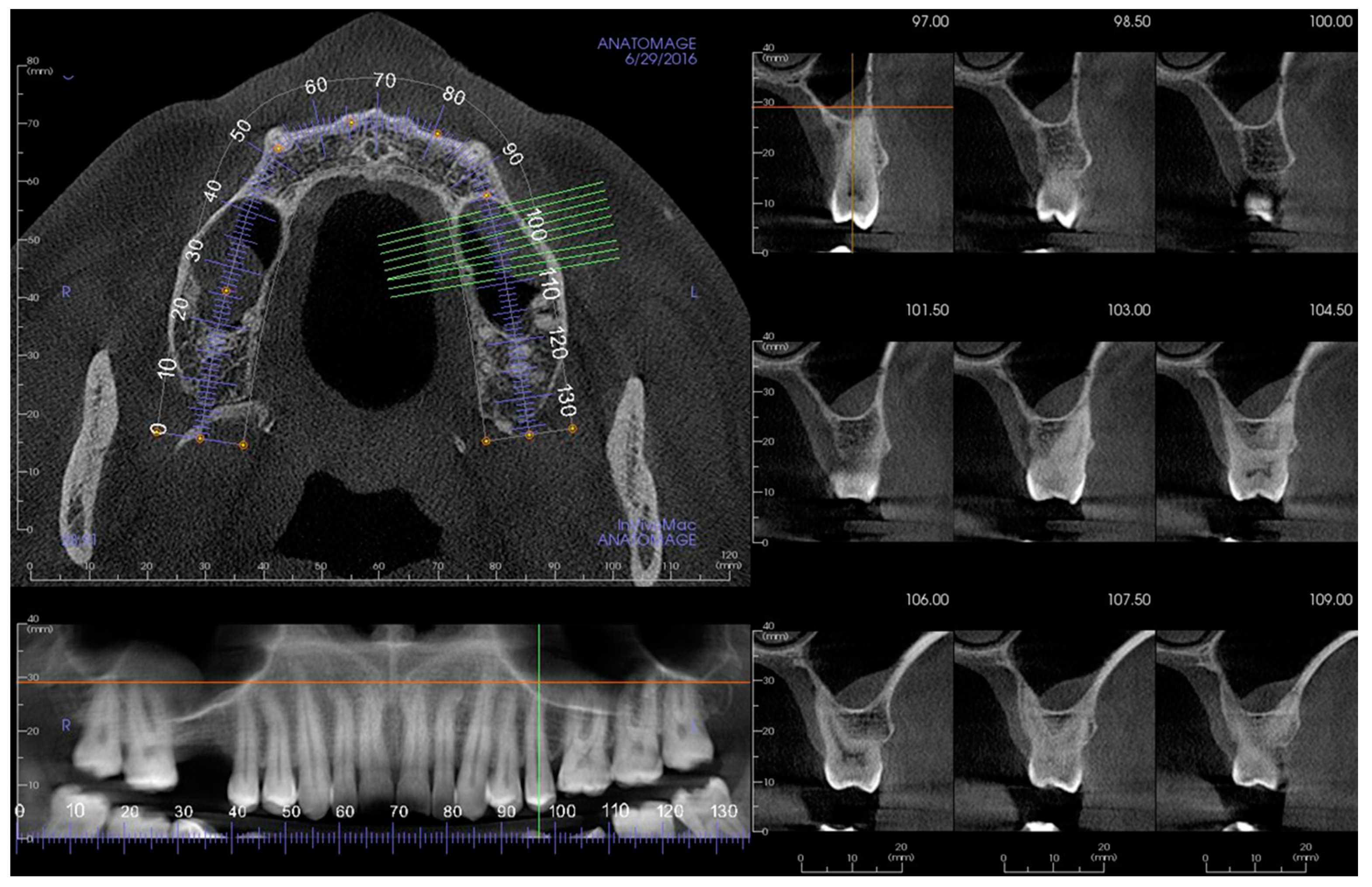
  • Group 1: root tips in contact with the sinus floor;
  • Group 2: root tips penetrating into the sinus;
  • Group 3: root tips below the sinus floor.
They reported that for the right side, 60% of the root tips were in Group 3, 30% were in Group 1, and 10% were in Group 2; for the left side, 68% were in Group 3, 21% were in Group 1, and 11% were in Group 2. There was no significant difference in the right or left. They concluded in the results that the distance from the root tip to the sinus floor was the longest for the first premolar root tip and the shortest for the second molar distobuccal root tip [25]. This close relationship may explain the results of the presence of periapical pathology and mucosal thickening with an odds ratio of 2.52 CI of having mucosal thickening with periapical pathology.
In other areas of medicine, it was recognized that infections in sinuses such as the frontal, ethmoidal, or maxillary sinuses could be sources of orbital infections and that spread potentially occurs by direct extension through the sinus wall and/or by local thrombophlebitis in areas where the bone is thin and in close proximity [26]. The reverse may also happen with infection within bone from apical disease, which causes the spread of infection to the sinus.
In relation to root-treated teeth, in this research, it was found that there was no association between the presence of root canal treatment and mucosal thickening (p = 0.38). One research looked at the association of dental condition and maxillary thickening. This retrospective research on CBCT found that 42.1% had mucosal thickening of >2 mm where periapical pathology was absent, and 53.6% when pathology was present (p < 0.05) [27]. In the present research, it was also found that teeth with periapical pathology had an influence on mucosal thickening and that the more posterior molars had an increased percentage of thickening (Figure 2). This reflects that the closer the association with the sinus, the more posterior position the tooth occupies [25].
Figure 2. Different tooth types and their relation to sinus pathology.
Thickening and periapical pathology were the highest in third molars, followed by second molars, first molars, and then first premolars. This reflects the prevalence of teeth with periapical pathology, and in other studies, it was found that the teeth most likely to have periapical pathology were molars, followed by first premolars [28]. In one research, the size of the lesion was compared using the periapical index, and it was concluded that the larger the periapical lesion, the greater the prevalence of mucosal thickening, and if more than one tooth had periapical pathology, the more severe the sinus disease recorded [29]. The fact that teeth have been root treated and the size of the lesion may be reduced or even healed would explain the results of no association with root canal treatment in the present research.
On exploring sinus floor perforation, 93.7% of teeth with periapical pathology had perforation of the sinus floor (p < 0.001). With the close relationship of the posterior teeth and the maxillary sinus [25], and the spread of infection in thin bone [26], this finding is what one would expect.

3. Conclusions

The reported incidence of the spread of infection from periapical tissues to the sinus is between 4.6 and 47% [30]. With the results of the present research, the close relationship of posterior teeth, the spread of infection, and the perforation of the sinus floor, the reported association with periapical disease and maxillary sinusitis can be understood.
The term endo-antral syndrome is used for the spread of periapical disease into the maxillary sinus [31][32][33]. All this shows there is a need to include dental investigation in cases of maxillary sinusitis, in particular where the etiology does not show ENT causes for the disease.
Computerized tomography is used for the evaluation of sinus disease within ENT. ENTs should routinely look for periapical pathology as a potential cause for sinusitis and regularly refer to dentists to assess these teeth before invasive sinus surgery.

References

  1. Garry, S.; O’Riordan, I.; James, D.; Corbett, M.; Barry, T.; Thornton, M. Odontogenic Sinusitis—Case Series and Review of Literature. J. Laryngol. Otol. 2021, 136, 49–54.
  2. Guerra-Pereira, I.; Vaz, P.; Faria-Almeida, R.; Braga, A.-C.; Felino, A. CT maxillary sinus evaluation—A retrospective cohort study. Med. Oral Patol. Oral Cir. Bucal 2015, 20, e419.
  3. Turfe, Z.; Ahmad, A.; Peterson, E.I.; Craig, J.R. Odontogenic sinusitis is a common cause of unilateral sinus disease with maxillary sinus opacification. Int. Forum Allergy Rhinol. 2019, 9, 1515–1520.
  4. Whyte, A.; Boeddinghaus, R. Imaging of odontogenic sinusitis. Clin. Radiol. 2019, 74, 503–516.
  5. Troeltzsch, M.; Pache, C.; Troeltzsch, M.; Kaeppler, G.; Ehrenfeld, M.; Otto, S.; Probst, F. Etiology and clinical characteristics of symptomatic unilateral maxillary sinusitis: A review of 174 cases. J. Cranio-Maxillofac. Surg. 2015, 43, 1522–1529.
  6. Hoskison, E.; Daniel, M.; Rowson, J.; Jones, N. Evidence of an increase in the incidence of odontogenic sinusitis over the last decade in the UK. J. Laryngol. Otol. 2012, 126, 43.
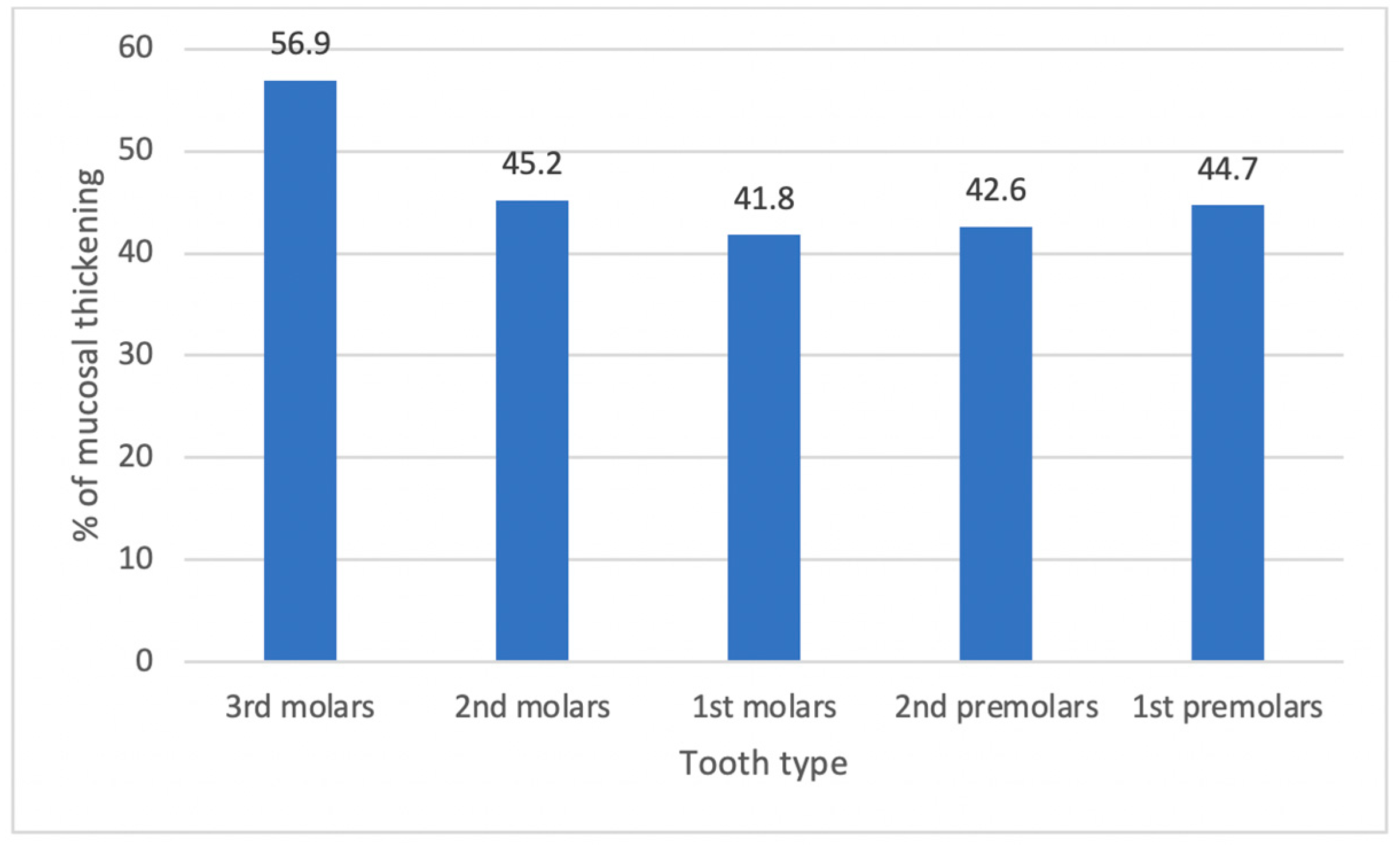
  7. Phothikhun, S.; Suphanantachat, S.; Chuenchompoonut, V.; Nisapakultorn, K. Cone-beam computed tomographic evidence of the association between periodontal bone loss and mucosal thickening of the maxillary sinus. J. Periodontol. 2012, 83, 557–564.
  8. Ugincius, P.; Kubilius, R.; Gervickas, A.; Vaitkus, S. Chronic odontogenic maxillary sinusitis. Stomatologija 2006, 8, 44–48.
  9. Arias-Irimia, O.; Barona-Dorado, C.; Santos-Marino, J.A.; Martínez-Rodríguez, N.; Martínez-González, J.M. Meta-analysis of the etiology of odontogenic maxillary sinusitis. Med. Oral Patol. Oral Cir. Bucal 2010, 15, e70–e73.
  10. Allevi, F.; Fadda, G.L.; Rosso, C.; Martino, F.; Pipolo, C.; Cavallo, G.; Felisati, G.; Saibene, A.M. Diagnostic criteria for odontogenic sinusitis: A systematic review. Am. J. Rhinol. Allergy 2021, 35, 713–721.
  11. Park, M.S.; Eo, M.Y.; Myoung, H.; Kim, S.M.; Lee, J.H. Early diagnosis of jaw osteomyelitis by easy digitalized panoramic analysis. Maxillofac. Plast. Reconstr. Surg. 2019, 41, 6.
  12. Workman, A.D.; Granquist, E.J.; Adappa, N.D. Odontogenic sinusitis: Developments in diagnosis, microbiology, and treatment. Curr. Opin. Otolaryngol. Head Neck Surg. 2018, 26, 27–33.
  13. Tataryn, R.; Lewis, M.; Horalek, A.; Thompson, C.; Cha, B.; Pokorny, A. Maxillary Sinusitis of Endodontic Origin: AAE Position Statement. 2018. Available online: https://www.aae.org/specialty/wp-content/uploads/sites/2/2018/04/AAE_PositionStatement_MaxillarySinusitis.pdf (accessed on 15 February 2022).
  14. Wang, R.-G.; Jiang, S.; Gu, R. The cartilaginous nasal capsule and embryonic development of human paranasal sinuses. J. Otolaryngol. 1994, 23, 239–243.
  15. Emirzeoglu, M.; Sahin, B.; Bilgic, S.; Celebi, M.; Uzun, A. Volumetric evaluation of the paranasal sinuses in normal subjects using computer tomography images: A stereological study. Auris Nasus Larynx 2007, 34, 191–195.
  16. Pirner, S.; Tingelhoff, K.; Wagner, I.; Westphal, R.; Rilk, M.; Wahl, F.; Bootz, F.; Eichhorn, K.W. CT-based manual segmentation and evaluation of paranasal sinuses. Eur. Arch. Oto-Rhino-Laryngol. 2009, 266, 507–518.
  17. Kucybała, I.; Janik, K.A.; Ciuk, S.; Storman, D.; Urbanik, A. Nasal Septal Deviation and Concha Bullosa—Do They Have an Impact on Maxillary Sinus Volumes and Prevalence of Maxillary Sinusitis? Pol. J. Radiol. 2017, 82, 126–133.
  18. Lana, J.P.; Carneiro, P.M.; Machado Vde, C.; de Souza, P.E.; Manzi, F.R.; Horta, M.C. Anatomic variations and lesions of the maxillary sinus detected in cone beam computed tomography for dental implants. Clin. Oral Implant. Res. 2012, 23, 1398–1403.
  19. Raghav, M.; Karjodkar, F.R.; Sontakke, S.; Sansare, K. Prevalence of incidental maxillary sinus pathologies in dental patients on cone-beam computed tomographic images. Contemp. Clin. Dent. 2014, 5, 361–365.
  20. Hsiao, Y.J.; Yang, J.; Resnik, R.R.; Suzuki, J.B. Prevalence of Maxillary Sinus Pathology Based on Cone-Beam Computed Tomography Evaluation of Multiethnicity Dental School Population. Implant. Dent. 2019, 28, 356–366.
  21. Hiari, M.; Hiari, M.A. Incidental paranasal sinus inflammatory changes in a Jordanian population. East. Mediterr. Health J. 1998, 4, 308–311.
  22. Eberhardt, J.A.; Torabinejad, M.; Christiansen, E.L. A computed tomographic study of the distances between the maxillary sinus floor and the apices of the maxillary posterior teeth. Oral Surg. Oral Med. Oral Pathol. 1992, 73, 345–347.
  23. Waite, D.E. Maxillary sinus. Dent. Clin. N. Am. 1971, 15, 349–368.
  24. Wallace, J.A. Transantral endodontic surgery. Oral Surg. Oral Med. Oral Pathol. Oral Radiol. Endod. 1996, 82, 80–83.
  25. Kilic, C.; Kamburoglu, K.; Yuksel, S.P.; Ozen, T. An assessment of the relationship between the maxillary sinus floor and the maxillary posterior teeth root tips using dental cone-beam computerized tomography. Eur. J. Dent. 2010, 4, 462–467.
  26. Mills, R.P.; Kartush, J.M. Orbital wall thickness and the spread of infection from the paranasal sinuses. Clin. Otolaryngol. Allied Sci. 1985, 10, 209–216.
  27. Aksoy, U.; Orhan, K. Association between odontogenic conditions and maxillary sinus mucosal thickening: A retrospective CBCT study. Clin. Oral Investig. 2019, 23, 123–131.
  28. van der Borden, W.G.; Wang, X.; Wu, M.-K.; Shemesh, H. Area and 3-dimensional volumetric changes of periapical lesions after root canal treatments. J. Endod. 2013, 39, 1245–1249.
  29. Lu, Y.; Liu, Z.; Zhang, L.; Zhou, X.; Zheng, Q.; Duan, X.; Zheng, G.; Wang, H.; Huang, D. Associations between maxillary sinus mucosal thickening and apical periodontitis using cone-beam computed tomography scanning: A retrospective study. J. Endod. 2012, 38, 1069–1074.
  30. Melén, I.; Lindahl, L.; Andréasson, L.; Rundcrantz, H. Chronic maxillary sinusitis: Definition, diagnosis and relation to dental infections and nasal polyposis. Acta Oto-Laryngol. 1986, 101, 320–327.
  31. Selden, H.S. The endo-antral syndrome. J. Endod. 1977, 3, 462–464.
  32. Selden, H.S. The endo-antral syndrome: An endodontic complication. J. Am. Dent. Assoc. 1989, 119, 397–398, 401–402.
  33. Selden, H.S. Endo-Antral syndrome and various endodontic complications. J. Endod. 1999, 25, 389–393.
More
Information
Contributor MDPI registered users' name will be linked to their SciProfiles pages. To register with us, please refer to https://encyclopedia.pub/register :
View Times: 600
Revisions: 3 times (View History)
Update Date: 21 Mar 2022
1000/1000
Video Production Service