Submitted Successfully!
To reward your contribution, here is a gift for you: A free trial for our video production service.
Thank you for your contribution! You can also upload a video entry or images related to this topic.
Version Summary Created by Modification Content Size Created at Operation
1 + 1925 word(s) 1925 2022-02-18 08:49:09 |
2 Format correct + 12 word(s) 1937 2022-03-07 02:13:11 | |
3 Format correct + 12 word(s) 1937 2022-03-07 02:15:32 |

Video Upload Options

Do you have a full video?

Confirm

Are you sure to Delete?
Cite
If you have any further questions, please contact Encyclopedia Editorial Office.
Oh, S. Isothermal Amplification Methods in Detecting Food-Borne Pathogens. Encyclopedia. Available online: https://encyclopedia.pub/entry/20263 (accessed on 27 July 2024).
Oh S. Isothermal Amplification Methods in Detecting Food-Borne Pathogens. Encyclopedia. Available at: https://encyclopedia.pub/entry/20263. Accessed July 27, 2024.
Oh, Se-Wook. "Isothermal Amplification Methods in Detecting Food-Borne Pathogens" Encyclopedia, https://encyclopedia.pub/entry/20263 (accessed July 27, 2024).
Oh, S. (2022, March 07). Isothermal Amplification Methods in Detecting Food-Borne Pathogens. In Encyclopedia. https://encyclopedia.pub/entry/20263
Oh, Se-Wook. "Isothermal Amplification Methods in Detecting Food-Borne Pathogens." Encyclopedia. Web. 07 March, 2022.
Isothermal Amplification Methods in Detecting Food-Borne Pathogens
Edit

The isothermal amplification method, a molecular-based diagnostic technology, such as loop-mediated isothermal amplification (LAMP) and recombinase polymerase amplification (RPA), is widely used as an alternative to the time-consuming and labor-intensive culture-based detection method. However, food matrices or other compounds can inhibit molecular-based diagnostic technologies, causing reduced detection efficiencies, and false-negative results. These inhibitors originating from food are polysaccharides and polyphenolic compounds in berries, seafood, and vegetables. Additionally, magnesium ions needed for amplification reactions can also inhibit molecular-based diagnostics. The successful removal of inhibitors originating from food and molecular amplification reaction is therefore proposed to enhance the efficiency of molecular-based diagnostics and allow accurate detection of food-borne pathogens. Among molecular-based diagnostics, polymerase chain reaction (PCR) inhibitors have been reported.

molecular amplification detection method food metrix bacteria inhibition

1. Introduction

Loop-mediated isothermal amplification (LAMP) is conducted in the presence of Bacillus stearothermophylus (Bst) DNA polymerase, deoxyribonucleotide triphosphate (dNTP), and 4–6 specific primers (inner and outer primers) that recognize 6–8 specific regions [1]. The Bst DNA polymerase removes the need for high thermocycling because of its activity at 50–70 °C, and the DNA polymerase is less sensitive to food-origin inhibitors [2][3]. The use of two outer primers (forward outer primer; F3, and backward outer primer; B3), two inner primers (forward inner primer; FIP, and backward inner primer; BIP), with additional loop primers (forward loop primer; LF, and backward loop primer; LB) also allows sequence-specific detection, improves specificity, and accelerates assay specificity to amplification targets [4]. However, although many long primers increase the reaction yield, a risk of primer dimerization owing to nonspecific interactions simultaneously exists [5][6]. To prevent primer dimerization, adding dimethyl sulfoxide (DMSO) and betaine is used as a strategy to decrease nonspecific interactions, thereby giving stability to oligonucleotides [7]. The use of additional compounds can amplify nucleic acids without non-specific amplification, and exponential nucleic acid amplification is achieved while forming loops within 40–60 min at a constant temperature of 60–65 °C [8]. Then, the amplified product can be detected using turbidity measurement, gel electrophoresis, colorimetry, electrochemiluminescence, lateral flow assay (LFA), and real-time monitoring [9][10].
Recombinase polymerase amplification (RPA) is conducted while a recombinase protein UvsX from T4-like bacteriophages forms a complex with primers in the presence of ATP and a crowding agent, such as polyethylene glycol (PEG) [11]. The crowding agent prevents spontaneous recombinase-primer degradation, thereby allowing amplification to begin. It also enhances the amplification efficiency by improving catalytic activity of the enzyme [12][13]. Therefore, using long primers (up to 45 nucleotides) can form secondary structures and potential primer artifacts; recommended length of RPA primers is 30–35 bases [11][14]. RPA is conducted at relatively low and constant temperatures of 37–42 °C for 20–40 min, and detection of amplicons are performed through gel electrophoresis, flocculation assay detection, LFA, electrochemical assay, chemiluminescent assay, and silicon microring resonator (SMR)-based photonic assay, and real-time monitoring [15][13][16].
HDA reacts with helicase, two primers, and two accessory proteins; methyl-directed mismatch repair (MutL), and single-stranded DNA (ssDNA) binding (SSB) proteins that stimulate UvrD helicase activity above ten-fold [17][18]. During the HDA replication process, the helicase unwinds double-stranded DNA (dsDNA) for the denaturation. In this step, the accessory proteins are required to bind and stabilize the ssDNA for the prevention of recombination of the complementary strand, thereby allowing primer hybridization [19]. The HDA reaction is divided into two systems: the mesophilic form of HDA (mHDA) and the thermophilic form of HDA (tHDA). The mHDA reacts at a medium temperature of 37 °C using UvrD helicase/Exo-Klenow polymerase; the tHDA reacts at a higher temperature (60–65 °C) using a thermostable Tte-UvrD helicase/Bst DNA polymerase [20]. Compared to mHDA, tHDA has a higher sensitivity and efficiency, and simplifies the reaction because it does not need MutL and SSB proteins to stabilize the DNA sequence [21]. The magnesium ion, which serves as a cofactor for the helicase and polymerase, is also used in HDA to increase the enzyme activity, thereby making the enzymes compatible with structurally modified primers [5][22]. Subsequently, amplification products are detected using gel electrophoresis, colorimetry assay, LFA, and real-time monitoring [21].
Nucleic acid sequence-based amplification (NASBA) targets 16s rRNA genes or mRNA transcripts for bacterial detection, enabling the analysis of bacterial viability [23][24]. NASBA amplifies single-stranded RNA using two primers and three enzymes (avian myeloblastosis virus reverse transcriptase (AMV-RT), RNase H, and T7 DNA-dependent RNA polymerase (DdRp)) [25]. While AVM-RT generates complementary complementary DNA by extending primers, dsDNA is formed through RNase H. However, T7 DdRP recognizes the exposed T7 promoter of the dsDNA and initiates transcription to initiate the reaction [25]. Since NASBA enzymes are heat labile, amplifications can be performed at a relatively low temperature, with optimal conditions of 41 °C for 1.5–2 h [26]. The low reaction temperature of NASBA, however, can produce to result in false-positive results because of nonspecific primer interactions. Nevertheless, adding DMSO and betaine can prevent this limitation [27][28]. Subsequently, amplification products are detected using gel electrophoresis, enzyme-linked immunosorbent assay (ELISA), enzyme-linked gel assay, electrochemiluminescent (ECL), and real-time monitoring with molecular beacons [5][29]. When the clustered regularly interspaced short palindromic repeats (CRISPR)/Cas system and NASBA are combined, RNA can be detected with high sensitivity through the NASBACC (NASBA-CRISPR Cleavage) system [30].
RCA has high specificity and sensitivity targeting RNA and DNA [31]. The RCA reaction is conducted using DNA/RNA polymerase (phi29 DNA polymerase or T7 RNA polymerase), short DNA or RNA linear single-stranded primer, circle template, and ligase [32]. DNA/RNA polymerase produces a long single-stranded RCA product (RCAP) complementary to a circular template. Unique oligonucleotide padlock probes (PLP) and T4 DNA ligase or special ssDNA ligase synthesize bacterial single- or double-stranded RNA/DNA templates into single-stranded circular DNA [5]. The various modified RCA systems have been developed for efficient amplification: a linear-RCA (LRCA) or an exponential-RCA (ERCA), such as multiply-primed RCA (MPRCA), hyperbranched RCA (HRCA), and primer-generation RCA (PG-RCA) [25]. The saltatory RCA (SRCA), a simplified form of RCA, has been developed, requiring ligase and PLP for cyclization [33][34]. RCA generally reacts for 1–1.5 h at 30–65 °C depending on the reaction system, and amplification products are detected using gel electrophoresis, colorimetry, and real-time monitoring [25].
MDA randomly and massively amplifies single-cell genomic DNA and is compatible with whole genome amplification (WGA) [35][36]. MDA is conducted using modified random hexamer primers, phi29 DNA polymerases (strand-displacing DNA polymerase from bacteriophage Ø29), denatured template DNA, and dNTPs [37]. The modified random hexamer primers eliminate the need to design target-specific primers are designed to anneal to random areas on each strand of the target DNA, thereby forming hyperbranched intermediates and dsDNA amplicons after exponential amplification [38]. The phi29 DNA polymerase enables amplification at a relatively low temperature (typically 30 °C) because of its high strand displacement activity; it has a higher replication fidelity and lower error rate than Taq DNA polymerases and Bst DNA polymerases [39][40]. Furthermore, the addition of PEG to the reaction for high-efficiency MDA causes molecular crowding, which enables sensitive allele detection in multiplex short tandem repeat genotyping [41].
Schematic diagram and summary of isothermal amplification techniques are shown in Figure 1 and Table 1.
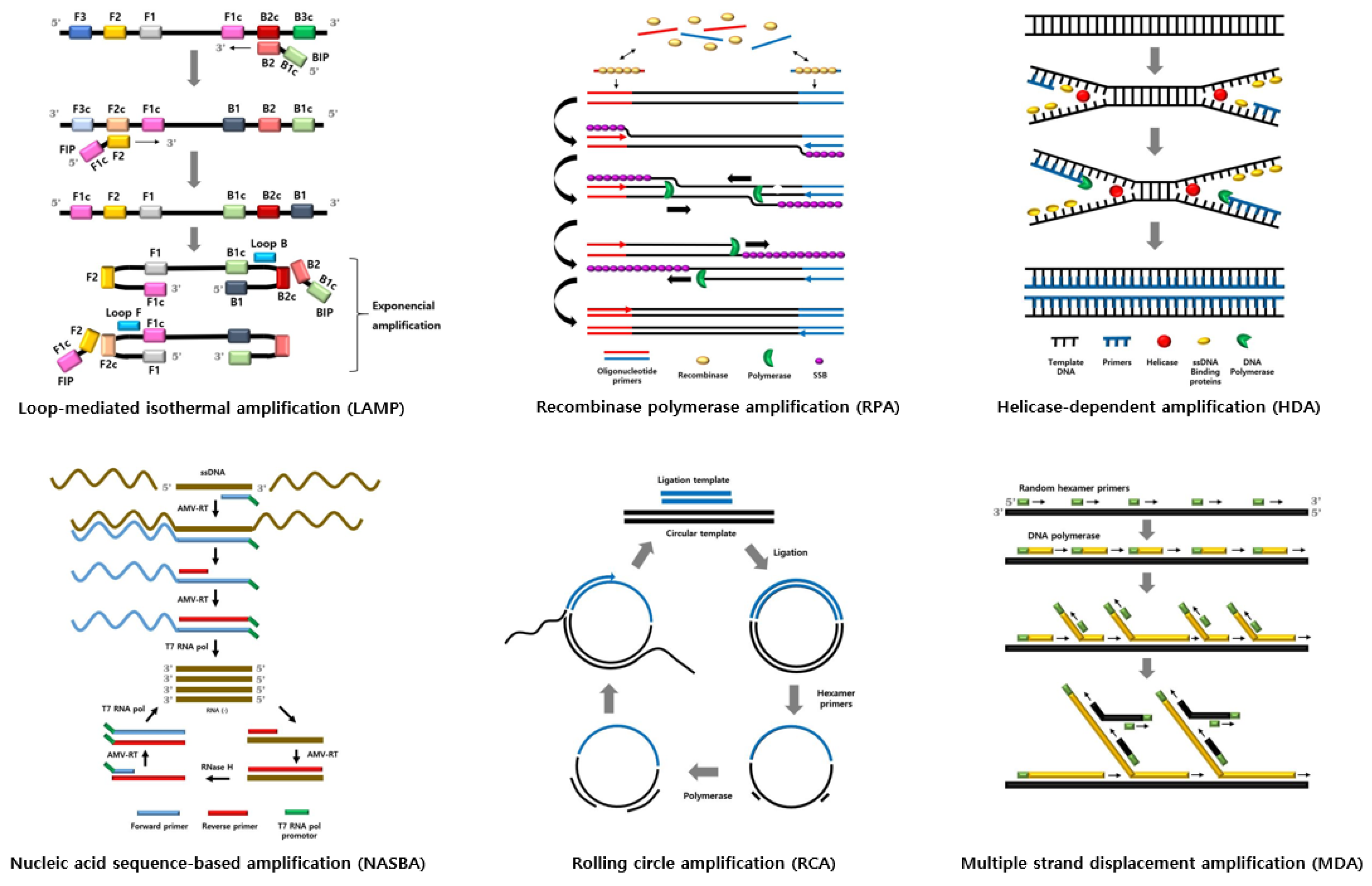
Figure 1. Schematic diagram of isothermal amplification techniques.
Table 1. Summary of comparison among various isothermal amplification methods.
Isothermal Amplification Methods Number of Primers Number of Enzymes Pre-Heating Working Temperature (°C) Reaction Time (min) Target Template Amplicon Resistance to Inhibitor Reference
LAMP 4–6 1 No 60–65 40–60 DNA DNA High [8]
RPA 2 2 No 37–42 20–40 DNA DNA Low [15]
HDA 2 1 (mHDA), 3 (tHDA) No 37 (mHDA), 60–65 (tHDA) 100–120 DNA DNA High [21]
NASBA 2 2–3 Yes 41 90–120 RNA RNA, DNA Low [26]
RCA 1 1 Yes 30–65 60–90 Circular DNA DNA Low [25]
MDA Random hexamer primers 1 No 35 270 Circular or linear DNAs Ramified double–stranded DNAs High [42]

Abbreviations used in the table: LAMP, loop-mediated isothermal amplification; RPA, recombinase polymerase amplification; HDA, helicase-dependent amplification; mHDA, mesophilic form of HDA; tHDA, thermophilic form of HDA; NASBA, nucleic acid sequence-based amplification; RCA, rolling circle amplification; MDA, multiple strand displacement amplification.

2. Inhibitors Originating from Isothermal Amplification Processes

Various inhibitors repress the isothermal amplification technique for detecting bacteria, including food-borne pathogens. These inhibitors originate from amplification or detection processes and the food metrix (Table 2).
Table 2. Inhibitors from the isothermal amplification reaction process.
Reaction Process Inhibitors Alleviation Strategies for Inhibition Amplification Methods Reference
Sample preparation and DNA extraction Residual food metrix Use the nucleic acid sample after dilution NASBA [43]
CTAB used as extraction buffer Use direct PCR buffers RPA [44]
Nucleic acid amplification Concentration Magnesium ions Increase the concentration of magnesium ions tHDA [45][46]
Add betaine, DMSO, and sorbitol to the reaction mixture [17]
Use 4–6 mM MgSO4, which is the optimal concentration for magnesium ions RCA [47]
Primer Optimize concentration of primer RPA [11]
Multi-RPA [48]
Template or background DNA Treat RNase A with pasteurization and 15 min incubation process before nucleic acid extraction Real-time NASBA [49]
Add the primer stability enhancer to the primer and beacon mixture NASBA [50]
Temperature Temperature fluctuations Optimize reaction temperature MDA [51]
Heat denaturation Substitute alkaline denaturation [52]
Detection method Colorimetric detection SYBR Green I Add fluorescent dyes after amplification LAMP [53]
Use wax capsules containing the dye, which react after amplification [52]
PEI Add PEI after amplification LAMP [54]
Calcein - -
Electrochemical detection Redox active compounds (e.g., MB and Hoechst 33258) Use other redox molecules (e.g., osmium redox and RuHex) LAMP [55]
Use voltammeric mode [53]
Use polydopamine-doped paper disks [56]
Abbreviations used in the table: NASBA, nucleic acid sequence-based amplification; CTAB, cetyltrimethyl ammonium bromide; PCR, polymerase chain reaction; RPA, recombinase polymerase amplification; DMSO, dimethyl sulfoxide; tHDA, thermophilic form of HDA; RCA, rolling circle amplification; MDA, multiple strand displacement amplification; LAMP, loop-mediated isothermal amplification; PEI, polyethyleneimine; MB, methylene blue; RuHex, ruthenium hexamine.
 
Bst DNA polymerase and helicase that enables isothermal amplification require a relatively high concentration of magnesium ions of about 4–8 mM compared to Taq DNA polymerase [18]. However, magnesium ions can inhibit molecular amplification depending on the concentration in the reaction mixture [47]. Murakami et al. [47] reported that by increasing the background signal amplification in RCA, the concentration of magnesium ions inhibited the signal during target gene amplification. Nb.BsmI, a nicking enzyme, needs magnesium ions to enhance amplification efficiency. Therefore, optimizing magnesium ion concentrations (4–6 mM MgSO4) and decreasing dNTPs and DNA polymerase concentrations can decrease background signal amplifications. Doseeva et al. [46] conducted a study to alleviate magnesium ion-dependent inhibition because the concentration of magnesium ions affected the amplification efficiency of tHDA. Results indicated that as the concentration of magnesium ions and that of the dATPs improved, the signal-to-noise ratio increased 1.5–2.0 times. It was also observed that the optimal concentrations of MgSO4, dATP, and dNTP were 4, 3, and 0.4 mmol/L, respectively. Additionally, betaine, DMSO, and sorbitol, which help the combined effect of magnesium ions and dATP during molecular amplification, were added to increase the efficiency and specificity of amplification in tHDA.
The inappropriate concentration of primers can inhibit molecular amplification processes. Thus, SSB proteins either inhibit the strand exchange activity of recombinase T4 UvsX or compete with recombinase proteins in RPA [57]. Additionally, primers for one target can inhibit the amplification of another target [11]. Therefore, it is essential to optimize primer concentration [11][16].

3. Conclusions

Inhibitors originating from food matrices, DNA extraction reactions and nucleic acid amplification reactions can adversely affect isothermal amplification techniques by inhibiting amplification to detect food-borne pathogens. Nevertheless, due to the complex nature of the food matrix, food-origin inhibitors, including inhibition mechanisms for molecular analysis, have not been fully characterized yet. Additionally, studies on the inhibitory mechanisms of inhibitors obtained during isothermal amplification processes are still insufficient and have not been clearly established. The above-mentioned removal strategies for inhibiting nucleic acid amplification are rather limited and uneconomical because they only target specific inhibitors, and have not been proven to be applicable to various inhibitors. Therefore, further studies on nucleic acid amplification inhibitors and inhibitor removal strategies are needed to detect food-borne pathogens in food using isothermal amplification technologies without inhibition of nucleic acid amplification inhibitors. In the future, these studies will lead to the manufacture of a ready-to-use kit that simultaneously purifies and removes inhibitors. In this case, efficiency is guaranteed for accurate detection of food-borne pathogens in complex food matrices and for the point-of-care testing (POCT) application of these isothermal amplification technologies.

References

  1. Becherer, L.; Borst, N.; Bakheit, M.; Frischmann, S.; Zengerle, R.; von Stetten, F.V. Loop-mediated isothermal amplification (LAMP)–review and classification of methods for sequence-specific detection. Anal. Methods 2020, 12, 717–746.
  2. Tao, Y.; Yun, J.; Wang, J.; Xu, P.; Li, C.; Liu, H.; Lan, Y.; Pan, J.; Du, W. High-performance detection of Mycobacterium bovis in milk using digital LAMP. Food Chem. 2020, 327, 126945.
  3. Meagher, R.J.; Priye, A.; Light, Y.K.; Huang, C.; Wang, E. Impact of primer dimers and self-amplifying hairpins on reverse transcription loop-mediated isothermal amplification detection of viral RNA. Analyst 2018, 143, 1924–1933.
  4. Fernández-Soto, P.; Mvoulouga, P.O.; Akue, J.P.; Abán, J.L.; Santiago, B.V.; Sánchez, M.C.; Muro, A. Development of a highly sensitive loop-mediated isothermal amplification (LAMP) method for the detection of Loa loa. PLoS ONE 2014, 9, e94664.
  5. Asadi, R.; Mollasalehi, H. The mechanism and improvements to the isothermal amplification of nucleic acids, at a glance. Anal. Biochem. 2021, 631, 114260.
  6. Shahbazi, E.; Mollasalehi, H.; Minai-Tehrani, D. Development and evaluation of an improved quantitative loop-mediated isothermal amplification method for rapid detection of Morganella morganii. Talanta 2019, 191, 54–58.
  7. Chen, Y.; Li, H.; Yang, L.; Wang, L.; Sun, R.; Shearer, J.E.; Sun, F. Rapid Detection of Clostridium botulinum in Food Using Loop-Mediated Isothermal Amplification (LAMP). Int. J. Environ. Res. Public Health 2021, 18, 4401.
  8. Ahuja, A.; Somvanshi, V.S. Diagnosis of plant-parasitic nematodes using loop-mediated isothermal amplification (LAMP): A review. J. Crop. Prot. 2021, 147, 105459.
  9. Leonardo, S.; Toldrà, A.; Campàs, M. Biosensors based on isothermal DNA amplification for bacterial detection in food safety and environmental monitoring. Sensors 2021, 21, 602.
  10. Lobato, I.M.; O’Sullivan, C.K. Recombinase polymerase amplification: Basics, applications and recent advances. Trends Anal. Chem. 2018, 98, 19–35.
  11. Lillis, L.; Siverson, J.; Lee, A.; Cantera, J.; Parker, M.; Piepenburg, O.; Lehman, D.A.; Boyle, D.S. Factors influencing recombinase polymerase amplification (RPA) assay outcomes at point of care. Mol. Cell Probes 2016, 30, 74–78.
  12. Zhai, J.; Wang, L.; Qiao, X.; Zhao, J.; Wang, X.; He, X. Detection of Neisseria gonorrhoeae and Chlamydia trachomatis infections in pregnant women by multiplex recombinase polymerase amplification. PLoS ONE 2021, 16, e0251119.
  13. Mayboroda, O.; Gonzalez Benito, A.; Sabaté del Rio, J.; Svobodova, M.; Julich, S.; Tomaso, H.; O’Sullivan, C.K.; Katakis, I. Isothermal solid-phase amplification system for detection of Yersinia pestis. Anal. Bioanal. Chem. 2016, 408, 671–676.
  14. Li, J.; Macdonald, J.; von Stetten, F. Review: A comprehensive summary of a decade development of the recombinase polymerase amplification. Analyst 2018, 144, 31–67.
  15. Hu, J.; Wang, Y.; Su, H.; Ding, H.; Sun, X.; Gao, H.; Geng, Y.; Wang, Z. Rapid analysis of Escherichia coli O157: H7 using isothermal recombinase polymerase amplification combined with triple-labeled nucleotide probes. Mol. Cell Probes 2020, 50, 101501.
  16. Du, X.J.; Zhou, T.J.; Li, P.; Wang, S. A rapid Salmonella detection method involving thermophilic helicase dependent amplification and a lateral flow assay. Mol. Cell Probes 2017, 34, 37–44.
  17. Glökler, J.; Lim, T.S.; Ida, J.; Frohme, M. Isothermal amplifications–a comprehensive review on current methods. Crit Rev. Biochem. Mol. Biol. 2021, 56, 543–586.
  18. Kumar, A.A.; Hennek, J.W.; Smith, B.S.; Kumar, S.; Beattie, P.; Jain, S.; Rolland, J.P.; Stossel, T.P.; Chunda-Liyoka, C.; Whitesides, G.M. From the bench to the field in low-cost diagnostics: Two case studies. Angew. Chem. Int. Ed. Engl. 2015, 54, 5836–5853.
  19. Kumar, Y. Isothermal amplification-based methods for assessment of microbiological safety and authenticity of meat and meat products. Food Control 2021, 121, 107679.
  20. Yang, Z.; McLendon, C.; Hutter, D.; Bradley, K.M.; Hoshika, S.; Frye, C.; Benner, S.A. Helicase dependent isothermal amplification of DNA and RNA using self-avoiding molecular recognition systems. ChemBioChem 2015, 16, 1365–1370.
  21. Barreda-García, S.; Miranda-Castro, R.; de-Los-Santos-Álvarez, N.; Miranda-Ordieres, A.J.; Lobo-Castañón, M.J. Helicase-dependent isothermal amplification: A novel tool in the development of molecular-based analytical systems for rapid pathogen detection. Anal. Bioanal. Chem. 2018, 410, 679–693.
  22. Thekisoe, O.M.; Bazie, R.S.; Coronel-Servian, A.M.; Sugimoto, C.; Kawazu, S.I.; Inoue, N. Stability of loop-mediated isothermal amplification (LAMP) reagents and its amplification efficiency on crude trypanosome DNA templates. J. Vet. Med. Sci. 2009, 71, 471–475.
  23. Zhai, L.; Liu, H.; Chen, Q.; Lu, Z.; Zhang, C.; Lv, F.; Bie, X. Development of a real-time nucleic acid sequence–based amplification assay for the rapid detection of Salmonella spp. from food. Braz. J. Microbiol. 2019, 50, 255–261.
  24. Xu, L.; Duan, J.; Chen, J.; Ding, S.; Cheng, W. Recent advances in rolling circle amplification-based biosensing strategies—A review. Anal. Chim. Acta 2021, 1148, 238187.
  25. Hønsvall, B.K.; Robertson, L.J. From research lab to standard environmental analysis tool: Will NASBA make the leap? Water Res. 2017, 109, 389–397.
  26. Brennan, D.; Coughlan, H.; Clancy, E.; Dimov, N.; Barry, T.; Kinahan, D.; Galvin, P. Development of an on-disc isothermal in vitro amplification and detection of bacterial RNA. Sens. Actuators B 2017, 239, 235–242.
  27. Takahashi, M.K.; Tan, X.; Dy, A.J.; Braff, D.; Akana, R.T.; Furuta, Y.; Collins, J.J. A low-cost paper-based synthetic biology platform for analyzing gut microbiota and host biomarkers. Nat. Commun. 2018, 9, 1–12.
  28. Li, J.; Macdonald, J. Advances in isothermal amplification: Novel strategies inspired by biological processes. Biosens. Bioelectron. 2015, 64, 196–211.
  29. Kellner, M.J.; Koob, J.G.; Gootenberg, J.S.; Abudayyeh, O.O.; Zhang, F. SHERLOCK: Nucleic acid detection with CRISPR nucleases. Nat. Protoc. 2019, 14, 2986–3012.
  30. Pardee, K.; Green, A.A.; Takahashi, M.K.; Braff, D.; Lambert, G.; Lee, J.W.; Ferrante, T.; Ma, D.; Donghia, D.; Fan, M.; et al. Rapid, low-cost detection of Zika virus using programmable biomolecular components. Cell 2016, 165, 1255–1266.
  31. van Emmerik, C.L.; Gachulincova, I.; Lobbia, V.R.; Daniëls, M.A.; Heus, H.A.; Soufi, A.; Nelissen, F.H.T.; van Ingen, H. Ramified Rolling Circle Amplification for efficient and flexible synthesis of nucleosomal DNA sequences. bioRxiv 2019, 676528.
  32. Wang, Z.; Yang, Q.; Zhang, Y.; Meng, Z.; Ma, X.; Zhang, W. Saltatory rolling circle amplification (SRCA): A novel nucleic acid isothermal amplification technique applied for rapid detection of Shigella spp. in vegetable salad. Food Anal. Methods 2018, 11, 504–513.
  33. Milton, A.A.P.; Momin, K.M.; Priya, G.B.; Ghatak, S.; Gandhale, P.N.; Angappan, M.; Das, S.; Sen, A. A novel in situ methodology for visual detection of Clostridium perfringens in pork harnessing saltatory rolling circle amplification. Anaerobe 2021, 69, 102324.
  34. Spits, C.; Le Caignec, C.; De Rycke, M.; Van Haute, L.; Van Steirteghem, A.; Liebaers, I.; Sermon, K. Optimization and evaluation of single-cell whole-genome multiple displacement amplification. Hum. Mutat. 2006, 27, 496–503.
  35. Tian, H.C.; Benitez, J.J.; Craighead, H.G. Single cell on-chip whole genome amplification via micropillar arrays for reduced amplification bias. PLoS ONE 2018, 13, e0191520.
  36. Forghani, F.; Li, S.; Zhang, S.; Mann, D.A.; Deng, X.; den Bakker, H.C.; Diez-Gonzalez, F. Detection and Serotyping of Salmonella and Escherichia coli in Wheat Flour by a Quasimetagenomic Approach Assisted by Magnetic Capture, Multiple Displacement Amplification and Real-Time Sequencing. Appl. Environ. Microbiol. 2020, 86, e00097-20.
  37. Wang, W.; Ren, Y.; Lu, Y.; Xu, Y.; Crosby, S.D.; Di Bisceglie, A.M.; Fan, X. Template-dependent multiple displacement amplification for profiling human circulating RNA. Biotechniques 2017, 63, 21–27.
  38. Jamal, R.; Li, X.; Weidhaas, J. Template length, concentration and guanidine and cytosine content influence on multiple displacement amplification efficiency. J. Microbiol. Methods 2021, 181, 106146.
  39. Huang, M.; Yang, F.; Fu, J.; Xiao, P.; Tu, J.; Lu, Z. Reaction parameter comparison and optimization of multiple displacement amplification. Anal. Methods 2020, 12, 46–53.
  40. Long, N.; Qiao, Y.; Xu, Z.; Tu, J.; Lu, Z. Recent advances and application in whole-genome multiple displacement amplification. Quant. Biol. 2020, 8, 1–16.
  41. Smith, T.J.; O’Connor, L.; Glennon, M.; Maher, M. Molecular diagnostics in food safety: Rapid detection of food-borne pathogens. Ir. J. Agric. Food Res. 2000, 39, 309–319.
  42. Santiago-Felipe, S.; Tortajada-Genaro, L.A.; Morais, S.; Puchades, R.; Maquieira, Á. Isothermal DNA amplification strategies for duplex microorganism detection. Food Chem. 2015, 174, 509–515.
  43. Lv, X.; Wang, L.; Zhang, J.; Zeng, H.; Chen, X.; Shi, L.; Cui, H.; He, X.; Zhao, L. Rapid and sensitive detection of VBNC Escherichia coli O157: H7 in beef by PMAxx and real-time LAMP. Food Control 2020, 115, 107292.
  44. Cai, Q.; Wang, R.; Qiao, Z.; Yang, W. Single-digit Salmonella detection with the naked eye using bio-barcode immunoassay coupled with recombinase polymerase amplification and a CRISPR-Cas12a system. Analyst 2021, 146, 5271–5279.
  45. Munawar, M.A.; Martin, F.; Toljamo, A.; Kokko, H.; Oksanen, E. RPA-PCR couple: An approach to expedite plant diagnostics and overcome PCR inhibitors. BioTechniques 2020, 69, 270–280.
  46. Doseeva, V.; Forbes, T.; Wolff, J.; Khripin, Y.; O’Neil, D.; Rothmann, T.; Nazarenko, I. Multiplex isothermal helicase-dependent amplification assay for detection of Chlamydia trachomatis and Neisseria gonorrhoeae. Diagn. Microbiol. Infect. Dis 2011, 71, 354–365.
  47. Murakami, T.; Sumaoka, J.; Komiyama, M. Sensitive isothermal detection of nucleic-acid sequence by primer generation-rolling circle amplification. Nucleic Acids Res. 2009, 37, e19.
  48. Paez, J.G.; Lin, M.; Beroukhim, R.; Lee, J.C.; Zhao, X.; Richter, D.J.; Gabriel, S.; Herman, P.; Sasaki, H.; Altshuler, D.; et al. Genome coverage and sequence fidelity of ϕ29 polymerase-based multiple strand displacement whole genome amplification. Nucleic Acids Res. 2004, 32, e71.
  49. Sidoti, F.; Bergallo, M.; Terlizzi, M.E.; Piasentin Alessio, E.P.; Astegiano, S.; Gasparini, G.; Cavallo, R. Development of a quantitative real-time nucleic acid sequence-based amplification assay with an internal control using molecular beacon probes for selective and sensitive detection of human rhinovirus serotypes. Mol. Biotechnol. 2012, 50, 221–228.
  50. Feng, J.; Dai, Z.; Tian, X.; Jiang, X. Detection of Listeria monocytogenes based on combined aptamers magnetic capture and loop-mediated isothermal amplification. Food Control 2018, 85, 443–452.
  51. Barreda-García, S.; Miranda-Castro, R.; de-Los-Santos-Álvarez, N.; Miranda-Ordieres, A.J.; Lobo-Castañón, M.J. Comparison of isothermal helicase-dependent amplification and PCR for the detection of Mycobacterium tuberculosis by an electrochemical genomagnetic assay. Anal. Bioanal. Chem. 2016, 408, 8603–8610.
  52. Zhang, X.; Qu, K.; Li, Q.; Cui, Z.; Zhao, J.; Sun, X. Recording the reaction process of loop-mediated isothermal amplification (LAMP) by monitoring the voltammetric response of 2′-deoxyguanosine 5′-triphosphate. Electroanalysis 2011, 23, 2438–2445.
  53. Rani, A.; Ravindran, V.B.; Surapaneni, A.; Shahsavari, E.; Haleyur, N.; Mantri, N.; Ball, A.S. Evaluation and comparison of recombinase polymerase amplification coupled with lateral-flow bioassay for Escherichia coli O157: H7 detection using different genes. Sci. Rep. 2021, 11, 1881.
  54. Wastling, S.L.; Picozzi, K.; Kakembo, A.S.; Welburn, S.C. LAMP for human African trypanosomiasis: A comparative study of detection formats. PLoS Negl. Trop. Dis. 2010, 4, e865.
  55. Deféver, T.; Druet, M.; Evrard, D.; Marchal, D.; Limoges, B. Real-time electrochemical PCR with a DNA intercalating redox probe. Anal. Chem. 2011, 83, 1815–1821.
  56. Sun, Y.; Quyen, T.L.; Hung, T.Q.; Chin, W.H.; Wolff, A.; Bang, D.D. A lab-on-a-chip system with integrated sample preparation and loop-mediated isothermal amplification for rapid and quantitative detection of Salmonella spp. in food samples. Lab Chip 2015, 15, 1898–1904.
  57. Nadal, A.; Coll, A.; Cook, N.; Pla, M. A molecular beacon-based real time NASBA assay for detection of Listeria monocytogenes in food products: Role of target mRNA secondary structure on NASBA design. J. Microbiol. Methods 2007, 68, 623–632.
More
Information
Contributor MDPI registered users' name will be linked to their SciProfiles pages. To register with us, please refer to https://encyclopedia.pub/register :
View Times: 642
Revisions: 3 times (View History)
Update Date: 07 Mar 2022
1000/1000
Video Production Service