Submitted Successfully!
To reward your contribution, here is a gift for you: A free trial for our video production service.
Thank you for your contribution! You can also upload a video entry or images related to this topic.
Version Summary Created by Modification Content Size Created at Operation
1 + 3786 word(s) 3786 2022-01-20 07:39:38 |
2 The format is correct Meta information modification 3786 2022-03-02 06:23:52 |

Video Upload Options

Do you have a full video?

Confirm

Are you sure to Delete?
Cite
If you have any further questions, please contact Encyclopedia Editorial Office.
Jiao, W. Design of Magnetic Nanoplatforms for Cancer Theranostics. Encyclopedia. Available online: https://encyclopedia.pub/entry/20079 (accessed on 12 September 2024).
Jiao W. Design of Magnetic Nanoplatforms for Cancer Theranostics. Encyclopedia. Available at: https://encyclopedia.pub/entry/20079. Accessed September 12, 2024.
Jiao, Wangbo. "Design of Magnetic Nanoplatforms for Cancer Theranostics" Encyclopedia, https://encyclopedia.pub/entry/20079 (accessed September 12, 2024).
Jiao, W. (2022, March 02). Design of Magnetic Nanoplatforms for Cancer Theranostics. In Encyclopedia. https://encyclopedia.pub/entry/20079
Jiao, Wangbo. "Design of Magnetic Nanoplatforms for Cancer Theranostics." Encyclopedia. Web. 02 March, 2022.
Design of Magnetic Nanoplatforms for Cancer Theranostics
Edit

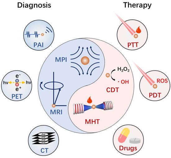
Cancer is the top cause of death globally. Developing smart nanomedicines that are capable of diagnosis and therapy (theranostics) in one–nanoparticle systems are highly desirable for improving cancer treatment outcomes. The magnetic nanoplatforms are the ideal system for cancer theranostics, because of their diverse physiochemical properties and biological effects. In particular, a biocompatible iron oxide nanoparticle based magnetic nanoplatform can exhibit multiple magnetic–responsive behaviors under an external magnetic field and realize the integration of diagnosis (magnetic resonance imaging, ultrasonic imaging, photoacoustic imaging, etc.) and therapy (magnetic hyperthermia, photothermal therapy, controlled drug delivery and release, etc.) in vivo. Furthermore, due to considerable variation among tumors and individual patients, it is a requirement to design iron oxide nanoplatforms by the coordination of diverse functionalities for efficient and individualized theranostics. 

iron oxide nanoparticles magnetotheranostics cancer Magnetic Nanoplatforms

1. Introduction

Due to the huge differences between individual patients, a tough question exists in the field of tumor diagnosis and therapy: when and where to apply what kind of treatment for a particular patient? Image–guided therapy, known as theranostics, provides a new solution for this problem. The integration of diagnosis and therapy means that treatment can be carried out under the guidance of images and monitored in real time to achieve precise and personalized medical treatment. Due to their diversified functions, nanomaterials provide a great opportunity for the integration of efficient diagnosis and therapy into a single nanoplatform [1]. There are several requirements for an ideal theranostics nanoplatform. Firstly, the nanoplatform should possess good diagnostic and/or therapeutic capabilities. Secondly, this nanoplatform must be able accumulate at the target area. Thirdly, the biocompatibility of this nanoplatform must be acceptable. Finally, it should have the ability to be integrated with other diagnostic and/or therapeutic technologies for multi–modality theranostics. Magnetotheranostics is a kind of advanced medical technology that utilizes the interaction between magnetic nanoplatforms and magnetic fields to realize the integration of therapy and diagnosis on a single nanoparticle. The magnetic nanoplatforms are known to have excellent biocompatibility [2][3], diversified diagnostic and therapeutic capabilities [4], as well as active/passive targeting capabilities [5][6], while the magnetic field is well recognized for no attenuation [7] and little damage to the tissue [8]. As a result, magnetotheranostics has received a great deal of attention in cancer research recently.
Figure 1. Diverse diagnosis and treatment technologies based on functionalized MIONs.

2. Controlled Synthesis of Magnetic Nanoplatforms

The magnetic function unit of magnetotheranostics mainly refers to MIONs. MIONs can be synthesized by physical, biological, and chemical means. Physical methods include ball milling, vapor deposition, photolithography and other technologies, but the properties of MIONs synthesized by physical methods are difficult to control [20]. The biosynthesis of MIONs has some advantages, such as better environmental friendliness and product biocompatibility, but it also faces the problems of low crystallinity and difficulty in controlling the size and morphology [21]. Chemical synthesis of MIONs is the most commonly used method. Starting from the initial co–precipitation method [22][23], researchers have successively developed thermal decomposition methods [24], hydrothermal methods [25], solvothermal methods [26], sol–gel methods [27], Micelle methods [28], and other methods to construct MIONs.
MIONs are a type of iron–based metal oxide nanoparticles with a spinel structure, whose composition can be expressed as MFe2O4, and M represents divalent metal ions, including Mn2+, Fe2+, Co2+, Ni2+, Zn2+, etc. (Figure 2a). In the most common Fe3O4 materials, M = Fe2+ and the Fe2+ occupies the octahedral (Oh) sites of the spinel structure, forming an inverse spinel structure. The antiferromagnetic coupling between Fe3+ makes the overall magnetic spin behave as 4 μB of Fe2+. The conditions of CoFe2O4 and NiFe2O4 are similar to those of Fe3O4 materials, and the total magnetic spins are 3 μB of Co2+ and 2 μB of Ni2+ respectively. When M = Mn2+, Mn2+ mainly occupies octahedral sites and partly occupies the tetrahedral (Td) sites, forming a mixed spinel structure. However, since the magnetic spins of Mn2+ and Fe3+ are both 5 μB, they always show a total magnetic spin of 5 μB in the end [29]. In ZnFe2O4, Zn2+ occupies a tetrahedral position to form a normal spinel structure. The magnetic spin of Zn2+ is 0 μB, and the magnetic spins of two Fe3+ cancel each other out, showing 0 μB overall, theoretically.
Figure 2. (a) Structure schematic of spinel structure and magnetic spin of MFe2O4. (b) Magnetic domain of ferrimagnetism/ferromagnetism (left) and superparamagnetism (right). (c) Magnetic spin states simulated using OOMMF program for nanosphere and nanocube [30] and vortex domain structure of nanoring simulated by the LLG Micromagnetics SimulatorTM package [31]. Reprint permission from [30][31]. Copyright 2012 American Chemical Society and 2012 American Institute of Physics.
In addition to the influence of the crystal structure itself, the magnetic properties of MIONs are greatly affected by their size. Excluding the influence of other factors, the smaller the MIONs, the lower the saturation magnetization (Ms) [32]. When the size is smaller than the critical size, the magnetic anisotropy of MIONs is not enough to resist the effects of thermal disturbance, resulting in the loss of its own remanence and hysteresis, but still maintaining a high initial magnetic susceptibility. This phenomenon is called superparamagnetic (Figure 2b). Due to its zero remanence, superparamagnetic iron oxide nanoparticles (SPIOs or SPIONs) have excellent colloidal dispersion and better stability than ferromagnetic or ferrimagnetic nanoparticles, and they have been approved for clinical use [10].
The morphology will also affect the magnetic properties of MIONs (Figure 2c). Compared with spherical MIONs, cubic MIONs have a higher Ms due to the less distributed spin disorder layer on the surface [30][33]. The ring–shaped MIONs possess a unique vortex magnetic domain [31], enabling it to have zero remanence and zero hysteresis while maintaining ferrimagnetism [34]. To synthesize MIONs with a specified morphology, which means to achieve anisotropic growth of MIONs, it is necessary to provide a near thermodynamically stable environment during crystal growth so that the interface energy of different crystal faces dominates the process. Therefore, the usage of thermal decomposition method or hydrothermal method are more appropriate choices.
The naked MIONs may not be suitable for direct biological application. During or after the preparation of MIONs, they need to be further modified to endow them with better stability for biological applications. Polymers are a kind of widely used coatings, including synthetic polyethylene glycol (PEG) [15][35], polyethyleneimine (PEI) [36], polyacrylic acid (PAA) [37], polyvinylpyrrolidone (PVP) [38] and natural dextran [16], chitosan [39], alginate [40], and so on.

3. Basis of Magnetic Nanomaterials Mediated Diagnosis and Therapy of Cancer

The magnetic properties of MIONs magnetic core can affect the relaxation process of protons, making it useful for MRI contrast agents. MIONs can absorb the energy of the magnetic field to generate in situ heating under the alternating magnetic field, and then can realize the magnetic hyperthermia of the tumor. The Fenton reaction by Fe2+ enables the generation of ROS to mediate tumor chemodynamic therapy. These physicochemical properties can be applied for cancer diagnosis and therapy. Together with the low toxicity [2] and clear degradation metabolism [3], MIONs have received increasing attention for theranostics.

3.1. Biosafety of Magnetic Nanoplatforms

MION formulations are generally considered to have excellent biological safety. Naked MIONs have strong antigenicity and are prone to cause allergic reactions. Surface modification such as dextran can significantly avoid side effects. There have been a large number of studies to evaluate the possible side effects of MIONs for clinical use so far. In the cell viability studies, most MIONs reported only showed cytotoxicity at particularly high concentrations [41][42][43].

3.2. Magnetic Resonance Imaging

Due to its high soft tissue contrast, high temporal and spatial resolution, and no ionizing radiation, MRI is widely used for imaging of soft tissues such as brain, heart, muscle, and tumor [44]. MRI signals are derived from nuclear magnetic resonance (NMR) signals from water protons in human tissues. Depending on the received proton longitudinal relaxation (T1) or transverse relaxation (T2) signals, MRI imaging methods are divided into two types: T1 weighting and T2 weighting. These relaxation signals can be affected by the magnetic properties of MIONs, thereby enhancing their signal strength and improving the contrast between diseased tissues and normal tissues. The contrast agents for these two imaging methods are thus called T1 contrast agents and T2 contrast agents, respectively, and their ability to enhance the corresponding relaxation rate is characterized by r1 and r2 values. The superparamagnetism of SPIONs makes them capable of disturbing the magnetic uniformity near itself under the high main magnetic field conditions of MRI, which can accelerate the lateral relaxation of surrounding protons, reduce the signal intensity in T2–weighted magnetic resonance images, and achieve negative image enhancement. Although some SPIONs were approved for clinical use as pure T2 contrast agents, they have been gradually withdrawn from clinical application in recent years, mainly due to shortcomings of poor imaging specificity (such as confusion with bleeding and calcification) [45]. Nevertheless, the T2 contrast enhancement of MIONs has been widely used in recent years for image tracking and therapy guidance of the spatiotemporal position of MIONs in vivo, such as the use of T2–weighted MRI to track cells marked by MIONs [46][47].
T1–weighted MRI can avoid the shortcomings of T2–weighted MRI, so it has better clinical usage. During the relaxation process, the protons can transfer energy with the T1 contrast agent to shorten its T1 relaxation time, especially for Gd3+, Fe3+, Mn2+, and other ions containing a large number of unpaired valence electrons. In comparison to the clinically used Gd–based contrast agents with biological safety problems [48][49], MIONs are well recognized for better biocompatibility, and Fe3+ grants them potential T1 imaging capabilities. However, large–sized MIONs have high Ms and T2 enhanced imaging performance. The high r2/r1 ratio limits their application in T1 contrast imaging. The emergence of ultrasmall SPIONs provides an opportunity to solve this problem. When the size of MIONs is reduced, their Ms decreases sharply. This reduces the r2 value, meanwhile the increased surface area increases its r1 value, leading to declined r2/r1 ratio to the range that allows T1 imaging. With the progress in new technologies for the large–scale synthesis of ultrasmall SPIONs [50][51], the clinical application of ultrasmall SPIONs as T1 contrast agents has also rapidly developed.

3.3. Other Diagnosis Applications

Magnetic particle imaging (MPI) has the advantages of no tissue signal attenuation, a linear correlation between the signal and tracer concentration, and no ionizing radiation during detection. It has become an emerging tomographic imaging technology that is expected to enter clinical applications, especially in lung and other organs that are difficult to be imaged by MRI [52]. The earliest tracer used in MPI technology is SPIONs, and so far it is still the dominant tracer materials [52]. The Ms of the MPI tracer is decisive for its imaging performance. The Ms of Fe@Fe3O4 NPs are reported to be as high as 176 emu/g, which enables good MPI performance [53]. Changes in the crystallinity of MIONs can also alter the Ms, thereby affecting their MPI performance [54]. Magnetomotive optical coherence tomography (MMOCT) is another type of imaging technology based on MIONs, and the image contrast is derived from dynamic magnetomotive force. Unlike MRI and MPI, MMOCT requires a magnetic field as low as 0.08 T [55] and can detect ultra–low concentrations of tracers. MMOCT has been shown to be able to image tumor models in animals [12]. Due to its ability to detect the movement state of MIONs particles, it has also recently been used for real–time monitoring of magnetic hyperthermia [56].

3.4. Magnetic Hyperthermia

Hyperthermia is a treatment with a long history. MIONs have the ability to convert the energy of an alternating magnetic field (AMF) into heat. Hyperthermia using this magnetothermal effect is called magnetic hyperthermia (MHT). As a means of in situ hyperthermia, magnetic hyperthermia can kill tumor tissues more accurately, and it is not limited by the depth of tissue penetration. It has developed many application scenarios in the field of tumor therapy [57][58][59]. The improvement of the efficiency of tumor magnetic hyperthermia depends on the improvement of specific absorption rate (SAR), and SAR is directly proportional to the Ms of MIONs [60]. It has been proven that the Ms of MIONs are strongly related to their size [32]. SPIONs with a diameter of less than 20 nm have a SAR in the range of hundreds of W/g [34]. In theory, the SAR of MIONs could be improved by increasing the size of MIONs. Paradoxically, larger–sized MIONs also exhibit ferromagnetic/ferrimagnetic properties. The existence of remanence disfavors colloidal stability of the MIONs, which in turn can decrease its SAR. In recent years, some MIONs with special magnetic domain structures have gradually shown their advantages. In one example, iron oxide nanorings of a specific size will exhibit vortex magnetic domains. The magnetic domains of this structure are closed loops connected end to end. While maintaining high ferrous hysteresis loss, the residual magnetization is kept at zero, achieving a SAR exceeding 2000 W/g while having excellent colloidal dispersion [34].

3.5. Chemodynamic Therapy

In an acidic environment, Fe2+ in MIONs can catalyze the Fenton reaction:
Fe3+ + H2O2 = Fe2+ + HO2• + H+
Fe2+ + H2O2 = Fe3+ + •OH + OH
Due to its similar behavior to peroxidases such as horseradish peroxidase (HRP), the ability of MIONs to catalyze the Fenton reaction is also called peroxidase–like activity of a nanozyme. This reaction can generate a large amount of ROS in the tumor cells, break the redox balance of tumor cells, and then damage tumor cells [61]. This phenomenon is known as chemodynamic therapy (CDT). However, since the pH in the tumor microenvironment does not meet the optimal conditions for the Fenton reaction [62][63], the direct application of MIONs in tumor CDT is limited. Hence, strategies to improve the Fenton reaction activity of MIONs are vital for their CDT efficacy.

4. Implementation of Magnetotheranostic Based on Magnetic Nanoplatforms

4.1. Magnetotheranostics Based on Magnetic Nanoplatforms Only

MIONs have their own therapeutic and diagnostic functions such as MRI, MPI, MHT, and CDT. Therefore, improvements on MIONs are beneficial for their magnetotheranostics performances. The Ms of MIONs received great attention from researchers at an earlier time [64], because the improvement of Ms will simultaneously enhance the T2–weighted imaging performance of MIONs and the thermal conversion efficiency of MHT. Recent work in this area has become more diversified, and one direction is T1–T2 dual–modality imaging combined with treatment. Liu et al. [60] synthesized wüstite Fe0.6Mn0.4O nanoflowers. Unlike the antiferromagnetic bulk wüstite, Fe0.6Mn0.4O nanoflowers exhibit ferromagnetism, which may be due to exchange coupling effect. The as–prepared nanoflowers exhibit excellent magnetic induction heating effects (SAR can reach 535 W/g), which could induce tumor regression in breast cancer through MHT. The longitudinal relaxation rate r1 and lateral relaxation rate r2 of Fe0.6Mn0.4O nanoflowers are as high as 4.9 and 61.2 mM−1 [Fe]+[Mn]·s−1, respectively. These nanoflowers showed both T1 and T2 enhancing properties in the mouse glioma model. Different from this static T1–T2 dual–modal contrast agent, another type is dynamic T1–T2 dual–modal contrast agents. They can present two states of T1 or T2 contrast, and certain events will prompt the transition between the two states. This is generally accomplished by disintegrating large particles into small particles (T2 to T1) or aggregation of small particles into large particles (T1 to T2). An example of the conversion of T2 enhancement to T1 enhancement is listed in Section 3.2 [65]. In the work by Zhou et al. [66], the ultrasmall SPIONs aggregated into clusters in the tumor in situ, resulting in the conversion of T1 enhancement to T2 enhancement. They used hyaluronic acid (HA) to encapsulate ultrasmall SPIONs, which showed T1 enhanced performance before penetrating into the tumor. After entering the tumor area, the surface–modified HA was degraded by the abundant hyaluronidase, which decreased the colloidal stability of ultrasmall SPIONs and caused aggregation of the nanoparticles into clusters, resulting in enhanced T2 imaging performance and weakened T1 imaging performance. Although the therapy performance of the designed ultrasmall SPIONs was not investigated in this work, its penetration–aggregation design still provided a strategy for future magnetotheranostics.

4.2. Integration of Magnetic Nanoplatforms with Phototheransotics

Similar to magnetotheranostics, comprehensive application of the optical properties of nanomaterials in therapeutic diagnostics are called phototheranostics, covering technologies such as photothermal therapy (PTT), photodynamic therapy (PDT), and photoacoustic imaging (PAI). Corresponding to the magnetic core in magnetotheranostics, the phototheranostics nanoplatform needs a photosensitizer as its core. Among the various photosensitizers, noble metal nanoparticles, especially Au nanoparticles [67] can efficiently complete energy conversion through the localized surface plasmon resonance (LSPR) effect, and are often used to form a hybrid magnetoptical theranostics nanoplatform with MIONs for therapeutic diagnostics. Liu et al. [68] assembled SiO2–coated Au nanowreaths (AuNWs) with ultrasmall SPIONs through molecules containing polycystamine blocks (Figure 3a). After sensing the GSH in the tumor cells, the disulfide bond of polycystamine was cleaved, causing the disassembly of ultrasmall SPIONs from the surface of AuNWs, and the MRI contrast performance of the ultrasmall SPIONs changed from T2 enhancement to T1 enhancement (Figure 3b). The released AuNWs were used for photothermal therapy and photoacoustic imaging (Figure 3c,d).
Figure 3. (a) Schematic illustration of the synthesis of magnetic gold nanowreath. (b) Corresponding T1–weighted images (top, disassembled; bottom, assembled) of magnetic AuNWs. Concentrations of Fe are 0.5, 0.25, 0.125, 0.0625, 0.03125, and 0.01563 mM (from left to right). (c) Ultrasonic (US), photoacoustic imaging (PA), and merged images of tumor before injection (0 h) and at 2, 4, 24, and 48 h after intravenous injection of magnetic AuNWs upon irradiation by an 808 nm pulsed laser. (d) Representative thermal images of U87MG tumor bearing mice after injection of magnetic AuNWs and PBS. The tumors were irradiated by an 808 nm CW laser at 0.75 W/cm2. Reprint with permission from [68]. Copyright 2018 American Chemical Society.

4.3. Integration of Magnetic Nanoplatforms with Fluorescence Imaging

Fluorescence imaging (FI) relies heavily on the penetration of light in tissue, which limits its application in the field of in vivo diagnosis and therapy. However, because fluorescent molecules can be designed to achieve highly specific responsiveness, fluorescence imaging is often used as an auxiliary imaging method for the magnetotheranostics nanoplatform to monitor its response behavior in the body. Zhou et al. [69] designed a nanoplatform capable of detecting tumor hypoxic environment, composed of ultrasmall SPIONs and assembly–responsive fluorescent dyes (NBD), and used nitroimidazole derivatives as hypoxia–sensitive detectors.

4.4. Integration of Magnetic Nanoplatforms with CT&PET/SPECT

Although iodine–based contrast agents for CT have been well developed, certain components of the magnetotheranostics nanoplatforms also have the ability to act as CT contrast agents. The use of CT to guide the diagnosis and therapy of the magnetic nanoplatforms has remarkable application prospects. A relatively common example is the gold–magnetic composites, wherein the high CT value of gold enables it to be traced by CT [70]. Liu et al. developed an ultrasonication–triggered interfacial assembly approach (Figure 4a,b) to synthesize magnetic Janus amphiphilic nanoparticles (MJANPs) for image–guided cancer MHT (Figure 4c) [71]. Au NPs–MIONs MJANPs made of Au NPs and MIONs could achieve MRI/CT dual–modality imaging and could be used to guide MHT (Figure 4d). Similarly, if CuInS/ZnS NPs and MIONs were used to make CuInS/ZnS NPs–MIONs MJANPs, MRI/FI dual–modality imaging can be used to guide MHT (Figure 4e).
Figure 4. (a) Schematics of the ultrasonatically triggered interfacial assembly process. (b) SIM image of the Pickering emulsion. Fluoresceinamine was used to label graphene oxide nanosheets (green), and rhodamine B isothiocyanate (RITC) was used to label Co cluster–embedded IONPs (red). (c) Photographs of pre– and post–MHT treatment mice. (d) In vivo T2–weighted MRI and CT images of tumor–bearing mice pre– and post–injection of Au NPs−MIONs MJANPs. Arrowheads indicate the tumor. (e) In vivo T2–weighted MRI and fluorescence images of tumor–bearing nude mice pre– and postinjection of CuInS/ZnS−MIONs MJANPs. Arrowheads indicate the tumor. Reprint with permission from [71]. Copyright 2019 American Chemical Society.

4.5. Magnetic Nanoplatforms Carrier Based Drug Delivery

The magnetotheranostic nanoplatform has become a representative delivery system due to its adjustable size, easy image tracking, and clear metabolic pathway. The delivery mechanism of MIONs can be divided into passive targeting and active targeting. Passive targeting mainly depends on the enhanced permeability and retention (EPR) effect of MIONs. Although the mechanism still needs to be further investigated [72][73], the EPR effect can indeed enhance the enrichment of nanoparticles (not just MIONs) in the tumor area [5]. In addition, the ligands of tumor characteristic markers can be employed to functionalize MIONs in order to give them the ability to actively target tumors [6], and for more efficient drug delivery. In a recent work, paclitaxel (PTX) and cisplatin (CDDP) have been loaded into the carboxymethyl dextran coating of the clinical iron supplement FMX, and actively target gliomas through HMC, which was an organic anion transport polypeptide targeting agent with near–infrared fluorescence. This system was used for MRI/FI visualized drug delivery of glioblastoma multiforme (GBM) [74]. Liu et al. [75] designed a delivery system with a Yolk–shell structure. The vesicles were composed of PEG–PPS–SS–PEG and loaded with ultrasmall SPIONs and DOX, which were encapsulated together by a polyacrylic acid coating. In tumor cells, vesicles were disintegrated under the influence of GSH. The complex of ultrasmall SPIONs and DOX was then separated, so that the drug release process could be monitored by an enhancement in T1 MRI. The complex microenvironment of the tumor tissue could limit the penetration of the nanoplatforms [76][77].

References

  1. Chen, H.; Zhang, W.; Zhu, G.; Xie, J.; Chen, X. Rethinking cancer nanotheranostics. Nat. Rev. Mater. 2017, 2, 17024.
  2. McCormack, P.L. Ferumoxytol In Iron Deficiency Anaemia in Adults with Chronic Kidney Disease. Drugs 2012, 72, 2013–2022.
  3. Lartigue, L.; Alloyeau, D.; Kolosnjaj–Tabi, J.; Javed, Y.; Guardia, P.; Riedinger, A.; Péchoux, C.; Pellegrino, T.; Wilhelm, C.; Gazeau, F. Biodegradation of Iron Oxide Nanocubes: High–Resolution In Situ Monitoring. ACS Nano 2013, 7, 3939–3952.
  4. Tong, S.; Zhu, H.; Bao, G. Magnetic iron oxide nanoparticles for disease detection and therapy. Mater. Today 2019, 31, 86–99.
  5. Fang, J.; Islam, W.; Maeda, H. Exploiting the dynamics of the EPR effect and strategies to improve the therapeutic effects of nanomedicines by using EPR effect enhancers. Adv. Drug Deliv. Rev. 2020, 157, 142–160.
  6. Mitchell, M.J.; Billingsley, M.M.; Haley, R.M.; Wechsler, M.E.; Peppas, N.A.; Langer, R. Engineering precision nanoparticles for drug delivery. Nat. Rev. Drug Discov. 2020, 20, 101–124.
  7. Park, J.; Jin, C.; Lee, S.; Kim, J.; Choi, H. Magnetically Actuated Degradable Microrobots for Actively Controlled Drug Release and Hyperthermia Therapy. Adv. Health Mater. 2019, 8, e1900213.
  8. Cazares–Cortes, E.; Cabana, S.; Boitard, C.; Nehlig, E.; Griffete, N.; Fresnais, J.; Wilhelm, C.; Abou–Hassan, A.; Ménager, C. Recent insights in magnetic hyperthermia: From the “hot–spot” effect for local delivery to combined magneto–photo–thermia using magneto–plasmonic hybrids. Adv. Drug Deliv. Rev. 2018, 138, 233–246.
  9. Ni, D.; Bu, W.; Ehlerding, E.B.; Cai, W.; Shi, J. Engineering of inorganic nanoparticles as magnetic resonance imaging contrast agents. Chem. Soc. Rev. 2017, 46, 7438–7468.
  10. Dadfar, S.M.; Roemhild, K.; Drude, N.; von Stillfried, S.; Knüchel, R.; Kiessling, F.; Lammers, T. Iron oxide nanoparticles: Diagnostic, therapeutic and theranostic applications. Adv. Drug Deliv. Rev. 2019, 138, 302–325.
  11. Lu, C.; Han, L.; Wang, J.; Wan, J.; Song, G.; Rao, J. Engineering of magnetic nanoparticles as magnetic particle imaging tracers. Chem. Soc. Rev. 2021, 50, 8102–8146.
  12. John, R.; Rezaeipoor, R.; Adie, S.G.; Chaney, E.J.; Oldenburg, A.L.; Marjanovic, M.; Haldar, J.P.; Sutton, B.P.; Boppart, S.A. In vivo magnetomotive optical molecular imaging using targeted magnetic nanoprobes. Proc. Natl. Acad. Sci. USA 2010, 107, 8085–8090.
  13. Teijeiro–Valiño, C.; Gómez, M.A.G.; Yañez–Villar, S.; García–Acevedo, P.; Arnosa–Prieto, A.; Belderbos, S.; Gsell, W.; Himmelreich, U.; Piñeiro, Y.; Rivas, J. Biocompatible magnetic gelatin nanoparticles with enhanced MRI contrast performance prepared by single–step desolvation method. Nano Express 2021, 2, 020011.
  14. Zhou, Y.; Fan, S.; Feng, L.; Huang, X.; Chen, X. Manipulating Intratumoral Fenton Chemistry for Enhanced Chemodynamic and Chemodynamic-Synergized Multimodal Therapy. Adv. Mater. 2021, 33, 2104223.
  15. Tromsdorf, U.I.; Bruns, O.; Salmen, S.C.; Beisiegel, U.; Weller, H. A Highly Effective, Nontoxic T1 MR Contrast Agent Based on Ultrasmall PEGylated Iron Oxide Nanoparticles. Nano Lett. 2009, 9, 4434–4440.
  16. Harisinghani, M.G.; Barentsz, J.; Hahn, P.F.; Deserno, W.M.; Tabatabaei, S.; Van De Kaa, C.H.; De La Rosette, J.; Weissleder, R. Noninvasive Detection of Clinically Occult Lymph–Node Metastases in Prostate Cancer. N. Engl. J. Med. 2003, 348, 2491–2499.
  17. Tang, L.; Casas, J.; Venkataramasubramani, M. Magnetic Nanoparticle Mediated Enhancement of Localized Surface Plasmon Resonance for Ultrasensitive Bioanalytical Assay in Human Blood Plasma. Anal. Chem. 2013, 85, 1431–1439.
  18. Li, B.; Gong, T.; Xu, N.; Cui, F.; Yuan, B.; Yuan, Q.; Sun, H.; Wang, L.; Liu, J. Improved Stability and Photothermal Performance of Polydopamine-Modified Fe3O4 Nanocomposites for Highly Efficient Magnetic Resonance Imaging-Guided Photothermal Therapy. Small 2020, 16, e2003969.
  19. Wang, P.; Shi, Y.; Zhang, S.; Huang, X.; Zhang, J.; Zhang, Y.; Si, W.; Dong, X. Hydrogen Peroxide Responsive Iron–Based Nanoplatform for Multimodal Imaging–Guided Cancer Therapy. Small 2018, 15, e1803791.
  20. Sodipo, B.K.; Aziz, A.A. Recent advances in synthesis and surface modification of superparamagnetic iron oxide nanoparticles with silica. J. Magn. Magn. Mater. 2016, 416, 275–291.
  21. Wu, W.; Wu, Z.; Yu, T.; Jiang, C.; Kim, W.-S. Recent progress on magnetic iron oxide nanoparticles: Synthesis, surface functional strategies and biomedical applications. Sci. Technol. Adv. Mater. 2015, 16, 023501.
  22. Pereira, C.; Pereira, A.M.; Fernandes, C.; Rocha, M.; Mendes, R.; Fernández–García, M.P.; Guedes, A.; Tavares, P.B.; Grenèche, J.-M.; Araújo, J.P.; et al. Superparamagnetic MFe2O4 (M = Fe, Co, Mn) Nanoparticles: Tuning the Particle Size and Magnetic Properties through a Novel One–Step Coprecipitation Route. Chem. Mater. 2012, 24, 1496–1504.
  23. Massart, R. Preparation of aqueous magnetic liquids in alkaline and acidic media. IEEE Trans. Magn. 1981, 17, 1247–1248.
  24. Park, J.; An, K.; Hwang, Y.; Park, J.G.; Noh, H.J.; Kim, J.Y.; Park, J.H.; Hwang, N.M.; Hyeon, T. Ultra–large–scale syntheses of mono disperse nanocrystals. Nat Mater 2004, 3, 891–895.
  25. Deng, H.; Li, X.; Peng, Q.; Wang, X.; Chen, J.; Li, Y. Monodisperse magnetic single–crystal ferrite microspheres. Angew. Chem. 2005, 117, 2842–2845.
  26. Hu, P.; Yu, L.; Zuo, A.; Guo, C.; Yuan, F. Fabrication of Monodisperse Magnetite Hollow Spheres. J. Phys. Chem. C 2008, 113, 900–906.
  27. Niederberger, M. Nonaqueous Sol–Gel Routes to Metal Oxide Nanoparticles. Accounts Chem. Res. 2007, 40, 793–800.
  28. Lee, Y.; Lee, J.; Bae, C.J.; Park, J.-G.; Noh, H.-J.; Hyeon, T. Large–Scale Synthesis of Uniform and Crystalline Magnetite Nanoparticles Using Reverse Micelles as Nanoreactors under Reflux Conditions. Adv. Funct. Mater. 2005, 15, 503–509.
  29. Lee, J.-H.; Huh, Y.-M.; Jun, Y.-W.; Seo, J.-W.; Jang, J.-T.; Song, H.-T.; Kim, S.; Cho, E.-J.; Yoon, H.-G.Y.; Suh, J.-S.; et al. Artificially engineered magnetic nanoparticles for ultra–sensitive molecular imaging. Nat. Med. 2006, 13, 95–99.
  30. Noh, S.-H.; Na, W.; Jang, J.-T.; Lee, J.-H.; Lee, E.J.; Moon, S.H.; Lim, Y.; Shin, J.-S.; Cheon, J. Nanoscale Magnetism Control via Surface and Exchange Anisotropy for Optimized Ferrimagnetic Hysteresis. Nano Lett. 2012, 12, 3716–3721.
  31. Yang, Y.; Liu, X.-L.; Yi, J.-B.; Yang, Y.; Fan, H.-M.; Ding, J. Stable vortex magnetite nanorings colloid: Micromagnetic simulation and experimental demonstration. J. Appl. Phys. 2012, 111, 044303.
  32. Ling, D.; Lee, N.; Hyeon, T. Chemical Synthesis and Assembly of Uniformly Sized Iron Oxide Nanoparticles for Medical Applications. Accounts Chem. Res. 2015, 48, 1276–1285.
  33. Liu, X.L.; Fan, H.M. Innovative magnetic nanoparticle platform for magnetic resonance imaging and magnetic fluid hyperthermia applications. Curr. Opin. Chem. Eng. 2014, 4, 38–46.
  34. Liu, X.L.; Yang, Y.; Ng, C.T.; Zhao, L.Y.; Zhang, Y.; Bay, B.H.; Fan, H.M.; Ding, J. Magnetic Vortex Nanorings: A New Class of Hyperthermia Agent for Highly Efficient In Vivo Regression of Tumors. Adv. Mater. 2015, 27, 1939–1944.
  35. Cole, A.J.; David, A.E.; Wang, J.; Galbán, C.J.; Yang, V.C. Magnetic brain tumor targeting and biodistribution of long–circulating PEG–modified, cross–linked starch–coated iron oxide nanoparticles. Biomaterials 2011, 32, 6291–6301.
  36. Liu, G.; Xie, J.; Zhang, F.; Wang, Z.-Y.; Luo, K.; Zhu, L.; Quan, Q.-M.; Niu, G.; Lee, S.; Ai, H.; et al. N–Alkyl–PEI–functionalized iron oxide nanoclusters for efficient siRNA delivery. Small 2011, 7, 2742–2749.
  37. Kang, X.-J.; Dai, Y.-L.; Ma, P.-A.; Yang, D.-M.; Li, C.-X.; Hou, Z.-Y.; Cheng, Z.-Y.; Lin, J. Poly(acrylic acid)–Modified Fe3O4Microspheres for Magnetic–Targeted and pH–Triggered Anticancer Drug Delivery. Chem. Eur. J. 2012, 18, 15676–15682.
  38. Riedinger, A.; Leal, M.P.; Deka, S.R.; George, C.; Franchini, I.R.; Falqui, A.; Cingolani, R.; Pellegrino, T. “Nanohybrids” Based on pH–Responsive Hydrogels and Inorganic Nanoparticles for Drug Delivery and Sensor Applications. Nano Lett. 2011, 11, 3136–3141.
  39. KC, R.B.; Lee, S.M.; Yoo, E.S.; Choi, J.H.; Ghim, H.D. Glycoconjugated chitosan stabilized iron oxide nanoparticles as a multifunctional nanoprobe. Mater. Sci. Eng. C 2009, 29, 1668–1673.
  40. Kim, J.; Arifin, D.R.; Muja, N.; Kim, T.; Gilad, A.A.; Kim, H.; Arepally, A.; Hyeon, T.; Bulte, J.W.M. Multifunctional Capsule–in–Capsules for Immunoprotection and Trimodal Imaging. Angew. Chem. Int. Ed. 2011, 50, 2317–2321.
  41. Li, Y.; Liu, J.; Zhong, Y.; Zhang, D.; Wang, Z.; An, Y.-L.; Lin, M.; Gao, Z.; Zhang, J. Biocompatibility of Fe3O4@Au composite magnetic nanoparticles in vitro and in vivo. Int. J. Nanomed. 2011, 6, 2805–2819.
  42. Khan, M.I.; Mohammad, A.; Patil, G.; Naqvi, S.; Chauhan, L.; Ahmad, I. Induction of ROS, mitochondrial damage and autophagy in lung epithelial cancer cells by iron oxide nanoparticles. Biomaterials 2012, 33, 1477–1488.
  43. Huang, D.-M.; Hsiao, J.-K.; Chen, Y.-C.; Chien, L.-Y.; Yao, M.; Chen, Y.-K.; Ko, B.-S.; Hsu, S.-C.; Tai, L.-A.; Cheng, H.-Y.; et al. The promotion of human mesenchymal stem cell proliferation by superparamagnetic iron oxide nanoparticles. Biomaterials 2009, 30, 3645–3651.
  44. Brito, B.; Price, T.W.; Gallo, J.; Bañobre–López, M.; Stasiuk, G.J. Smart magnetic resonance imaging–based theranostics for cancer. Theranostics 2021, 11, 8706–8737.
  45. Wahsner, J.; Gale, E.M.; Rodríguez–Rodríguez, A.; Caravan, P. Chemistry of MRI Contrast Agents: Current Challenges and New Frontiers. Chem. Rev. 2018, 119, 957–1057.
  46. Wu, C.; Xu, Y.; Yang, L.; Wu, J.; Zhu, W.; Li, D.; Cheng, Z.; Xia, C.; Guo, Y.; Gong, Q.; et al. Negatively Charged Magnetite Nanoparticle Clusters as Efficient MRI Probes for Dendritic Cell Labeling and In Vivo Tracking. Adv. Funct. Mater. 2015, 25, 3581–3591.
  47. Karimian–Jazi, K.; Münch, P.; Alexander, A.; Fischer, M.; Pfleiderer, K.; Piechutta, M.; Karreman, M.A.; Solecki, G.M.; Berghoff, A.S.; Friedrich, M.; et al. Monitoring innate immune cell dynamics in the glioma microenvironment by magnetic resonance imaging and multiphoton microscopy (MR–MPM). Theranostics 2020, 10, 1873–1883.
  48. Marckmann, P.; Skov, L.; Rossen, K.; Dupont, A.; Damholt, M.B.; Heaf, J.G.; Thomsen, H.S. Nephrogenic systemic fibrosis: Suspected causative role of gadodiamide used for contrast–enhanced magnetic resonance imaging. J. Am. Soc. Nephrol. 2006, 17, 2359–2362.
  49. Sieber, M.A.; Lengsfeld, P.; Walter, J.; Schirmer, H.; Frenzel, T.; Siegmund, F.; Weinmann, H.-J.; Pietsch, H. Gadolinium–based contrast agents and their potential role in the pathogenesis of nephrogenic systemic fibrosis: The role of excess ligand. J. Magn. Reson. Imaging 2008, 27, 955–962.
  50. Zhang, H.; Li, L.; Liu, X.L.; Jiao, J.; Ng, C.-T.; Yi, J.; E Luo, Y.; Bay, B.-H.; Zhao, L.Y.; Peng, M.L.; et al. Ultrasmall Ferrite Nanoparticles Synthesized via Dynamic Simultaneous Thermal Decomposition for High–Performance and Multifunctional T1 Magnetic Resonance Imaging Contrast Agent. ACS Nano 2017, 11, 3614–3631.
  51. Kim, B.H.; Lee, N.; Kim, H.; An, K.; Park, Y.I.; Choi, Y.; Shin, K.; Lee, Y.; Kwon, S.G.; Bin Na, H.; et al. Large–Scale Synthesis of Uniform and Extremely Small–Sized Iron Oxide Nanoparticles for High–Resolution T1 Magnetic Resonance Imaging Contrast Agents. J. Am. Chem. Soc. 2011, 133, 12624–12631.
  52. Chandrasekharan, P.; Tay, Z.W.; Hensley, D.; Zhou, X.Y.; Fung, B.K.; Colson, C.; Lu, Y.; Fellows, B.D.; Huynh, Q.; Saayujya, C.; et al. Using magnetic particle imaging systems to localize and guide magnetic hyperthermia treatment: Tracers, hardware, and future medical applications. Theranostics 2020, 10, 2965–2981.
  53. Gloag, L.; Mehdipour, M.; Ulanova, M.; Mariandry, K.; Nichol, M.A.; Hernández–Castillo, D.J.; Gaudet, J.; Qiao, R.; Zhang, J.; Nelson, M.; et al. Zero valent iron core–iron oxide shell nanoparticles as small magnetic particle imaging tracers. Chem. Commun. 2020, 56, 3504–3507.
  54. Song, G.; Chen, M.; Zhang, Y.; Cui, L.; Qu, H.; Zheng, X.; Wintermark, M.; Liu, Z.; Rao, J. Janus Iron Oxides @ Semiconducting Polymer Nanoparticle Tracer for Cell Tracking by Magnetic Particle Imaging. Nano Lett. 2017, 18, 182–189.
  55. Oldenburg, A.L.; Crecea, V.; Rinne, S.A.; Boppart, S.A. Phase–resolved magnetomotive OCT for imaging nanomolar concentrations of magnetic nanoparticles in tissues. Opt. Express 2008, 16, 11525–11539.
  56. Huang, P.-C.; Chaney, E.J.; Aksamitiene, E.; Barkalifa, R.; Spillman, D.R.; Bogan, B.J.; Boppart, S.A. Biomechanical sensing of in vivo magnetic nanoparticle hyperthermia–treated melanoma using magnetomotive optical coherence elastography. Theranostics 2021, 11, 5620–5633.
  57. Xu, C.; Zheng, Y.; Gao, W.; Xu, J.; Zuo, G.; Chen, Y.; Zhao, M.; Li, J.; Song, J.; Zhang, N.; et al. Magnetic Hyperthermia Ablation of Tumors Using Injectable Fe3O4/Calcium Phosphate Cement. ACS Appl. Mater. Interfaces 2015, 7, 13866–13875.
  58. Yin, P.; Shah, S.; Pasquale, N.J.; Garbuzenko, O.B.; Minko, T.; Lee, K. –B. Stem cell–based gene therapy activated using magnetic hyperthermia to enhance the treatment of cancer. Biomaterials 2015, 81, 46–57.
  59. Moise, S.; Byrne, J.M.; El Haj, A.J.; Telling, N.D. The potential of magnetic hyperthermia for triggering the differentiation of cancer cells. Nanoscale 2018, 10, 20519–20525.
  60. Liu, X.L.; Ng, C.T.; Chandrasekharan, P.; Yang, H.T.; Zhao, L.Y.; Peng, E.; Lv, Y.B.; Xiao, W.; Fang, J.; Yi, J.; et al. Synthesis of Ferromagnetic Fe0.6Mn0.4O Nanoflowers as a New Class of Magnetic Theranostic Platform for In Vivo T1–T2Dual–Mode Magnetic Resonance Imaging and Magnetic Hyperthermia Therapy. Adv. Health Mater. 2016, 5, 2092–2104.
  61. Zhou, Z.; Song, J.; Tian, R.; Yang, Z.; Yu, G.; Lin, L.; Zhang, G.; Fan, W.; Zhang, F.; Niu, G.; et al. Activatable Singlet Oxygen Generation from Lipid Hydroperoxide Nanoparticles for Cancer Therapy. Angew. Chem. Int. Ed. 2017, 56, 6492–6496.
  62. Du, J.; Bao, J.; Fu, X.; Lu, C.; Kim, S.H. Mesoporous sulfur–modified iron oxide as an effective Fenton–like catalyst for degradation of bisphenol A. Appl. Catal. B Environ. 2016, 184, 132–141.
  63. Zhao, S.; Yu, X.; Qian, Y.; Chen, W.; Shen, J. Multifunctional magnetic iron oxide nanoparticles: An advanced platform for cancer theranostics. Theranostics 2020, 10, 6278–6309.
  64. Jang, J.-T.; Nah, H.; Lee, J.-H.; Moon, S.H.; Kim, M.G.; Cheon, J. Critical Enhancements of MRI Contrast and Hyperthermic Effects by Dopant–Controlled Magnetic Nanoparticles. Angew. Chem. Int. Ed. 2009, 48, 1234–1238.
  65. Liang, H.; Guo, J.; Shi, Y.; Zhao, G.; Sun, S.; Sun, X. Porous yolk–shell Fe/Fe3O4 nanoparticles with controlled exposure of highly active Fe(0) for cancer therapy. Biomaterials 2020, 268, 120530.
  66. Zhou, H.; Tang, J.; Li, J.; Li, W.; Liu, Y.; Chen, C. In vivo aggregation–induced transition between T1and T2relaxations of magnetic ultra–small iron oxide nanoparticles in tumor microenvironment. Nanoscale 2017, 9, 3040–3050.
  67. Xu, C.; Pu, K. Second near–infrared photothermal materials for combinational nanotheranostics. Chem. Soc. Rev. 2020, 50, 1111–1137.
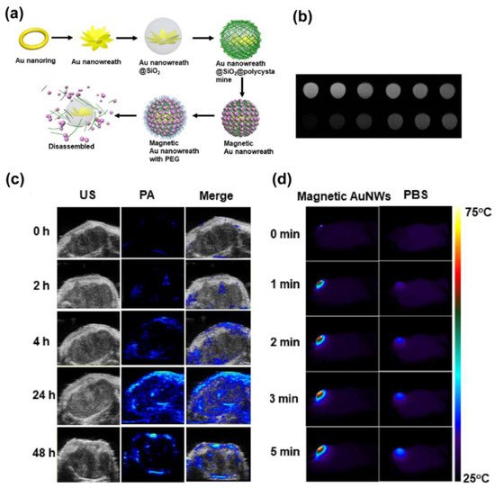
  68. Liu, Y.; Yang, Z.; Huang, X.; Yu, G.; Wang, S.; Zhou, Z.; Shen, Z.; Fan, W.; Liu, Y.; Davission, M.; et al. Glutathione–Responsive Self–Assembled Magnetic Gold Nanowreath for Enhanced Tumor Imaging and Imaging–Guided Photothermal Therapy. ACS Nano 2018, 12, 8129–8137.
  69. Zhou, H.; Guo, M.; Li, J.; Qin, F.; Wang, Y.; Liu, T.; Liu, J.; Sabet, Z.F.; Wang, Y.; Liu, Y.; et al. Hypoxia–Triggered Self–Assembly of Ultrasmall Iron Oxide Nanoparticles to Amplify the Imaging Signal of a Tumor. J. Am. Chem. Soc. 2021, 143, 1846–1853.
  70. Ma, J.; Li, P.; Wang, W.; Wang, S.; Pan, X.; Zhang, F.; Li, S.; Liu, S.; Wang, H.; Gao, G.; et al. Biodegradable Poly(amino acid)–Gold–Magnetic Complex with Efficient Endocytosis for Multimodal Imaging–Guided Chemo–photothermal Therapy. ACS Nano 2018, 12, 9022–9032.
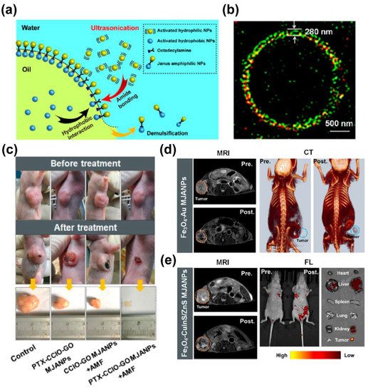
  71. Liu, X.; Peng, M.L.; Li, G.; Miao, Y.Q.; Luo, H.; Jing, G.; He, Y.; Zhang, C.; Zhang, F.; Fan, H. Ultrasonication–Triggered Ubiquitous Assembly of Magnetic Janus Amphiphilic Nanoparticles in Cancer Theranostic Applications. Nano Lett. 2019, 19, 4118–4125.
  72. Shi, Y.; Van Der Meel, R.; Chen, X.; Lammers, T. The EPR effect and beyond: Strategies to improve tumor targeting and cancer nanomedicine treatment efficacy. Theranostics 2020, 10, 7921–7924.
  73. Wilhelm, S.; Tavares, A.J.; Dai, Q.; Ohta, S.; Audet, J.; Dvorak, H.F.; Chan, W.C.W. Analysis of nanoparticle delivery to tumours. Nat. Rev. Mater. 2016, 1, 16014.
  74. Reichel, D.; Sagong, B.; Teh, J.; Zhang, Y.; Wagner, S.; Wang, H.; Chung, L.W.K.; Butte, P.; Black, K.L.; Yu, J.S.; et al. Near Infrared Fluorescent Nanoplatform for Targeted Intraoperative Resection and Chemotherapeutic Treatment of Glioblastoma. ACS Nano 2020, 14, 8392–8408.
  75. Liu, D.; Zhou, Z.; Wang, X.; Deng, H.; Sun, L.; Lin, H.; Kang, F.; Zhang, Y.; Wang, Z.; Yang, W.; et al. Yolk–shell nanovesicles endow glutathione–responsive concurrent drug release and T1 MRI activation for cancer theranostics. Biomaterials 2020, 244, 119979.
  76. Zhou, Q.; Shao, S.; Wang, J.; Xu, C.; Xiang, J.; Piao, Y.; Zhou, Z.; Yu, Q.; Tang, J.; Liu, X.; et al. Enzyme–activatable polymer–drug conjugate augments tumour penetration and treatment efficacy. Nat. Nanotechnol. 2019, 14, 799–809.
  77. Dai, Q.; Wilhelm, S.; Ding, D.; Syed, A.; Sindhwani, S.; Zhang, Y.; Chen, Y.Y.; MacMillan, P.; Chan, W.C.W. Quantifying the Ligand–Coated Nanoparticle Delivery to Cancer Cells in Solid Tumors. ACS Nano 2018, 12, 8423–8435.
More
Information
Contributor MDPI registered users' name will be linked to their SciProfiles pages. To register with us, please refer to https://encyclopedia.pub/register :
View Times: 416
Revisions: 2 times (View History)
Update Date: 03 Mar 2022
1000/1000
ScholarVision Creations