You're using an outdated browser. Please upgrade to a modern browser for the best experience.
Submitted Successfully!
Thank you for your contribution! You can also upload a video entry or images related to this topic. For video creation, please contact our Academic Video Service.
Version Summary Created by Modification Content Size Created at Operation
1 Clara I Rodriguez + 3237 word(s) 3237 2022-02-22 10:18:04 |
2 Format correct Vicky Zhou + 3236 word(s) 3236 2022-02-22 13:19:24 | |
3 Format correct Vicky Zhou + 2 word(s) 3238 2022-02-22 13:20:23 | |
4 Format correct Vicky Zhou + 17 word(s) 3253 2022-02-22 13:23:22 |

Video Upload Options

We provide professional Academic Video Service to translate complex research into visually appealing presentations. Would you like to try it?
Cite
If you have any further questions, please contact Encyclopedia Editorial Office.
Rodriguez, C.I. Educating Extracellular Vesicles to Improve Bone Regeneration. Encyclopedia. Available online: https://encyclopedia.pub/entry/19741 (accessed on 18 December 2025).
Rodriguez CI. Educating Extracellular Vesicles to Improve Bone Regeneration. Encyclopedia. Available at: https://encyclopedia.pub/entry/19741. Accessed December 18, 2025.
Rodriguez, Clara I. "Educating Extracellular Vesicles to Improve Bone Regeneration" Encyclopedia, https://encyclopedia.pub/entry/19741 (accessed December 18, 2025).
Rodriguez, C.I. (2022, February 22). Educating Extracellular Vesicles to Improve Bone Regeneration. In Encyclopedia. https://encyclopedia.pub/entry/19741
Rodriguez, Clara I. "Educating Extracellular Vesicles to Improve Bone Regeneration." Encyclopedia. Web. 22 February, 2022.
Educating Extracellular Vesicles to Improve Bone Regeneration
Edit

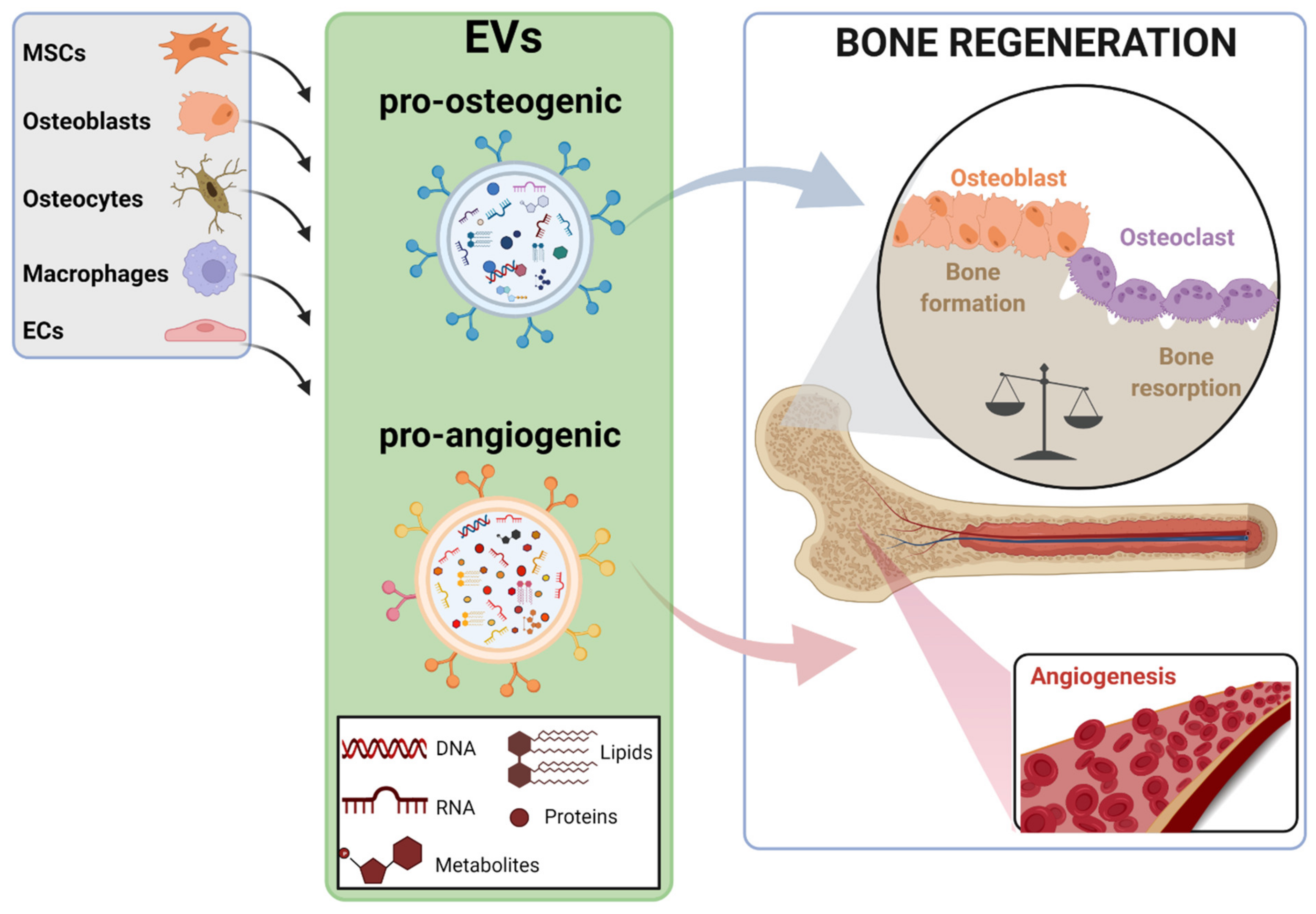
The incidence of bone-related disorders is continuously growing as the aging of the population in developing countries continues to increase. Although therapeutic interventions for bone regeneration exist, their effectiveness is questioned, especially under certain circumstances, such as critical size defects. This gap of curative options has led to the search for new and more effective therapeutic approaches for bone regeneration; among them, the possibility of using extracellular vesicles (EVs) is gaining ground. EVs are secreted, biocompatible, nano-sized vesicles that play a pivotal role as messengers between donor and target cells, mediated by their specific cargo. Evidence shows that bone-relevant cells secrete osteoanabolic EVs, whose functionality can be further improved by several strategies. This, together with the low immunogenicity of EVs and their storage advantages, make them attractive candidates for clinical prospects in bone regeneration. However, before EVs reach clinical translation, a number of concerns should be addressed. Unraveling the EVs’ mode of action in bone regeneration is one of them; the molecular mediators driving their osteoanabolic effects in acceptor cells are beginning to be uncovered. Increasing the functional and bone targeting abilities of EVs are also matters of intense research.

extracellular vesicles osteoanabolic bone regeneration MSCs miRNAs

1. Introduction

Bone is a dynamic organ that is constantly remodeling to ensure a constant net bone mass within an organism. This is achieved by two opposite and balanced phases: bone formation and bone resorption, carried out by osteoblasts and osteoclasts, respectively. Moreover, bone has a self-repairing ability, and therefore when an injury occurs, the damaged part regains its original structure and mechanical strength by the activation of the bone healing process. However, under certain situations, such as critical-sized bone defects (defined as those that will not heal spontaneously during the life of a patient), bone loss due to bone-related diseases, lack of vascularization, infections, and tumors, this ability is not enough and requires clinical intervention [1]. In the case of osteoporosis (OP), a highly prevalent metabolic disease affecting more than 200 million of patients worldwide, each year more than 8.9 million fractures are reported globally [2]. In the EU, the cost of OP in 2019, including pharmacological intervention, entailed more than €56 billion, doubling that needed in 2010 [3][4].
Bone tissue is highly demanded in clinics, and after blood, is the second most transplanted tissue worldwide [5]. Thus, currently, the most successful intervention to treat bone defects is still bone grafting, a strategy first outlined in the early 1900s [6]. Bone autografts are the gold standard in bone regeneration procedures since they avoid rejection from the patient’s immune system. Unfortunately, autologous bone supply is limited and the need of additional surgery for bone extraction increases the risk of infections and morbidity. Another option is the use of allografts (from a human donor) or xenografts (from large animals like pigs or bovines), which nevertheless entail some risk of pathogen transmission; more importantly, these bone implants have shown poor bone regeneration abilities [7]. All in all, there is an urgent need to discover new, effective therapies to boost bone regeneration to satisfy the growing world population (progressively more aged) affected by bone conditions.
On this basis, the field of bone tissue engineering has emerged, focused on developing “bone substitutes” that mimic the bone tissue features, usually formed by a 3D scaffold and bone-relevant cell types, which are able to promote osteogenic differentiation in host tissue without any adverse inflammatory response [8]. The features of mesenchymal stem cells (MSCs), such as their capacity to undergo osteogenic differentiation, immunomodulatory potential, and trophic effects, make them quite attractive components for these bone constructs [8][9]. In fact, the use of MSCs-based therapies in clinics is gaining interest in the field of bone regeneration due to the clinical improvements exhibited by patients affected by bone-related diseases, such as Osteogenesis Imperfecta (OI) after MSCs administrations [8][10][11][12]. However, there are still some limitations to the clinical translation of MSCs, such as the large number of cells that are required as well as the high cellular heterogeneity, even within populations of MSCs from the same donor. In addition, other factors, such as cell culture conditions, cell source, and donor age, have determined the variable outcomes shown in different clinical trials using MSCs [13]. Regarding the mode of action of MSCs therapy in bone regeneration, quite revealing findings point to the paracrine mechanisms elicited by these cells, rather than the initially expected cell engraftment and subsequent osteogenic differentiation [10][14]. Moreover, a set of experiments performed in OI animal models, suggested that the extracellular vesicles (EVs) secreted by MSCs could be mediating the recovery of bone phenotypes observed in OI patients subjected to MSCs therapy [15].
EVs are small, lipid membrane delimited particles secreted by most cell types and present in several biological fluids, such as blood and urine, that play a key role in cell-to-cell communication [16]. This paracrine crosstalk is mediated by the EVs’ cargo, an array of bioactive molecules including proteins, lipids, and nucleic acids, that interestingly exhibit a parent-cell-specific signature [17][18]. Thus, through specific surface molecule interaction, EVs can be uptaken by target cells and modify their biology/fate [19]. Attending to their size, the EVs population can be divided into exosomes (diameters of 30–200 nm), microvesicles (diameters of 200–1000 nm), and apoptotic bodies (diameters > 1000 nm) [19][20]. The biogenesis of EVs is a complex process and currently it is accepted that each EV subtype can be originated by two predominant pathways: the endosomal pathway, through multivesicular endosome fusion, or by the outward budding and fission of the plasmatic membrane [21]. The fact that EVs carry functional molecules that can modulate target cell responses opens the possibility of using EVs as next-generation drug delivery platforms, a vision strongly supported by their low immunogenicity. Thus, EVs are known to escape from immune clearance when systemically administered, due to the expression of surface molecules, such as CD47, which mediates the “don’t eat me” signal that blocks phagocytosis [22]. Bone-relevant cell types have been shown to secrete EVs that regulate bone homeostasis, and in addition, recent evidences suggest that EV therapy is at least as efficient as cell therapy in eliciting bone regeneration in large bone defect animal models [19][23][24]. Thus, the use of EVs as osteoanabolic delivery systems could be a reliable clinical approach for bone regeneration [25].

2. EV Sources for Bone Regeneration and Mechanisms of Action

From a clinical perspective, most research in the field of bone regeneration and EVs, which has grown exponentially in the last years, has focused on either exosomes or microvesicles. However, these EV populations are not fully characterized, in spite of the remarkable efforts made by the International Society of Extracellular Vesicles (ISEV) to determine the minimal information for studies regarding EVs (Minimal information for studies of extracellular vesicles 2018 (MISEV2018): a position statement of the International Society for Extracellular Vesicles and update of the MISEV2014 guidelines, doi:10.1080/20013078.2018.1535750). Following these recommendations, herein will use EVs as a broad term encompassing all the vesicles secreted by cells [26].
Bone resident cells, including bone-cell types (MSCs, osteoblasts, osteoclasts, and osteocytes) as well as other cell populations present in the bone microenvironment, such as endothelial cells (ECs) and macrophages, secrete EVs that mediate the continuous bone remodeling process (Figure 1) [23]. Interestingly, the cargo contained in the EVs reflects the biological function of parent cells. Thus, osteoclast-secreted EVs can inhibit osteoblast activity and therefore suppress bone formation, whereas the EVs secreted by osteoblastic-lineage cells, such as MSCs and osteoblasts, enhance osteoblast differentiation in vitro and promote bone regeneration in vivo [27][28]. Moreover, osteoblast-derived EVs also can be uptaken by osteoclasts, but the consequences triggered in these cells are conflicting. Thus, the promotion of osteoclasts’ differentiation in vitro and in vivo has been described, but also the inhibition of osteoclastogenesis, and therefore of bone resorption [29][30][31]. Current evidence, mainly obtained from basic and preclinical experimentation, strongly suggest that the specific cargo of EVs determines the signaling triggered in recipient cells. In this line, miRNAs contained in EVs are known to be transferred to target cells as a mechanism of genetic exchange between cells, playing a key role in regulating bone homeostasis [32]. Actually, a specific, dysregulated miRNAs signature has been described in plasma/serum EVs of some pathological conditions, including bone disease. This is quite valuable in silent pathologies, such as OP, which has no clinical manifestation until a fracture occurs, being that these circulating miRNAs, proposed as potential biomarkers, are capable of predicting the risk of fracture [33][34].
Ijms 23 01865 g001
Figure 1. Illustration of the bone-relevant cell types known to secrete EVs that promote bone regeneration. Pro-osteogenic and pro-angiogenic EVs, determined by their specific cargo, are produced by cell types present in bone microenvironment, such as mesenchymal stem cells (MSCs), osteoblasts, osteocytes, macrophages, and endothelial cells (ECs). Pro-osteogenic EVs stimulate MSCs and osteoblasts differentiation, inducing the bone formation process, while pro-angiogenic EVs elicit the formation of new blood vessels in bone tissue. Both processes are essential to conduct a successful regeneration of bone tissue. The figure was created with BioRender.com (accessed on 1 December 2021).
Herein will discuss the current knowledge about the bone-relevant cell types that secrete osteoanabolic EVs and the current identified mechanisms, mainly driven by miRNAs (and some proteins, to a lesser extent) (Table 1), by which these EVs exert their function in target cells.
Table 1. Main cell sources that secrete osteoanabolic EVs and the identified cargo mediating bone regeneration in different mice models of bone disease.
EVs Source Bioactive Cargo Disease Model Target Molecule-Pathway Target Process Ref.
Bone marrow (BM)-MSCs miR-335 Bone fracture VAPB-WNT/β-CATENIN ↓ Osteoclastogenesis
&
↑ Osteogenesis
[35]
miR-25 Bone fracture SMURF1-RUNX2 ↑ Osteogenesis [36]
NID1 Femoral defects Myosin-10 ↑ Angiogenesis [37]
mir-29a Wild type mice VASH1 ↑ Angiogenesis
&
↑ Osteogenesis
[38]
Aged BM-MSCs mir-128-3p Bone fracture SMAD5 ↓ Osteogenesis [39]
Umbilical cord (UC)-MSCs miR-1263 Disuse osteoporosis (OP) MOB1-HIPPO ↓ Apoptosis [40]
miR-21 Glucocorticoid-induced osteonecrosis of the femoral head (GIONFH) PTEN-PI3K/AKT ↓ Apoptosis [41]
miR-365a-5p GIONFH SAV1-HIPPO ↑ Osteogenesis [42]
miR-3960 Senile OP unknown ↑ Osteogenesis
&
↑ Osteoclastogenesis
[43]
CLEC11A Ovariectomized (OVX)-OP, Disuse OP, Senile OP unknown ↑ Osteogenesis
&
↓ Osteoclastogenesis
[44]
Hypoxia-UC-MSCs mir-126 Bone fracture SPRED-1 ↑ Angiogenesis [45]
ECs miR-126 Distraction osteogenesis SPRED-1 ↑ Osteogenesis
&
↑ Angiogenesis
[24]
miR-155 OVX-OP Spi1, Mitf, Socs1 ↓ Osteoclastogenesis [46]

3. Novel Strategies to Improve the Bone Regenerative Potential of EVs

The bone regenerative potential of EVs, especially if they come from MSCs and ECs, is an undeniable fact nowadays, supported (as been mentioned above) by a considerable number of basic and preclinical studies [47]. Nevertheless, the use of EVs as an advanced therapy for bone regeneration has been hampered mainly by two observations inherent to EV biology. First, the osteoanabolic potential of EVs is far from being optimum to achieve complete bone regeneration, and therefore these osteoanabolic abilities should be improved; second, osteoanabolic EVs do not mainly target bone tissue. Thus, upon intravenous administration in mice, EVs show a rapid (within the first hour) tissue distribution, accumulating mainly in the spleen, liver, lung, and kidneys [48][49]. Even so, there are also studies indicating the accumulation of MSCs-derived EVs in bone tissue, although to a lesser extent [50]. On the contrary, EVs coming from osteoclasts, known to negatively regulate bone formation by targeting and inhibiting osteoblasts, have shown acceptable intra-osseous accumulation in injected mice [28]. Therefore, current attempts pursuing the production of EVs with the maximum bone regenerative potential mainly rely on the enhancement of the EVs’ osteoanabolic abilities as well as on their bone cell targeting and, therefore, bone tissue.

3.1. Enhancing the Osteoanabolic Potential of EVs

MSCs undergoing osteogenic differentiation, especially those isolated from bone marrow (BM), have demonstrated acceptable bone regeneration properties due to two facts: their EVs show increased bone targeting potential and exhibit osteoanabolic-specific cargo [51][52][53]. Hence, it is not surprising that the vast variety of investigations focus on enhancing the osteogenesis of parent MSCs in order to achieve innate EVs with maximum osteoanabolic and bone targeting abilities.

3.1.1. Preconditioning of Parent Cells

Preconditioning of MSCs’ culture conditions, either by the addition of exogenous molecules (cytokines, growth factors, drugs) or by the optimization of physical factors (hypoxia or shear stress), has been proposed, as these strategies induce a robust osteogenic differentiation in MSCs in order to obtain highly osteoanabolic EVs (Figure 2) [54]. Along this line, the mimicking of the bone healing signaling milieu, such as that occurring in the inflammatory phase upon bone injury, has been demonstrated to be effective. Thus, when priming AT-MSCs with TNF-α, a specific pro-inflammatory molecule, the secreted EVs showed enhanced abilities in promoting the proliferation and osteogenic differentiation of human primary osteoblastic cells. Interestingly, an increase in WNT3a protein, a known inducer of osteogenesis, was detected in the cargo of these EVs [55].
Ijms 23 01865 g002
Figure 2. Preconditioning strategies to enhance the osteoanabolic potential of EVs. Pre-treatment of MSCs with inflammatory factors or histone deacetylase inhibitors enhance their osteogenic differentiation, whereas hypoxia conditions elicit pro-angiogenic responses in these cells, leading to the secretion of pro-osteogenic or pro-angiogenic EVs, respectively. Mechanistically, the mimicking of the bone inflammatory microenvironment after bone injury triggers the expression of the pro-osteogenic protein WNT3a in MSCs, which in turn, is enriched in the EVs secreted by these cells. The inhibition of histone deacetylases, such as via the use of thrichostatin A (TSA), elicits an epigenetic reprogramming of MSCs, ensuring an open conformation of chromatin and promoting the transcription of pro-osteogenic genes. The hypoxia simulation in MSCs, achieved by low oxygen cell culture or by chemical compounds (for instance dimethyloxaylglycine (DMOG)), induces the activation of the HIF-1α transcription factor, which drives the cell responses to hypoxia, among them being hypoxia-induced angiogenesis. The figure was created with BioRender.com (accessed on 1 December 2021).

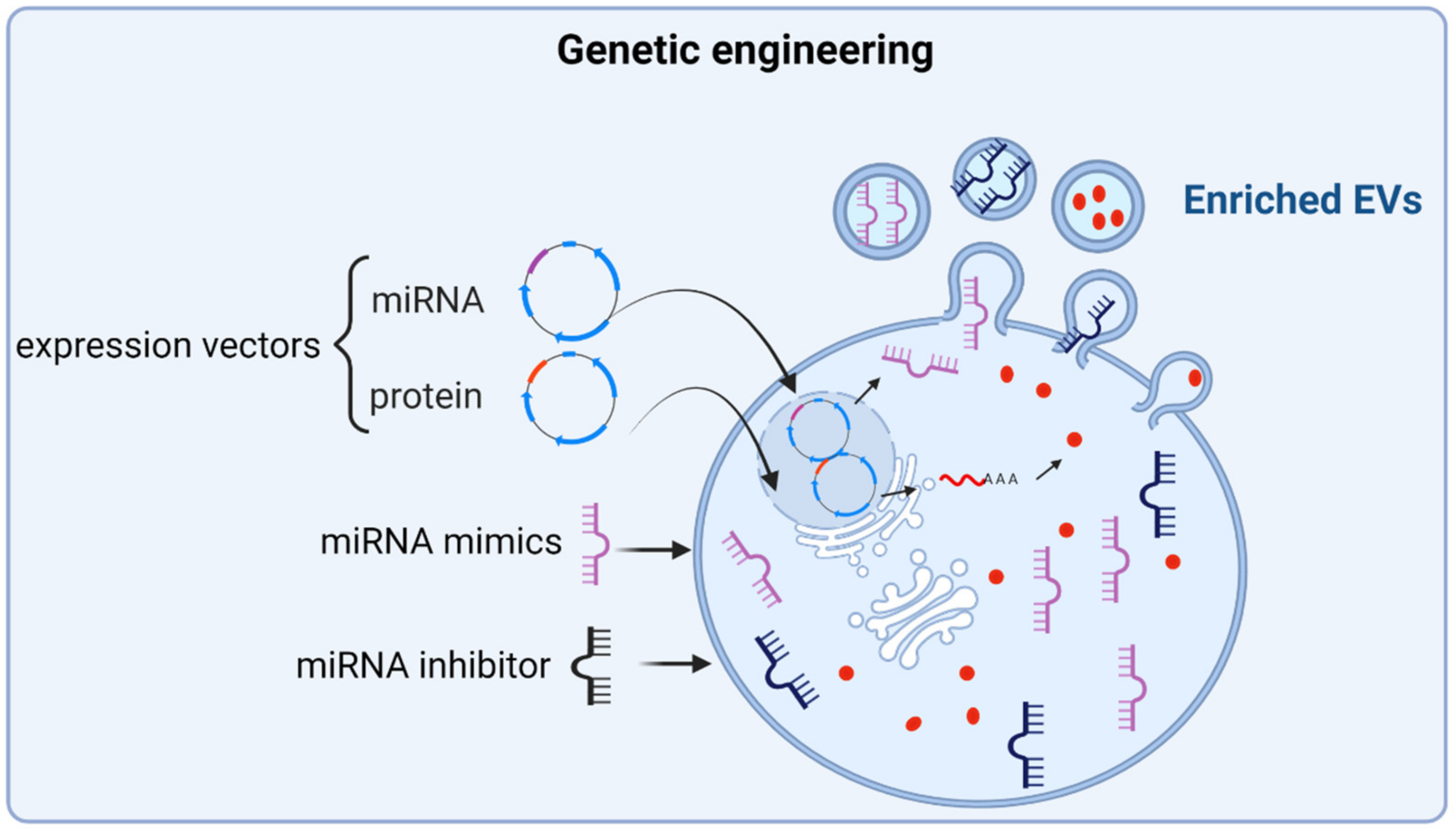
3.1.2. Engineering of Parent Cells

MSCs can be genetically modified to increase the expression of certain pro-osteogenic molecules with the assumption that, this way, their secreted EVs would also be enriched in those induced molecules, enhancing their osteoanabolic properties (Figure 3). In fact, this hypothesis has been validated by overexpressing pro-osteogenic miRNAs in MSCs, such as miR-375 and miR-101. The authors demonstrated that EVs could be enriched in these miRNAs when overexpressed in parent MSCs, without affecting distinctive features of EVs such as morphology, size, and the expression of surface proteins CD9 and CD63, which are used as EV markers. Moreover, these EVs improved the osteogenic differentiation of MSCs and enhanced bone regeneration in animal models of bone defects [56][57].
Ijms 23 01865 g003
Figure 3. Genetic engineering as an approach to enrich EVs with osteoanabolic factors. The induced expression of known osteoanabolic miRNAs, proteins, or inhibitors of anti-osteogenic miRNAs in MSCs, by using expression vectors or direct transfection approaches of these molecules, yields EVs enriched in these molecules. The figure was created with BioRender.com (accessed on 1 December 2021).

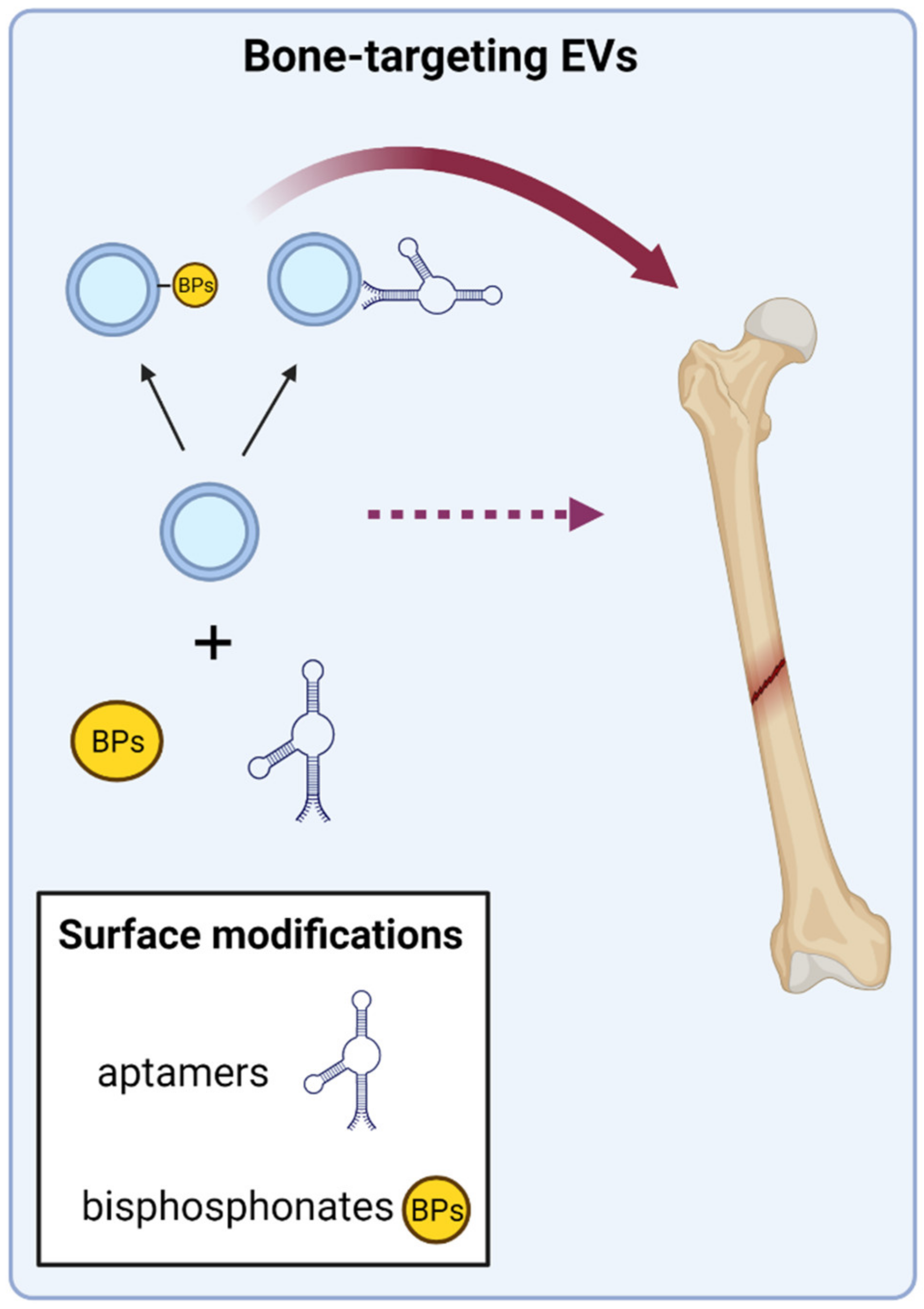
3.2. Directing EVs to Target Bone Tissue

EV therapy to treat skeletal conditions can be delivered by local or intravenous administration, and each one has their specifications and advantages/disadvantages. The local administration, most suitable for concrete bone fractures or defects, ensures the bone targeting of EVs, but requires the concomitant use of scaffolds, such hydrogels, in order to maintain the EVs in the site of injury. On the contrary, when considering global skeletal conditions, such as OP or OI, the intravenous administration of EVs is the considered option; nevertheless a major drawback of this administration route is the low bone tropism that EVs show. Some innovative strategies are currently under intensive research in order to achieve and enhance the bone targeting of EVs; especially promising are those that directly modify the EVs’ surface with specific bone-targeting molecules (Figure 4).
Ijms 23 01865 g004
Figure 4. Functionalization of EVs’ surface to improve bone targeting. The surface modification of EVs with molecules showing affinity for bone cells has been described. This is the case of specific aptamers, DNA/RNA molecules with affinity for a desired target, and in this case MSCs and osteoblasts. Anti-resorptive drugs, such as bisphosphonates (BPs), which show high affinity for the mineralized bone matrix, have also been covalently bound to the surface of EVs. Both approaches have demonstrated increased bone targeting of functionalized EVs. The figure was created with BioRender.com (accessed on 1 December 2021).

4. Conclusions

The increasing knowledge about EV biology has strengthened the idea that EVs hold great potential to be applied with therapeutic purposes, mainly due to their ability to transfer diverse bioactive molecules modifying the fate of recipient cells. Thus, EVs may offer a promising “cell free” advanced therapy as next-generation biocompatible vehicles delivering therapeutic factors.
However, before EVs move forward to the clinic, it is mandatory to address several requirements that challenge their claimed therapeutic abilities, including standardization and scalability production, their full molecular characterization, and bioengineering improvements that increase their therapeutic potency. Moreover, the inherent biology of the target tissue plays a key role in the success of EV-based therapies. When intended for bone regeneration purposes, EV therapeutics have to overcome two main limitations, both matters of intense research: the osteoanabolic properties of EVs, which should be enhanced in order to achieve robust, in vivo bone regeneration and the limited tropism for bone tissue that the osteoanabolic EVs show upon administration. Therefore, to achieve bone regeneration, the ideal EVs should combine features aiming to counteract these two limitations.
EVs isolated from a wide range of bone-relevant cells have demonstrated osteoanabolic potential. However, the fact that the majority of studies only rely on EVs isolated from a single cell type hinders the comparison of their osteoanabolic capacity. Therefore, the systematic analysis of different EVs isolated from different cell types abdcomparing their osteogenic capacity should be a prerequisite to identify those EVs, or their combination, with the maximum osteoanabolic potential. This knowledge will come along with the understanding of the mode of action of EVs, and to achieve it, essential requirements should be considered, such as deciphering the molecular players driving the downstream signaling of EVs in target cells. Accordingly, comprehensive multi-omic technologies have enabled a deep characterization of EV cargo, but the identification of those molecular drivers in EVs conducting bone regeneration is just beginning to emerge. So far, the majority of the current research has identified several single molecules, especially miRNAs and some proteins, as drivers of the EVs downstream regulation in the recipient cells. However, considering that EVs carry an array of molecules and that EVs from different cell sources achieve the induction of bone regeneration, it is more likely for a synergistic collaboration of different molecules in target cells to occur, as opposed to a single upstream molecular regulator. In fact, recent evidence point to this observation: Lee and collaborators reported that AT-MSCs-EVs attenuated bone loss in OVX mice by the simultaneous transfer of proteins and miRNAs targeting osteoclasts. Thus, the inhibition of osteoclastogenesis elicited by AT-MSCs-EVs and the subsequent restoration of bone mass in OVX mice was mediated by the transfer of osteoprotegerin (OPG), a decoy receptor for RANK ligands that inhibits osteoclasts differentiation, and miR-21-5p and let-7b-5p, which reduced osteoclast differentiation [50]. Liu and coworkers also identified a multi-component pro-osteogenic miRNAs cargo in BM-MSCs-EVs: let-7a-5p, let-7c-5p, miR-328a-5p, and miR-31a-5p. These miRNAs were shown to synergistically mediate the osteoanabolic properties of BM-MSC-EVs by promoting the activation of the canonical BMP signaling pathway [58].
The increasing knowledge about the most suitable EV cell source and the bioengineering approaches under development will address the aforementioned limitations facilitating the development of EV-based therapeutics that will transform the pharmaceutical scene for bone regeneration. Currently (as of December 2021), there is one clinical trial testing EVs, specifically exosomes, as therapeutic drugs applied for a bone disease: a phase I trial evaluating intra-articular injections of a single dose allogenic MSCs-derived exosomes for knee osteoarthritis (ExoOA-1; NCT05060107). It is anticipated that, as different approaches demonstrate improvements in the osteoanabolic potential and bone-targeting abilities of EVs, there will be increasing clinical trials evaluating the safety and potential of this advanced therapy for bone regenerative purposes in the not-so-distant future.

References

  1. Schemitsch, E.H. Size Matters: Defining Critical in Bone Defect Size! J. Orthop. Trauma 2017, 31, S20–S22.
  2. Barnsley, J.; Buckland, G.; Chan, P.E.; Ong, A.; Ramos, A.S.; Baxter, M.; Laskou, F.; Dennison, E.M.; Cooper, C.; Patel, H.P. Pathophysiology and treatment of osteoporosis: Challenges for clinical practice in older people. Aging Clin. Exp. Res. 2021, 33, 759–773.
  3. Kanis, J.A.; Cooper, C.; Rizzoli, R.; Reginster, J.Y. European guidance for the diagnosis and management of osteoporosis in postmenopausal women. Osteoporos. Int. 2019, 30, 3–44.
  4. Kanis, J.A.; Norton, N.; Harvey, N.C.; Jacobson, T.; Johansson, H.; Lorentzon, M.; McCloskey, E.V.; Willers, C.; Borgström, F. SCOPE 2021: A new scorecard for osteoporosis in Europe. Arch. Osteoporos. 2021, 16, 82.
  5. Campana, V.; Milano, G.; Pagano, E.; Barba, M.; Cicione, C.; Salonna, G.; Lattanzi, W.; Logroscino, G. Bone substitutes in orthopaedic surgery: From basic science to clinical practice. J. Mater. Sci. Mater. Med. 2014, 25, 2445–2461.
  6. Donati, D.; Zolezzi, C.; Tomba, P.; Viganò, A. Bone grafting: Historical and conceptual review, starting with an old manuscript by Vittorio Putti. Acta Orthop. 2007, 78, 19–25.
  7. Oryan, A.; Alidadi, S.; Moshiri, A.; Maffulli, N. Bone regenerative medicine: Classic options, novel strategies, and future directions. J. Orthop. Surg. Res. 2014, 9, 18.
  8. Macías, I.; Alcorta-Sevillano, N.; Infante, A.; Rodríguez, C.I. Cutting Edge Endogenous Promoting and Exogenous Driven Strategies for Bone Regeneration. Int. J. Mol. Sci. 2021, 22, 7724.
  9. Li, L.; Li, J.; Zou, Q.; Zuo, Y.; Cai, B.; Li, Y. Enhanced bone tissue regeneration of a biomimetic cellular scaffold with co-cultured MSCs-derived osteogenic and angiogenic cells. Cell Prolif. 2019, 52, e12658.
  10. Infante, A.; Gener, B.; Vázquez, M.; Olivares, N.; Arrieta, A.; Grau, G.; Llano, I.; Madero, L.; Bueno, A.M.; Sagastizabal, B.; et al. Reiterative infusions of MSCs improve pediatric osteogenesis imperfecta eliciting a pro-osteogenic paracrine response: TERCELOI clinical trial. Clin. Transl. Med. 2021, 11, e265.
  11. Horwitz, E.M.; Prockop, D.J.; Fitzpatrick, L.A.; Koo, W.W.; Gordon, P.L.; Neel, M.; Sussman, M.; Orchard, P.; Marx, J.C.; Pyeritz, R.E.; et al. Transplantability and therapeutic effects of bone marrow-derived mesenchymal cells in children with osteogenesis imperfecta. Nat. Med. 1999, 5, 309–313.
  12. Götherström, C.; Westgren, M.; Shaw, S.W.; Aström, E.; Biswas, A.; Byers, P.H.; Mattar, C.N.; Graham, G.E.; Taslimi, J.; Ewald, U.; et al. Pre- and postnatal transplantation of fetal mesenchymal stem cells in osteogenesis imperfecta: A two-center experience. Stem Cells Transl. Med. 2014, 3, 255–264.
  13. Levy, O.; Kuai, R.; Siren, E.M.J.; Bhere, D.; Milton, Y.; Nissar, N.; De Biasio, M.; Heinelt, M.; Reeve, B.; Abdi, R.; et al. Shattering barriers toward clinically meaningful MSC therapies. Sci. Adv. 2020, 6, eaba6884.
  14. Otsuru, S.; Gordon, P.L.; Shimono, K.; Jethva, R.; Marino, R.; Phillips, C.L.; Hofmann, T.J.; Veronesi, E.; Dominici, M.; Iwamoto, M.; et al. Transplanted bone marrow mononuclear cells and MSCs impart clinical benefit to children with osteogenesis imperfecta through different mechanisms. Blood 2012, 120, 1933–1941.
  15. Otsuru, S.; Desbourdes, L.; Guess, A.J.; Hofmann, T.J.; Relation, T.; Kaito, T.; Dominici, M.; Iwamoto, M.; Horwitz, E.M. Extracellular vesicles released from mesenchymal stromal cells stimulate bone growth in osteogenesis imperfecta. Cytotherapy 2018, 20, 62–73.
  16. Monguió-Tortajada, M.; Morón-Font, M.; Gámez-Valero, A.; Carreras-Planella, L.; Borràs, F.E.; Franquesa, M. Extracellular-Vesicle Isolation from Different Biological Fluids by Size-Exclusion Chromatography. Curr. Protoc. Stem Cell Biol. 2019, 49, e82.
  17. Van Niel, G.; D’Angelo, G.; Raposo, G. Shedding light on the cell biology of extracellular vesicles. Nat. Rev. Mol. Cell Biol. 2018, 19, 213–228.
  18. Dutra Silva, J.; Su, Y.; Calfee, C.S.; Delucchi, K.L.; Weiss, D.; McAuley, D.F.; O’Kane, C.; Krasnodembskaya, A.D. Mesenchymal stromal cell extracellular vesicles rescue mitochondrial dysfunction and improve barrier integrity in clinically relevant models of ARDS. Eur. Respir. J. 2021, 58.
  19. Teng, F.; Fussenegger, M. Shedding Light on Extracellular Vesicle Biogenesis and Bioengineering. Adv. Sci. 2020, 8, 2003505.
  20. Li, M.; Liao, L.; Tian, W. Extracellular Vesicles Derived From Apoptotic Cells: An Essential Link Between Death and Regeneration. Front. Cell Dev. Biol. 2020, 8, 573511.
  21. Herrmann, I.K.; Wood, M.J.A.; Fuhrmann, G. Extracellular vesicles as a next-generation drug delivery platform. Nat. Nanotechnol. 2021, 16, 748–759.
  22. Parada, N.; Romero-Trujillo, A.; Georges, N.; Alcayaga-Miranda, F. Camouflage strategies for therapeutic exosomes evasion from phagocytosis. J. Adv. Res. 2021, 31, 61–74.
  23. Gao, M.; Gao, W.; Papadimitriou, J.M.; Zhang, C.; Gao, J.; Zheng, M. Exosomes-the enigmatic regulators of bone homeostasis. Bone Res. 2018, 6, 36.
  24. Jia, Y.; Zhu, Y.; Qiu, S.; Xu, J.; Chai, Y. Exosomes secreted by endothelial progenitor cells accelerate bone regeneration during distraction osteogenesis by stimulating angiogenesis. Stem Cell Res. Ther. 2019, 10, 12.
  25. Liu, S.; Xu, X.; Liang, S.; Chen, Z.; Zhang, Y.; Qian, A.; Hu, L. The Application of MSCs-Derived Extracellular Vesicles in Bone Disorders: Novel Cell-Free Therapeutic Strategy. Front. Cell Dev. Biol. 2020, 8, 619.
  26. Théry, C.; Witwer, K.W.; Aikawa, E.; Alcaraz, M.J.; Anderson, J.D.; Andriantsitohaina, R.; Antoniou, A.; Arab, T.; Archer, F.; Atkin-Smith, G.K.; et al. Minimal information for studies of extracellular vesicles 2018 (MISEV2018): A position statement of the International Society for Extracellular Vesicles and update of the MISEV2014 guidelines. J. Extracell Vesicles 2018, 7, 1535750.
  27. Qin, Y.; Wang, L.; Gao, Z.; Chen, G.; Zhang, C. Bone marrow stromal/stem cell-derived extracellular vesicles regulate osteoblast activity and differentiation in vitro and promote bone regeneration in vivo. Sci. Rep. 2016, 6, 21961.
  28. Li, D.; Liu, J.; Guo, B.; Liang, C.; Dang, L.; Lu, C.; He, X.; Cheung, H.Y.; Xu, L.; He, B.; et al. Osteoclast-derived exosomal miR-214-3p inhibits osteoblastic bone formation. Nat. Commun. 2016, 7, 10872.
  29. Kobayashi-Sun, J.; Yamamori, S.; Kondo, M.; Kuroda, J.; Ikegame, M.; Suzuki, N.; Kitamura, K.I.; Hattori, A.; Yamaguchi, M.; Kobayashi, I. Uptake of osteoblast-derived extracellular vesicles promotes the differentiation of osteoclasts in the zebrafish scale. Commun. Biol 2020, 3, 190.
  30. Cappariello, A.; Loftus, A.; Muraca, M.; Maurizi, A.; Rucci, N.; Teti, A. Osteoblast-Derived Extracellular Vesicles Are Biological Tools for the Delivery of Active Molecules to Bone. J. Bone Min. Res. 2018, 33, 517–533.
  31. Minamizaki, T.; Nakao, Y.; Irie, Y.; Ahmed, F.; Itoh, S.; Sarmin, N.; Yoshioka, H.; Nobukiyo, A.; Fujimoto, C.; Niida, S.; et al. The matrix vesicle cargo miR-125b accumulates in the bone matrix, inhibiting bone resorption in mice. Commun. Biol. 2020, 3, 30.
  32. Valadi, H.; Ekström, K.; Bossios, A.; Sjöstrand, M.; Lee, J.J.; Lötvall, J.O. Exosome-mediated transfer of mRNAs and microRNAs is a novel mechanism of genetic exchange between cells. Nat. Cell Biol. 2007, 9, 654–659.
  33. Xu, J.; Chen, Y.; Yu, D.; Zhang, L.; Dou, X.; Wu, G.; Wang, Y.; Zhang, S. Evaluation of the cargo contents and potential role of extracellular vesicles in osteoporosis. Aging 2021, 13, 19282–19292.
  34. Shao, J.L.; Li, H.; Zhang, X.R.; Zhang, X.; Li, Z.Z.; Jiao, G.L.; Sun, G.D. Identification of Serum Exosomal MicroRNA Expression Profiling in Menopausal Females with Osteoporosis by High-throughput Sequencing. Curr. Med. Sci. 2020, 40, 1161–1169.
  35. Hu, H.; Wang, D.; Li, L.; Yin, H.; He, G.; Zhang, Y. Role of microRNA-335 carried by bone marrow mesenchymal stem cells-derived extracellular vesicles in bone fracture recovery. Cell Death Dis. 2021, 12, 156.
  36. Jiang, Y.; Zhang, J.; Li, Z.; Jia, G. Bone Marrow Mesenchymal Stem Cell-Derived Exosomal miR-25 Regulates the Ubiquitination and Degradation of Runx2 by SMURF1 to Promote Fracture Healing in Mice. Front. Med. 2020, 7, 577578.
  37. Chen, Q.; Shou, P.; Zheng, C.; Jiang, M.; Cao, G.; Yang, Q.; Cao, J.; Xie, N.; Velletri, T.; Zhang, X.; et al. Fate decision of mesenchymal stem cells: Adipocytes or osteoblasts? Cell Death Differ. 2016, 23, 1128–1139.
  38. Lu, G.D.; Cheng, P.; Liu, T.; Wang, Z. BMSC-Derived Exosomal miR-29a Promotes Angiogenesis and Osteogenesis. Front. Cell Dev. Biol. 2020, 8, 608521.
  39. Xu, T.; Luo, Y.; Wang, J.; Zhang, N.; Gu, C.; Li, L.; Qian, D.; Cai, W.; Fan, J.; Yin, G. Exosomal miRNA-128-3p from mesenchymal stem cells of aged rats regulates osteogenesis and bone fracture healing by targeting Smad5. J. Nanobiotechnol. 2020, 18, 47.
  40. Yang, B.C.; Kuang, M.J.; Kang, J.Y.; Zhao, J.; Ma, J.X.; Ma, X.L. Human umbilical cord mesenchymal stem cell-derived exosomes act via the miR-1263/Mob1/Hippo signaling pathway to prevent apoptosis in disuse osteoporosis. Biochem. Biophys. Res. Commun. 2020, 524, 883–889.
  41. Kuang, M.J.; Huang, Y.; Zhao, X.G.; Zhang, R.; Ma, J.X.; Wang, D.C.; Ma, X.L. Exosomes derived from Wharton’s jelly of human umbilical cord mesenchymal stem cells reduce osteocyte apoptosis in glucocorticoid-induced osteonecrosis of the femoral head in rats via the miR-21-PTEN-AKT signalling pathway. Int. J. Biol. Sci. 2019, 15, 1861–1871.
  42. Kuang, M.J.; Zhang, K.H.; Qiu, J.; Wang, A.B.; Che, W.W.; Li, X.M.; Shi, D.L.; Wang, D.C. Exosomal miR-365a-5p derived from HUC-MSCs regulates osteogenesis in GIONFH through the Hippo signaling pathway. Mol. Ther. Nucleic Acids 2021, 23, 565–576.
  43. Hu, Y.; Xu, R.; Chen, C.Y.; Rao, S.S.; Xia, K.; Huang, J.; Yin, H.; Wang, Z.X.; Cao, J.; Liu, Z.Z.; et al. Extracellular vesicles from human umbilical cord blood ameliorate bone loss in senile osteoporotic mice. Metabolism 2019, 95, 93–101.
  44. Hu, Y.; Zhang, Y.; Ni, C.Y.; Chen, C.Y.; Rao, S.S.; Yin, H.; Huang, J.; Tan, Y.J.; Wang, Z.X.; Cao, J.; et al. Human umbilical cord mesenchymal stromal cells-derived extracellular vesicles exert potent bone protective effects by CLEC11A-mediated regulation of bone metabolism. Theranostics 2020, 10, 2293–2308.
  45. Liu, W.; Li, L.; Rong, Y.; Qian, D.; Chen, J.; Zhou, Z.; Luo, Y.; Jiang, D.; Cheng, L.; Zhao, S.; et al. Hypoxic mesenchymal stem cell-derived exosomes promote bone fracture healing by the transfer of miR-126. Acta Biomater. 2020, 103, 196–212.
  46. Song, H.; Li, X.; Zhao, Z.; Qian, J.; Wang, Y.; Cui, J.; Weng, W.; Cao, L.; Chen, X.; Hu, Y.; et al. Reversal of Osteoporotic Activity by Endothelial Cell-Secreted Bone Targeting and Biocompatible Exosomes. Nano Lett. 2019, 19, 3040–3048.
  47. Wang, X.; Thomsen, P. Mesenchymal stem cell-derived small extracellular vesicles and bone regeneration. Basic Clin. Pharmacol. Toxicol. 2021, 128, 18–36.
  48. Lai, C.P.; Mardini, O.; Ericsson, M.; Prabhakar, S.; Maguire, C.; Chen, J.W.; Tannous, B.A.; Breakefield, X.O. Dynamic biodistribution of extracellular vesicles in vivo using a multimodal imaging reporter. ACS Nano 2014, 8, 483–494.
  49. Kang, M.; Jordan, V.; Blenkiron, C.; Chamley, L.W. Biodistribution of extracellular vesicles following administration into animals: A systematic review. J. Extracell Vesicles 2021, 10, e12085.
  50. Lee, K.S.; Lee, J.; Kim, H.K.; Yeom, S.H.; Woo, C.H.; Jung, Y.J.; Yun, Y.E.; Park, S.Y.; Han, J.; Kim, E.; et al. Extracellular vesicles from adipose tissue-derived stem cells alleviate osteoporosis through osteoprotegerin and miR-21-5p. J. Extracell Vesicles 2021, 10, e12152.
  51. Huang, C.C.; Kang, M.; Lu, Y.; Shirazi, S.; Diaz, J.I.; Cooper, L.F.; Gajendrareddy, P.; Ravindran, S. Functionally engineered extracellular vesicles improve bone regeneration. Acta Biomater. 2020, 109, 182–194.
  52. Li, W.; Liu, Y.; Zhang, P.; Tang, Y.; Zhou, M.; Jiang, W.; Zhang, X.; Wu, G.; Zhou, Y. Tissue-Engineered Bone Immobilized with Human Adipose Stem Cells-Derived Exosomes Promotes Bone Regeneration. ACS Appl Mater. Interfaces 2018, 10, 5240–5254.
  53. Wei, Y.; Tang, C.; Zhang, J.; Li, Z.; Zhang, X.; Miron, R.J.; Zhang, Y. Extracellular vesicles derived from the mid-to-late stage of osteoblast differentiation markedly enhance osteogenesis in vitro and in vivo. Biochem. Biophys. Res. Commun. 2019, 514, 252–258.
  54. García-Sánchez, D.; Fernández, D.; Rodríguez-Rey, J.C.; Pérez-Campo, F.M. Enhancing survival, engraftment, and osteogenic potential of mesenchymal stem cells. World J. Stem Cells 2019, 11, 748–763.
  55. Lu, Z.; Chen, Y.; Dunstan, C.; Roohani-Esfahani, S.; Zreiqat, H. Priming Adipose Stem Cells with Tumor Necrosis Factor-Alpha Preconditioning Potentiates Their Exosome Efficacy for Bone Regeneration. Tissue Eng. Part A 2017, 23, 1212–1220.
  56. Li, Y.; Wang, J.; Ma, Y.; Du, W.; Feng, K.; Wang, S. miR-101-loaded exosomes secreted by bone marrow mesenchymal stem cells requires the FBXW7/HIF1α/FOXP3 axis, facilitating osteogenic differentiation. J. Cell Physiol. 2021, 236, 4258–4272.
  57. Chen, S.; Tang, Y.; Liu, Y.; Zhang, P.; Lv, L.; Zhang, X.; Jia, L.; Zhou, Y. Exosomes derived from miR-375-overexpressing human adipose mesenchymal stem cells promote bone regeneration. Cell Prolif. 2019, 52, e12669.
  58. Liu, A.; Lin, D.; Zhao, H.; Chen, L.; Cai, B.; Lin, K.; Shen, S.G. Optimized BMSC-derived osteoinductive exosomes immobilized in hierarchical scaffold via lyophilization for bone repair through Bmpr2/Acvr2b competitive receptor-activated Smad pathway. Biomaterials 2021, 272, 120718.
More
Upload a video for this entry
Information
Contributor MDPI registered users' name will be linked to their SciProfiles pages. To register with us, please refer to https://encyclopedia.pub/register : Clara I Rodriguez
View Times: 675
Revisions: 4 times (View History)
Update Date: 22 Feb 2022
Notice
You are not a member of the advisory board for this topic. If you want to update advisory board member profile, please contact office@encyclopedia.pub.
OK
Confirm
Only members of the Encyclopedia advisory board for this topic are allowed to note entries. Would you like to become an advisory board member of the Encyclopedia?
Yes
No
${ textCharacter }/${ maxCharacter }
Submit
Cancel
There is no comment~
${ textCharacter }/${ maxCharacter }
Submit
Cancel
${ selectedItem.replyTextCharacter }/${ selectedItem.replyMaxCharacter }
Submit
Cancel
Confirm
Are you sure to Delete?
Yes No
Academic Video Service