Your browser does not fully support modern features. Please upgrade for a smoother experience.
Submitted Successfully!
Thank you for your contribution! You can also upload a video entry or images related to this topic. For video creation, please contact our Academic Video Service.
Version Summary Created by Modification Content Size Created at Operation
1 Daniela B Vera + 3087 word(s) 3087 2021-12-29 04:57:53 |
2 update references and layout Amina Yu + 1 word(s) 3088 2022-01-20 02:35:50 | |
3 update layout Amina Yu + 1 word(s) 3088 2022-01-20 02:39:27 |

Video Upload Options

We provide professional Academic Video Service to translate complex research into visually appealing presentations. Would you like to try it?
Cite
If you have any further questions, please contact Encyclopedia Editorial Office.
Vera, D. Role of Mitochondria in NGF/TRKA/miR-145 in Ovarian Cancer. Encyclopedia. Available online: https://encyclopedia.pub/entry/18504 (accessed on 12 February 2026).
Vera D. Role of Mitochondria in NGF/TRKA/miR-145 in Ovarian Cancer. Encyclopedia. Available at: https://encyclopedia.pub/entry/18504. Accessed February 12, 2026.
Vera, Daniela. "Role of Mitochondria in NGF/TRKA/miR-145 in Ovarian Cancer" Encyclopedia, https://encyclopedia.pub/entry/18504 (accessed February 12, 2026).
Vera, D. (2022, January 19). Role of Mitochondria in NGF/TRKA/miR-145 in Ovarian Cancer. In Encyclopedia. https://encyclopedia.pub/entry/18504
Vera, Daniela. "Role of Mitochondria in NGF/TRKA/miR-145 in Ovarian Cancer." Encyclopedia. Web. 19 January, 2022.
Role of Mitochondria in NGF/TRKA/miR-145 in Ovarian Cancer
Edit

Ovarian cancer is the most lethal gynecological neoplasm, and epithelial ovarian cancer (EOC) accounts for 90% of ovarian malignancies. Nerve growth factor (NGF) and tropomyosin kinase A (TRKA), its high-affinity receptor, play a crucial role in pathogenesis through cell proliferation, angiogenesis, invasion, and migration. NGF/TRKA increase their expression during the progression of EOC by upregulation of oncogenic proteins as vascular endothelial growth factor (VEGF) and c-Myc. Otherwise, the expression of most oncoproteins is regulated by microRNAs (miRs). Our laboratory group reported that the tumoral effect of NGF/TRKA depends on the regulation of miR-145 levels in EOC. Currently, mitochondria have been proposed as new therapeutic targets to activate the apoptotic pathway in the cancer cell. 

mitochondria oxidative phosphorylation (OXPHOS) neurotrophins (NTs) nerve growth factor (NGF) tropomyosin receptor kinase A (TRKA) epithelial ovarian cancer (EOC) miRs miR-145 chemoresistance antitumoral complementary therapies

1. Introduction

Epithelial ovarian cancer (EOC), representing almost 90% of ovarian malignancies[1][2]. EOC comprises several subtypes, such as serous, endometrioid, mucinous, clear cell, and mixed. OC has been diagnosed in advanced stages due to a lack of specific symptoms and biomarkers that make it difficult for the early detection of the disease [3]. Five-year relative survival in patients with OC is below 45%, and the proportion of women who die from this disease has not improved substantially over time, unlike other common cancer types [4]. Standard treatment consists of cytoreductive surgery and, after, chemotherapy based on platinum and taxol compounds. Punctually, chemotherapy considers cycles of intravenous carboplatin plus paclitaxel [5][6]. In general, patients who receive therapy in early stages of OC tolerate the treatment well and go into remission, but cancer relapse is frequent[4][5] . Consequently, there is a need for new therapeutic approaches to prevent recurrence and avoid death by OC.

Among the different histological types of OC, the most common is epithelial ovarian cancer (EOC), representing almost 90% of ovarian malignancies [1][2]. EOC comprises several subtypes, such as serous, endometrioid, mucinous, clear cell, and mixed. High-grade serous ovarian cancer (HGSOC) is the most frequent histological subtype[2][6]. At the same time, cytoreductive surgery and chemotherapy are first-line therapies [8]. Eventually, the patients experience chemoresistance, which represents a problem and decreases the chances of eradicating cancer cells [7][8]. Unfortunately, current therapies are not as effective as expected because 70–80% of patients with OC suffer a relapse within the first 2 years [9]. Hence, it is essential to understand the physiopathology of this disease to develop new therapeutic strategies or enhance the existing ones.

Cancer cells amass metabolic alterations that allow them to access unusual nutrient sources to sustain accelerated proliferation. More recent studies revealed that mitochondria play a crucial role in carcinogenesis by modulating cell proliferation and resistance to apoptosis in cancer cells [9][10]. Evidence showed that mitochondrial activity still participates in tumor energy production[11][12] . Hence, cancer cells adapt their metabolism to acquire energy from the nutrient-poor environment to survive and proliferate. Accordingly, these adaptations in the tumor cells favor them to survive and proliferate, a phenomenon known as metabolic reprogramming [10][11][12] and considered one of the hallmarks in cancer cells [13].

2. Role of Nerve Growth Factor and microRNAs in EOC

2.1. Role of Nerve Growth Factor in Epithelial Ovarian Cancer

In the ovary, neurotrophins (NTs) play an essential role in ovarian performance [14]. NTs belong to a family of growth factors that promote neuronal survival and differentiation and display crucial functions in non-neuronal cells, such as the ovary. They are involved in folliculogenesis, steroidogenesis, and angiogenesis [15][16][17][18][19]. In EOC biopsies, Nerve Growth Factor (NGF) and its high-affinity receptor tropomyosin kinase A (TRKA) increase during EOC progression [20]. The active form of the receptor (p-TRKA) showed the most significant increase in EOC biopsies, which suggests that p-TRKA could be a potential marker of malignancy of EOC [20]. In vitro and ex vivo experiments have shown that NGF/TRKA play a key role in EOC pathogenesis, promoting essential processes such as cell proliferation, invasion, migration, and angiogenesis by increasing several oncogenic proteins, such as vascular endothelial growth factor (VEGF) [19][20][21][22][23]. In this context, NGF acts as a direct and indirect angiogenic factor, promoting an increase of VEGF expression in EOC cells [20][21] and stimulating endothelial cells directly to enhance tumoral angiogenesis [24].
Angiogenesis involves releasing mitogenic growth factors to the endothelium, a process depending on a pro- and antiangiogenic balance [25][26]. Increased angiogenesis occurs in premalignant events that may lead to cancer [23]. Our previous studies using microarrays’ analysis found that EOC explants stimulated with NGF overexpress most genes related to cellular proliferation [27]. Additionally, global gene expression profiles of serous EOC human samples were obtained with DNA oligonucleotides microarray, resulting in a list of 22 genes related to various features of the NGF/VEGF signaling pathway. Therefore, functional enrichment analysis of upregulated genes identified predominant GO terms: apoptosis, NGF receptor signaling pathway, transcription factor activity, and steroid binding [27].
In turn, specific genes were evaluated in the NGF signaling involved in cellular proliferation, such as TRKA, PI3K, AKT2, MAPK, and FOXL-2. Additionally, aiming to assess the pro- or anti-apoptotic effect of NGF, the proteins Bcl-2 and BAX were determined. Both proteins regulate apoptosis downstream of the master regulator p53 [28]. Bcl-2 prevents apoptosis upon several stimuli, while BAX forms a heterodimer with Bcl-2, thus exerting a proapoptotic effect [29]. EOC cells are intrinsically resistant to cell death, and NGF stimulation produces an increase in the Bcl-2/BAX ratio, which supports the anti-apoptotic effects of NGF in EOC cells [27].
Similar to NGF, some researchers have described the pro-tumoral role of other NTs, such as brain derivate neurotrophic factor (BDNF) and its high-affinity receptor TRKB [30]. BDNF from follicular fluid stimulates the TRKB receptor of fallopian tube epithelium cells, promoting their survival, migration, and attachment, which are critical stages in the EOC tumorigenesis [31]. Direct stimulation with BDNF enhances cell proliferation, migration, invasion, and angiogenesis potential [32]. Besides, BDNF/TRKB could directly induce endothelial cell migration [32], indicating that NTs are a critical component in EOC angiogenesis. These studies show that NTs contribute to the tumoral progression of EOC, acting as autocrine growth factors and angiogenic factors.
On the other hand, NTs could contribute to other critical processes for cancer adaptation, such as increasing chemoresistance in EOC cells. NTs-mediated chemoresistance is favored by activation of key signaling pathways, such as the Wnt/β-catenin pathway [33]. This signaling pathway promotes the increase of epithelial–mesenchymal transition (EMT) proteins and favors EOC cells’ more migratory and chemoresistant phenotype [34]. In addition, NTs and their receptors could be involved in the modulation of efflux transporters in EOC cells. The tyrosine kinase inhibitors, which inhibit TRK receptors’ signaling, reverse multidrug resistance by directly inhibiting the function of ABC transporters and enhancing the efficacy of conventional chemotherapeutic drugs in different models of cancers [35].
NGF, through TRKA activation, can alter the expression of several molecules associated with cancer development and progression [19][36]. There is also evidence that NGF could control the expression of microRNAs (miRs) [36]. In EOC, deregulation in expression has been described, including alterations of the miR-200 family, cluster-17-92, and miR-23b, among others [37]. Some miRs could be associated with NGF/TRKA activation, modifying protein levels needed for EOC progression.

2.2. Role of microRNAs in EOC

The miRs belong to a group of non-coding RNA ranging between 18 to 25 nucleotides, involved in post-transcriptional regulation of messenger RNAs (mRNAs). They promote gene silencing through mRNA degradation or preventing the translation of proteins involved in critical cellular processes [38]. These mRNA post-transcriptionally promote gene silencing through mRNA degradation or prevent the translation of proteins involved in critical cellular processes [38]. The miRs are synthesized from long primary miR (pri-miR) in a process carried out by RNA polymerase II from precursor genes located in different parts of DNA [39]. The pri-miR are cleaved in the nucleus, resulting in precursor miR (pre-miR) being exported from the nucleus to the cytoplasm by exportin-5 [39]. In the cytoplasm, pre-miRs are enzymatically cleaved, releasing a miRNA duplex. This duplex comprises two strands. One of the strands corresponds to the “passing” strand, which can be degraded. The other strand is the mature miRNA, which will bind to the target mRNA, regulating the expression post-transcriptionally of proteins involved in processes such as proliferation, migration, and invasion [40].
In this way, deregulation of miRs can lead to the development of different pathologies, including cancer [41]. It has been identified that many miRs are deregulated in EOC compared to non-cancer ovarian cells [37]. Some miRs that increase their levels during the progression of this pathology are miR-200, miR-182, and miR-21 [42][43][44]. In contrast, miR-493, miR-23b, and miR-145 can be found within the miRs that decrease their levels [45][46][47].
Upon binding to its high-affinity receptor, TRKA, NGF activates the MAPKs and PI3K/Akt signaling pathways, inducing cell survival, proliferation, migration, and invasion [41]. Interestingly, NGF stimulation decreases miR-23b and miR-145 in EOC cells, both oncosuppressor miRs [36]. These miRs decrease the translation of several oncogenic proteins, including the EMT protein ZEB-1 and the efflux transporter ABCB1 [48]. Therefore, the decrease of these miRs by NGF could increase these oncoproteins and increase the chemoresistance of EOC cells.
Nonetheless, it was observed that when EOC cells are stimulated with NGF, they decrease miR-145 levels, suggesting that NGF regulates miR-145 in EOC cells [36]. Through its interaction with the TRKA receptor, it was proposed that NGF decreases transcription of miR-145 levels in EOC cells and increases oncogenic proteins involved in proliferation, migration, and angiogenesis [36]. Additionally, recent evidence suggests that decreased miR-145 levels would favor increased proteins related to chemoresistance, such as Multidrug Resistance protein 1 (MDR1) [48][49]. The miR-145 has been proposed as a suppressor of drug resistance in different cancers, regulating this resistance through different mechanisms and target mRNAs. In the case of EOC, studies have shown that miR-145 negatively regulates the ABCB1 transporter, also known as multidrug MDR1 or P-glycoprotein (P-gp). ABCB1 is related to resistance to paclitaxel by increasing the flow or expulsion of chemotherapeutics; so, it is suggested that miR-145 may regulate sensitivity to paclitaxel in EOC cells through drug accumulation [48].
In this respect, a decrease in miR-145 leads to an increase in the Programmed death-1 ligand (PD-L1) in a mechanism mediated by c-Myc, which induces immunological tolerance through the apoptosis of T cells helping tumor cells evade the immune system. Cisplatin provokes a rise in the PD-L1 level. The miR-145/c-Myc/PD-L1 axis contributes to resistance to cisplatin in EOC [49][50]. The above mentioned is consistent with an in silico analysis previously carried out by the group, which observed that miR-145 could decrease the expression of c-Myc, which was demonstrated with in vitro studies [36].
In addition, another chemoresistance mechanism in which miR-145 is participating is in the regulation of apoptosis. Several studies have indicated the relationship between miR-145 and the Bcl-2 family of proteins regulating apoptosis via the mitochondrial [49]. The laboratory group determined by in silico analysis that Bcl-2 is a target of miR-145 [36]. In esophageal squamous cell carcinoma and cell lung cancer, overexpression of miR-145 led to a decrease in Bcl-2, the anti-apoptotic protein, and an increase in BAX in proapoptotic protein, caspase 3 excised and inducing apoptosis [51][52].

3. Mitochondria in Epithelial Ovarian Cancer: The Importance of Oxidative Phosphorylation, Chemoresistance, and miR-145 on NGF Regulation

3.1. Importance of Oxidative Phosphorylation in Epithelial Ovarian Cancer

In OC, the tumor develops in the same place, the abdominal cavity, which becomes a particular TME characterized by ascites and scarcity of oxygen and nutrients. Under these conditions, cancer cells shift to OXPHOS, which produces higher levels of ATP and help their survival in cancer cells. Therefore, OXPHOS could be an ideal metabolic target for EOC. The reports have shown that aerobic glycolysis adaptation does not involve a complete closure of OXHPHOS in tumors [53]. Recently, it was reported that in OC, chemoresistant cell types significantly depend on OXPHOS and, therefore, high sensitivity to OXPHOS inhibitors [54]. The authors further indicated that the preservation of functional mitochondria in EOC could be due to an augmented mitochondrial turnover rate, suggesting mitophagy inhibition as a potential strategy to tackle cisplatin-resistant OC progression [54].
Recent evidence shows mitochondrial deregulation or mitochondrial dysfunction in tumorigenesis processes that mirror alterations in biogenesis, morphology, and mitochondrial dynamics’ protein levels. In EOC, the increased mitochondrial number is associated with increased PGC1-α, Transcription factor A mitochondrial (TFAM) protein levels, and mtDNA content [55].
Signorile and collaborators reported that the OC mitochondria presented an augmented maximum length, and a decreased cristae width and junction diameter were associated with increased OPA1 and Prohibitin 2 (PBH2) protein levels. The modification of mitochondrial structure was associated with an increased level of OPA1 and PHB2 proteins. The authors reported an increased level of PHB2 related to an increased level of OPA1, thus representing another element of resistance to apoptosis in ovarian cancer cells [55].
Newly, Bindra and coworkers reported that gene expression of mitochondrial proteins in samples of ovarian tumors was associated with survival in women [56]. Additionally, the authors demonstrated that ovarian tumors showed higher mitochondrial content and ETC enzymatic activities than non-cancerous tissues. However, differences in mtDNA levels were not found. Interestingly, ETC activity was associated with interleukin-6 levels in samples with ascites. The authors proposed altered mitochondrial functional phenotypes in ovarian tumors [56].

3.2. Role of Mitochondria in Chemoresistance

The chemotherapy for EOC usually consists of treatment with carboplatin and paclitaxel to eradicate cancer cells but leaves behind cell populations that can cause the disease to reoccur [57]. Cancer patients with disseminated metastases have a lower response to chemotherapy treatment and, therefore, a lower probability of survival. Chemoresistance in OC is due to ovarian cancer cells’ intrinsic ability to resist chemotherapy and TME [57]. Effective treatment of EOC remains a significant challenge.
The metabolic differences revealed that resistant cells to apoptosis undergo a shift toward OXPHOS [57]. Generally, the changes are accompanied by a reorganization in the mitochondrial network through increasing the mitochondrial ultrastructure [58]. The TME conditions with limited oxygen and glucose restrict metabolic plasticity in cancer cells, which has recently played a critical role in cancer progression and chemoresistance.
Zampieri and collaborators reported that chemosensitive EOC cell lines (A2780 and PEO1) displayed a glycolytic phenotype. Moreover, their chemoresistant counterparts (C200 and PEO4) exhibited a highly metabolically active phenotype with the ability to switch between OXPHOS or glycolysis [54]. The authors aimed to understand why cisplatin-resistant ovarian cancer cells had fitter mitochondria than cisplatin-sensitive ovarian cancer cells. Therefore, the authors proposed that resistant cells could recycle mitochondria faster in cisplatin-resistant ovarian cancer cells [54].
On the other hand, the direct impact of cisplatin on mitochondria induces ROS production that dictates the cell fate of ovarian cancer cells. The authors found that cisplatin-sensitive HGSOC cell lines contain higher mitochondrial content and levels of mitochondrial ROS (mtROS) than cells resistant to cisplatin-induced cell death [59]. The authors reported that mitochondria are in two ways critical for cisplatin sensitivity with knock-down of BAX/BAK, glutathione, and ROS scavenger, which reduced cisplatin-induced apoptosis [59].
Recently, Wang studied the regulation by Galectin-3 (Gal-3), a carbohydrate-binding protein related to cell migration, cell adhesion, and cell–cell interaction in cancer cells [60]. Gal-3 has been implied in the tumor progression and chemoresistance of EOC cells. Results demonstrated that overexpression of Gal-3 reduces apoptosis in the cisplatin-treated OVCAR-3 cells. In conclusion, Gal-3 diminishes the sensitivity of ovarian cancer cells to cisplatin via regulating cisplatin-induced mitochondrial dysfunction [60].
Dar and collaborators examined cellular bioenergetic profiling in EOC cell lines and demonstrated differences between sensitive and chemosensitive EOC cells [61]. Interestingly, A2780 and PEO1 (chemosensitive EOC cells) showed a glycolytic phenotype. In comparison, C200 and PEO4 (chemoresistant EOC cells) exhibited a highly metabolically active phenotype with the ability to switch between OXPHOS or glycolysis. The authors demonstrated that the chemosensitive cancer cells could not survive glucose deprivation, while the chemoresistant cells displayed adaptability. Thus, EOC cells seem to show the possibility of using glycolysis or OXPHOS as an energy source. The authors proposed that the flexibility for using OXPHOS or glycolysis may indicate an adaptation to achieve a higher “cellular fitness” that may also be associated with chemoresistance [61].
Lately, it has been reported that mitochondrial chaperone protein HSP60 is associated with decreased overall survival of EOC patients. The researchers determined if targeting HSP60 could induce cytotoxicity in sensitive and chemoresistant ovarian cancer cells, whether synergistic when combined with chemotherapeutic drugs [62]. Basal HSP60 mRNA levels were increased in chemoresistant EOC cells as compared with their sensitive counterparts. Moreover, the combination of the HSP60 antibody with cisplatin was significantly synergistic in both sensitive and chemoresistant EOC cells. The authors identified a novel target that may serve not only for OC treatment but also for sensitization of patients to chemotherapy. In this respect, the role of mitochondria in chemoresistance could be a new therapeutic approach for cancer therapy to decrease adverse effects and strengthen anti-tumor efficacy.

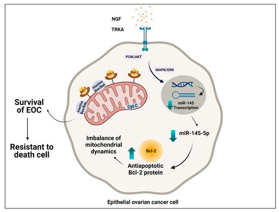
3.3. Interplay between NGF/TRKA and miR-145 Levels: Possible Implications for Mitochondria

Recent studies have established that miR-145 overexpression regulates mitochondrial metabolic reprogramming in ovarian cancer cells by decreasing ADP-ribosylation factor-like 5B (ARL5B), resulting in decreased mtDNA copy number, ATP levels, membrane potential, and mitochondrial markers, along with an increase in Cyt C, leading to the inhibition of mitochondrial function [63]. It has been reported that the Cyt C release is involved in apoptosis intrinsically in ovarian cancer cells [63][64].
Considering that NGF induces cellular proliferation and an anti-apoptotic condition in EOC cells [65], our research group demonstrated that NGF decreases the transcriptional activity and cellular levels of miR-145, which may increase mRNA and expression of Bcl-2, which leads to the inhibition of the intrinsic apoptotic pathway and the survival of the EOC cell (Figure 1). However, the implications that NGF would have on mitochondrial dynamics are not entirely clear. In HGSOC, the increase of DRP1, one protein involved in mitochondrial fission, as well as in cell survival and resistance to platinum compounds of OC cells, has been described [34]. Interestingly, Bcl-2 would be mediating fission mitochondrial and inducing apoptosis through DRP1 [66]. This finding is in agreement with recent studies of OC cells where the inhibition of DRP1 by mitochondrial division inhibitor 1 (Mdivi-1) inhibited mitochondrial fission and intrinsic apoptosis [67].
Figure 1. Possible interplay between mitochondria and regulation of NGF/TRKA and miR-145. Epithelial ovarian cancer cells are intrinsically resistant to cell death. Through its interaction with the TRKA receptor, NGF decreases transcription of miR-145 levels, causing an increase in oncogenic proteins involved in processes such as proliferation, migration, and angiogenesis. Additionally, NGF induces a decrease of transcriptional activity and cellular levels of miR-145, with an increase of the Bcl-2, expression of which leads to the inhibition of the intrinsic apoptotic pathway and the survival of the EOC cell.
However, this apparent contradiction seems to have an explanation in the tumor environment. In hypoxic OC cells, an increase in mitochondrial fission and also resistance to cisplatin therapy occur. Nevertheless, when DRP1 and mitochondrial fission are inhibited in hypoxic OC cells, sensitization to therapy occurs, which allows determining that mitochondrial dynamics also depend on the tumor microenvironment [68].

References

  1. Torre, L.A.; Trabert, B.; DeSantis, C.E.; Miller, K.D.; Samimi, G.; Runowicz, C.D.; Gaudet, M.M.; Jemal, A.; Siegel, R.L. Ovarian cancer statistics, 2018. CA Cancer J. Clin. 2018, 68, 284–296. [Google Scholar] [CrossRef]
  2. Lheureux, S.; Braunstein, M.; Oza, A.M. Epithelial ovarian cancer: Evolution of management in the era of precision medicine. CA Cancer J. Clin. 2019, 69, 280–304. [Google Scholar] [CrossRef]
  3. Arend, R.; Martinez, A.; Szul, T.; Birrer, M.J. Biomarkers in ovarian cancer: To be or not to be. Cancer 2019, 125, 4563–4572. [Google Scholar] [CrossRef] [PubMed]
  4. Reid, B.M.; Permuth, J.B.; Seller, T.A. Epidemiology of ovarian cancer: A review. Cancer Biol. Med. 2017, 14, 9–32. [Google Scholar] [CrossRef] [PubMed]
  5. Kuroki, L.; Guntupalli, S.R. Treatment of epithelial ovarian cancer. BMJ 2020, 371, m3773. [Google Scholar] [CrossRef]
  6. Kurnit, K.C.; Fleming, G.F.; Lengyel, E. Updates and new options in advanced epithelial ovarian cancer treatment. Obstet. Gynecol. 2021, 137, 108–121. [Google Scholar] [CrossRef]
  7. Lee, J.; Minasian, L.; Kohn, E.C. New strategies in ovarian cancer treatment. Cancer 2019, 125, 4623–4629. [Google Scholar] [CrossRef]
  8. Lengyel, E. Ovarian cancer development and metastasis. Am. J. Pathol. 2010, 177, 1053–1064. [Google Scholar] [CrossRef]
  9. Pistritto, G.; Trisciuoglio, D.; Ceci, C.; Garufi, A.; D’Orazi, G. Apoptosis as anticancer mechanism: Function and dysfunction of its modulators and targeted therapeutic strategies. Aging 2016, 8, 603–619. [Google Scholar] [CrossRef] [PubMed]
  10. Avolio, R.; Matassa, D.S.; Criscuolo, D.; Landriscina, M.; Esposito, F. Modulation of mitochondrial metabolic reprogramming and oxidative stress to overcome chemoresistance in cancer. Biomolecules 2020, 10, 135. [Google Scholar] [CrossRef]
  11. Pillai, S.R.; Damaghi, M.; Marunaka, Y.; Spugnini, E.P.; Fais, S.; Gillies, R.J. Causes, consequences, and therapy of tumors acidosis. Cancer Metastasis Rev. 2019, 38, 205–222. [Google Scholar] [CrossRef] [PubMed]
  12. Sies, H.; Berndt, C.; Jones, D.P. Oxidative stress. Ann. Rev. Biochem. 2017, 86, 715–748. [Google Scholar] [CrossRef] [PubMed]
  13. Pavlova, N.N.; Thompson, C.B. The emerging hallmarks of cancer metabolism. Cell Metab. 2016, 23, 27–47. [Google Scholar] [CrossRef]
  14. Skaper, S.D. The biology of neurotrophins, signalling pathways, and functional peptide mimetics of neurotrophins and their receptors. Drug Targets 2008, 7, 46–62.
  15. Dissen, G.A.; Romero, C.; Hirshfield, A.N.; Ojeda, S.R. Nerve growth factor is required for early follicular development in the mammalian ovary. Endocrinology 2001, 142, 9.
  16. Kawamura, K.; Kawamura, N.; Mulders, S.M.; Sollewijn Gelpke, M.D.; Hsueh, A.J.W. Ovarian brain-derived neurotrophic factor (BDNF) promotes the development of oocytes into preimplantation embryos. Proc. Natl. Acad. Sci. USA 2005, 102, 9206–9211.
  17. Anderson, R.A. Brainwork in the ovary: Kisspeptin and BDNF signaling converge to ensure oocyte survival. Endocrinology 2014, 155, 2751–2753.
  18. Streiter, S.; Fisch, B.; Sabbah, B.; Ao, A.; Abir, R. The importance of neuronal growth factors in the ovary. Mol. Hum. Reprod. 2016, 22, 3–17.
  19. Vera, C.; Tapia, V.; Vega, M.; Romero, C. Role of nerve growth factor and its TRKA receptor in normal ovarian and epithelial ovarian cancer angiogenesis. J. Ovarian Res. 2014, 7, 8.
  20. Tapia, V.; Gabler, F.; Muñoz, M.; Yazigi, R.; Paredes, A.; Selman, A.; Vega, M.; Romero, C. Tyrosine kinase A receptor (TrkA): A potential marker in epithelial ovarian cancer. Gynecol. Oncol. 2011, 121, 13–23.
  21. Julio-Pieper, M.; Lara, H.E.; Bravo, J.A.; Romero, C. Effects of nerve growth factor (NGF) on blood vessels area and expression of the angiogenic factors VEGF and TGFbeta I in the rat ovary. Reprod. Biol. Endocrinol. 2006, 4, 57.
  22. Campos, X.; Muñoz, Y.; Selman, A.; Yazigi, R.; Moyano, L.; Weinstein-Oppenheimer, C.; Lara, H.E.; Romero, C. Nerve growth factor and its high-affinity receptor TrkA participate in the control of vascular endothelial growth factor expression in epithelial ovarian cancer. Gynecol. Oncol. 2007, 104, 168–175.
  23. Garrido, M.P.; Torres, I.; Vega, M.; Romero, C. Angiogenesis in gynecological cancers: Role of neurotrophins. Front. Oncol. 2019, 9, 913.
  24. Garrido, M.P.; Hurtado, I.; Valenzuela-Valderrama, M.; Salvatierra, R.; Hernández, A.; Vega, M.; Selman, A.; Quest, A.F.G.; Romero, C. NGF-enhanced vasculogenic properties of epithelial ovarian cancer cells are reduced by inhibition of the COX-2/PGE2 signaling axis. Cancers 2019, 11, 1970.
  25. Reichard, A.; Asosingh, K. The role of mitochondria in angiogenesis. Mol. Biol. Rep. 2019, 46, 1393–1400.
  26. Lim, D.; Do, Y.; Kwon, B.S.; Chang, W.; Lee, M.-S.; Kim, J.; Cho, J.G. Angiogenesis and vasculogenic mimicry as therapeutic targets in ovarian cancer. BMB Rep. 2020, 53, 291–298.
  27. Urzúa, U.; Tapia, V.; Geraldo, M.; Selman, A.; Vega, M.; Romero, C. Nerve growth factor stimulates cellular proliferation of human epithelial ovarian cancer. Horm. Metab. Res. 2012, 44, 656–661.
  28. Xintaropoulou, C.; Ward, C.; Wise, A.; Queckborner, S.; Turnbull, A.; Michie, C.O.; Williams, A.R.W.; Rye, T.; Gourley, C.; Langdon, S.P. Expression of glycolytic enzymes in ovarian cancers and evaluation of the glycolytic pathway as a strategy for ovarian cancer treatment. BMC Cancer 2018, 18, 636.
  29. Martinou, I.; Desagher, S.; Eskes, R.; Antonsson, B.; André, E.; Fakan, S.; Martinou, J.-C. The release of cytochrome c from mitochondria during apoptosis of NGF-deprived sympathetic neurons is a reversible event. J. Cell Biol. 1999, 144, 883–889.
  30. Siu, M.K.; Wong, O.G.; Cheung, A.N. TrkB as a therapeutic target for ovarian cancer. Expert Opin. Ther. Targets 2009, 13, 1169–1178.
  31. Kang, M.; Chong, K.Y.; Hartwich, T.M.P.; Bi, F.; Witham, A.K.; Patrick, D.; Morrisson, M.J.; Cady, S.L.; Cerchia, A.P.; Kelk, D.; et al. Ovarian BDNF promotes survival, migration, and attachment of tumor precursors originated from P53 mutant fallopian tube epithelial cells. Oncogenesis 2020, 9, 55.
  32. Matsuda, S.; Fujita, T.; Kajiya, M.; Takeda, K.; Shiba, H.; Kawaguchi, H.; Kurihara, H. Brain-derived neurotrophic factor induces migration of endothelial cells through a TrkB-ERK-integrin AVβ3-FAK cascade. J. Cell. Physiol. 2012, 227, 2123–2129.
  33. Li, B.; Cai, S.; Zhao, Y.; He, Q.; Yu, X.; Cheng, L.; Zhang, Y.; Hu, X.; Ke, M.; Chen, S.; et al. Nerve growth factor modulates the tumor cells migration in ovarian cancer through the WNT/β-catenin pathway. Oncotarget 2016, 7, 81026–81048.
  34. Tsuyoshi, H.; Orisaka, M.; Fujita, Y.; Asare-Werehene, M.; Tsang, B.K.; Yoshida, Y. Prognostic impact of Dynamin related protein 1 (Drp1) in epithelial ovarian cancer. BMC Cancer 2020, 20, 467.
  35. Peng, Y.; Croce, C.M. The role of MicroRNAs in human cancer. Sig. Transduct. Target Ther. 2016, 1, 15004.
  36. Garrido, M.P.; Torres, I.; Avila, A.; Chnaiderman, J.; Valenzuela-Valderrama, M.; Aramburo, J.; Oróstica, L.; Durán-Jara, E.; Lobos-Gonzalez, L.; Romero, C. NGF/TRKA decrease MiR-145-5p levels in epithelial ovarian cancer cells. Int. J. Mol. Sci. 2020, 21, 7657.
  37. Deb, B.; Uddin, A.; Chakraborty, S. MiRNAs and ovarian cancer: An overview. J. Cell Physiol. 2018, 233, 3846–3854.
  38. Bartel, D.P. MicroRNAs: Genomics, biogenesis, mechanism, and function. Cell 2004, 116, 281–297.
  39. Ha, M.; Kim, V.N. Regulation of MicroRNA biogenesis. Nat. Rev. Mol. Cell Biol. 2014, 15, 509–524.
  40. Matsuyama, H.; Suzuki, H.I. Systems and synthetic MicroRNA biology: From biogenesis to disease pathogenesis. Int. J. Mol. Sci. 2020, 21, 132.
  41. Vera, C.; Retamales-Ortega, R.; Garrido, M.; Vega, M.; Romero, C. Signaling pathways related to nerve growth factor and MiRNAs in epithelial ovarian cancer. In Ovarian Cancer—From Pathogenesis to Treatment; Devaja, O., Papadopoulos, A., Eds.; IntechOpen: London, UK, 2018.
  42. Zuberi, M.; Mir, R.; Das, J.; Ahmad, I.; Javid, J.; Yadav, P.; Masroor, M.; Ahmad, S.; Ray, P.C.; Saxena, A. Expression of serum MiR-200a, MiR-200b, and MiR-200c as candidate biomarkers in epithelial ovarian cancer and their association with clinicopathological features. Clin. Transl. Oncol. 2015, 17, 779–787.
  43. Wang, A.; Jin, C.; Li, H.; Qin, Q.; Li, L. LncRNA ADAMTS9-AS2 regulates ovarian cancer progression by targeting MiR-182-5p/FOXF2 signaling pathway. Int. J. Biol. Macromol. 2018, 120, 1705–1713.
  44. Báez-Vega, P.M.; Vargas, I.M.E.; Valiyeva, F.; Encarnación-Rosado, J.; Roman, A.; Flores, J.; Marcos-Martínez, M.J.; Vivas-Mejía, P.E. Targeting MiR-21-3p inhibits proliferation and invasion of ovarian cancer cells. Oncotarget 2016, 7, 36321–36337.
  45. Kleemann, M.; Schneider, H.; Unger, K.; Bereuther, J.; Fischer, S.; Sander, P.; Marion Schneider, E.; Fischer-Posovszky, P.; Riedel, C.U.; Handrick, R.; et al. Induction of apoptosis in ovarian cancer cells by MiR-493-3p directly targeting AKT2, STK38L, HMGA2, ETS1 and E2F5. Cell. Mol. Life Sci. 2019, 76, 539–559.
  46. Wu, H.; Xiao, Z.; Wang, K.; Liu, W.; Hao, Q. MiR-145 is downregulated in human ovarian cancer and modulates cell growth and invasion by targeting P70S6K1 and MUC1. Biochem. Biophys. Res. Commun. 2013, 441, 693–700.
  47. Yan, J.; Jiang, J.; Meng, X.-N.; Xiu, Y.-L.; Zong, Z.-H. MiR-23b targets cyclin G1 and suppresses ovarian cancer tumorigenesis and progression. J. Exp. Clin. Cancer Res. 2016, 35, 31.
  48. Zhu, X.; Li, Y.; Xie, C.; Yin, X.; Liu, Y.; Cao, Y.; Fang, Y.; Lin, X.; Xu, Y.; Xu, W.; et al. MiR-145 sensitizes ovarian cancer cells to paclitaxel by targeting Sp1 and Cdk6: MiR-145 sensitizes cancer cells to paclitaxel. Int. J. Cancer 2014, 135, 1286–1296.
  49. Xu, W.; Hua, Y.; Deng, F.; Wang, D.; Wu, Y.; Zhang, W.; Tang, J. MiR-145 in cancer therapy resistance and sensitivity: A comprehensive review. Cancer Sci. 2020, 111, 3122–3131.
  50. Sheng, Q.; Zhang, Y.; Wang, Z.; Ding, J.; Song, Y.; Zhao, W. Cisplatin-mediated down-regulation of MiR-145 contributes to up-regulation of PD-L1 via the C-Myc transcription factor in cisplatin-resistant ovarian carcinoma cells. Clin. Exp. Immunol. 2020, 200, 45–52.
  51. Chen, Q.; Hou, J.; Wu, Z.; Zhao, J.; Ma, D. MiR-145 Regulates the sensitivity of esophageal squamous cell carcinoma cells to 5-FU via targeting REV3L. Pathol. Res. Pract. 2019, 215, 152427.
  52. Pan, Y.; Ye, C.; Tian, Q.; Yan, S.; Zeng, X.; Xiao, C.; Wang, L.; Wang, H. MiR-145 suppresses the proliferation, invasion and migration of NSCLC cells by regulating the BAX/BCL-2 ratio and the caspase-3 cascade. Oncol. Lett. 2018, 15, 4337–4343.
  53. Lim, H.Y.; Ho, Q.S.; Low, J.; Choolani, M.; Wong, K.P. Respiratory competent mitochondria in human ovarian and peritoneal cancer. Mitochondrion 2011, 11, 437–443.
  54. Zampieri, L.X.; Grasso, D.; Bouzin, C.; Brusa, D.; Rossignol, R.; Sonveaux, P. Mitochondria participate in chemoresistance to cisplatin in human ovarian cancer cells. Mol. Cancer Res. 2020, 18, 1379–1391.
  55. Signorile, A.; De Rasmo, D.; Cormio, A.; Musicco, C.; Rossi, R.; Fortarezza, F.; Palese, L.; Loizzi, V.; Resta, L.; Scillitani, G.; et al. Human ovarian cancer tissue exhibits increase of mitochondrial biogenesis and cristae remodeling. Cancers 2019, 11, 1350.
  56. Bindra, S.; McGill, M.A.; Triplett, M.K.; Tyagi, A.; Thaker, P.H.; Dahmoush, L.; Goodheart, M.J.; Ogden, R.T.; Owusu-Ansah, E.; Karan, K.; et al. Ovarian tumor mitochondria exhibit abnormal phenotypes and blunted associations with biobehavioral factors. Sci. Rep. 2021, 11, 11595.
  57. Emmings, E.; Mullany, S.; Chang, Z.; Landen, C.N.; Linder, S.; Bazzaro, M. Targeting mitochondria for treatment of chemoresistant ovarian cancer. Int. J. Mol. Sci. 2019, 20, 229.
  58. Grieco, J.P.; Allen, M.E.; Perry, J.B.; Wang, Y.; Song, Y.; Rohani, A.; Compton, S.L.E.; Smyth, J.W.; Swami, N.S.; Brown, D.A.; et al. Progression-mediated changes in mitochondrial morphology promotes adaptation to hypoxic peritoneal conditions in serous ovarian cancer. Front. Oncol. 2021, 10, 600113.
  59. Kleih, M.; Böpple, K.; Dong, M.; Gaißler, A.; Heine, S.; Olayioye, M.A.; Aulitzky, W.E.; Essmann, F. Direct impact of cisplatin on mitochondria induces ROS production that dictates cell fate of ovarian cancer cells. Cell Death Dis. 2019, 10, 851.
  60. Wang, D.; You, D.; Li, L. Galectin-3 regulates chemotherapy sensitivity in epithelial ovarian carcinoma via regulating mitochondrial function. J. Toxicol. Sci. 2019, 44, 47–56.
  61. Dar, S.; Chhina, J.; Mert, I.; Chitale, D.; Buekers, T.; Kaur, H.; Giri, S.; Munkarah, A.; Rattan, R. Bioenergetic adaptations in chemoresistant ovarian cancer cells. Sci. Rep. 2017, 7, 8760.
  62. Harper, A.K.; Fletcher, N.M.; Fan, R.; Morris, R.T.; Saed, G.M. Heat shock protein 60 (HSP60) serves as a potential target for the sensitization of chemoresistant ovarian cancer cells. Reprod. Sci. 2020, 27, 1030–1036.
  63. Zhao, S.; Zhang, Y.; Pei, M.; Wu, L.; Li, J. MiR-145 inhibits mitochondrial function of ovarian cancer by targeting ARL5B. J. Ovarian Res. 2021, 14, 8.
  64. Vaux, D.L. Apoptogenic factors released from mitochondria. Biochim. Biophys. Acta 2011, 1813, 546–550.
  65. Jin, Z.; Gu, J.; Xin, X.; Li, Y.; Wang, H. Expression of hexokinase 2 in epithelial ovarian tumors and its clinical significance in serous ovarian cancer. Eur. J. Gynaecol. Oncol. 2014, 35, 519–524.
  66. Gross, A.; Katz, S.G. Non-apoptotic functions of BCL-2 family proteins. Cell Death Differ. 2017, 24, 1348–1358.
  67. Yu, Y.; Xu, L.; Qi, L.; Wang, C.; Xu, N.; Liu, S.; Li, S.; Tian, H.; Liu, W.; Xu, Y.; et al. ABT737 induces mitochondrial pathway apoptosis and mitophagy by regulating DRP1-dependent mitochondrial fission in human ovarian cancer cells. Biomed. Pharmacother. 2017, 96, 22–29.
  68. Han, Y.; Kim, B.; Cho, U.; Park, I.S.; Kim, S.I.; Dhanasekaran, D.N.; Tsang, B.K.; Song, Y.S. Mitochondrial fission causes cisplatin resistance under hypoxic conditions via ROS in ovarian cancer cells. Oncogene 2019, 38, 7089–7105.
More
Upload a video for this entry
Information
Contributor MDPI registered users' name will be linked to their SciProfiles pages. To register with us, please refer to https://encyclopedia.pub/register : Daniela Vera
View Times: 642
Revisions: 3 times (View History)
Update Date: 29 Mar 2022
Notice
You are not a member of the advisory board for this topic. If you want to update advisory board member profile, please contact office@encyclopedia.pub.
OK
Confirm
Only members of the Encyclopedia advisory board for this topic are allowed to note entries. Would you like to become an advisory board member of the Encyclopedia?
Yes
No
${ textCharacter }/${ maxCharacter }
Submit
Cancel
There is no comment~
${ textCharacter }/${ maxCharacter }
Submit
Cancel
${ selectedItem.replyTextCharacter }/${ selectedItem.replyMaxCharacter }
Submit
Cancel
Confirm
Are you sure to Delete?
Yes No
Academic Video Service