Submitted Successfully!
To reward your contribution, here is a gift for you: A free trial for our video production service.
Thank you for your contribution! You can also upload a video entry or images related to this topic.
Version Summary Created by Modification Content Size Created at Operation
1 + 2635 word(s) 2635 2022-01-04 09:17:40 |
2 format correct Meta information modification 2635 2022-01-17 02:13:22 |

Video Upload Options

Do you have a full video?

Confirm

Are you sure to Delete?
Cite
If you have any further questions, please contact Encyclopedia Editorial Office.
Dyakin, V. Non-Equilibrium Thermodynamics and Psychology in Biological Aging. Encyclopedia. Available online: https://encyclopedia.pub/entry/18279 (accessed on 23 July 2024).
Dyakin V. Non-Equilibrium Thermodynamics and Psychology in Biological Aging. Encyclopedia. Available at: https://encyclopedia.pub/entry/18279. Accessed July 23, 2024.
Dyakin, Victor. "Non-Equilibrium Thermodynamics and Psychology in Biological Aging" Encyclopedia, https://encyclopedia.pub/entry/18279 (accessed July 23, 2024).
Dyakin, V. (2022, January 14). Non-Equilibrium Thermodynamics and Psychology in Biological Aging. In Encyclopedia. https://encyclopedia.pub/entry/18279
Dyakin, Victor. "Non-Equilibrium Thermodynamics and Psychology in Biological Aging." Encyclopedia. Web. 14 January, 2022.
Non-Equilibrium Thermodynamics and Psychology in Biological Aging
Edit

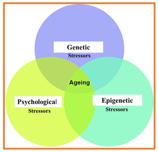
In humans, biological aging (age-associated degrading changes), widely observed in molecular and cellular processes, underly the time-dependent decline in spatial navigation, time perception, cognitive and psychological abilities, and memory. Cross-talk of biological, cognitive, and psychological clocks provides an integrative contribution to healthy and advanced aging. At the molecular level, genome, proteome, and lipidome instability are widely recognized as the primary causal factors in aging.

Through stress response systems (SRS), the environmental and psychological stressors contribute to the age-associated “collapse” of protein homochirality. The role of prevalent protein chirality and entropy of protein folding in biological aging is mainly overlooked. In a more generalized context, the time-dependent shift from enzymatic to the nonenzymatic transformation of biochirality might represent an important and yet underappreciated hallmark of aging. We provide the experimental arguments in support of the racemization theory of aging.

spontaneous non-enzymatic post translational modifications racemization biological clock racemization theory of aging psychological aging psychological stress stress tress response system

1. Introduction

The roots of stress research lie in the belief that stress can accelerate biological aging
[1]
Molecular psychology and psychiatry represent an emerging modern research trend. However, the role of prevalent bio-molecular chirality and molecular aging in the function of the stress response system (SRS) is practically neglected. Indeed, the consequences of psychological stresses linked to biological aging are evident at the organism, system, cellular, and molecular levels [2][3]. Under stress conditions, self-perception is contributed to by the sense of embodiment [4] and by conscious control of your own thoughts [5].
No doubt, the age-dependent biological processes are influenced by the subject’s state of mind and psychological state [6][7]. The functioning of an organism at the molecular and cellular levels underlies humans’ perception of environmental challenges, self-perception, and age-associated modification of physiological and cognitive functions. No doubt that all hierarchical domains of biological events—including molecular–molecular, molecular–cellular, cellular–organismic, organismic–cognitive/behavioral—exhibit bi-directional impact [6][8]. The understanding of such bidirectionality constitutes the solid ground for pharmacological and psychological treatment. From the thermodynamic perspective, the organism, as a whole, as well as its major constituents (biomacromolecules, cellular organelles, cells, tissues, and organs) maintains themselves in the entropy-driven non-equilibrium state [9][10][11].
We will focus on the thermodynamics of protein folding in the age-dependent association with the function of the stress response system (SRS), and provide the experimental arguments in support of the racemization theory of aging.

2. Aging, Entropy, and Aging Defense System

It is by avoiding the rapid decay into the inert state of ‘equilibrium’ that an organism appears so enigmatic
[9]
Conceptualization of the underlying mechanisms of aging is in high demand due to many age-related diseases. Significant progress is made from the view that the variable transient fluctuating entropy (from the Greek entropia = transformation) in the non-equilibrium (NE) state of a biomolecular ensemble is under permanent impact by the thermodynamic tendency toward the high-entropy state of equilibrium. NE thermodynamic of living systems is closely associated with the concepts of biochirality, entropy, biological information processing, and aging [12]. NE thermodynamic theories, in particular, Classical Irreversible Thermodynamics (CIT) [13], complemented by the concept of fluctuating entropy [14][15], provide a valuable formalism for understanding the dynamics of living systems, including the origin of life, cell differentiation, as well as the synthesis of the homochiral population of proteins and the spontaneous loss of conformational entropy during folding [16][17][18].
Energy-consuming ribosomal protein synthesis (the human body requires production of 1021 ATP molecules per second) continuously creates the pool of homochiral; high entropy unfolded biomolecules in the thermodynamically NE state. Prevalent chirality of nascent proteins is transferred to all levels of their structural hierarchy during spontaneous condensation (folding) to native (i.e., functional) state (NS) [19][20]. The relaxation, conformation, and formation of protein assemblies constitute large many-body systems that operate in a fluctuating, out-of-equilibrium environment.
Changes in the residual conformational entropy at the protein level are significant components of the thermodynamics of folding, binding, enzyme-substrate recognition, and time-dependent protein dysfunctions [21][22]. Spontaneous protein folding (3-D transformation) to NS is accompanied by free energy loss and reduction of configurational entropy.
NS of homochiral proteins (trapped in a local energy minimum) is thermodynamically only relatively stable and, therefore, is prone to further spontaneous relaxation to aberrant conformation and aggregative states. Such spontaneous processes can be triggered by amino acid (AAs) racemization [23][24]. Spontaneous racemization is known as one of the unavoidable degrading forces of the functional state of proteins. More (than native) thermodynamically stable states of proteins such as amyloids, fibrils, and aggregates are associated with protein aging translated to the cellular and organism levels. However, even recent advances in the understanding of protein folding and miss-folding ignore the homochirality of the NS and, correspondently, overlook the contribution of spontaneous racemization to protein aging [25] (Figure 1). Furthermore, protein aggregation is known to slow down the rates of structural conversions (leading to kinetic trapping), extend the cellular lifetime of trapped protein, and make them prone to the impact of spontaneous (molecular environment-specific) racemization.
Figure 1. Funnel-shaped potential free-energy landscape. Impact of spontaneous racemization. Adopted with alterations from [25].
In terms of quantitative thermodynamic variables, spontaneous protein fold from the initial chain of AAs to the NS is accompanied by the loss of Gibbs energy (ΔG) and minimizing the entropy (ΔS Fold). From the thermodynamic perspective (1), some other processes should compensate for the loss of Δ S Fold.
ΔG = ΔH − T × ΔS
and
S = k × ln(W)
where k is Boltzmann’s constant and W is the number of microstates that give rise to the macro-state of interest.
One of them is the gain of enthalpy under forces of the binding (condensation). Two others are the increase in the entropy of solvent and increment in the entropy of protein due to loss of protein homochirality (racemization) [9]. Therefore, aging can be considered as an exchange between the accumulation of damage and compensatory mechanisms. The complexity of compensatory events comprises the aging defense mechanism, which, unfortunately, also is vulnerable to the impact of spontaneous biological reactions. Age-associated degrading changes are observed in psychological and neurobiological processes underlying the decline in basic functions of the organism, including spatial navigation [26][27] and time processing [28], contributing to the perception of the virtual reality environment [29]. Evolutionary biologists conclude that the force of selection declines as a function of age due to the decreased likelihood of reproduction. The fact that the force of selection declines as a function of age promotes the appearance of two main hypotheses formulated to explain why organisms age: the mutation accumulation (MA) and the antagonistic pleiotropy (AP) hypotheses [30][31][32][33]. MA theory proposed by Peter Medawar in 1952 [34] suggests that mutations with deleterious late-life effects can accumulate if such products are confined to late life, when selection against them is weak. Williams AP theory proposed by George C. Williams in 1957 [35] is based on the fitness trade-offs and assumes the ability of a gene to control more than one trait, with part of these traits being beneficial to the fitness of the organism early on in life, whereas other traits are detrimental to the fitness later on.
The racemization hypothesis of aging (RHA) helps integrate the evolutionary and molecular genetics of aging. Experimental evidence suggests that both aging and psychological stress affect the immune, hormonal, and neuro-transmitting systems [36][37][38][39]. Stress is recognized as a fundamental physiological phenomenon essential to survival and related to several psychiatric [35] and neurodegenerative [40] disorders. A common view that the response to stressful stimuli is triggered and exacerbated by the SRS that integrates a wide diversity of brain structures, neuronal circuits, as well as perceptual and cognitive functions. In other words, the mechanisms of stress response integrate brain and body activity at the molecular, cellular, and neuronal network levels. At the molecular level, the functions and age-related decline of the STS are linked to the maintenance or deterioration of the prevalent chirality of proteins. Protein aging is a trade-off between the maintenance of prevalent chirality and spontaneous racemization. From this integrative view, it is evident that the STS mediates the physiological and psychological outcomes of aging individuals.

3. Psychological and Physical Stressor

The discrimination of psychological and physical stressors is essential for understanding the complex mechanism of the human SRS (Figure 2) [41][42][43]. Psychological stressors are defined as stimuli that threaten the current state and are perceived in an anticipatory condition, e.g., aversive environmental stimuli, predator-related cues, and failure to satisfy internal drives. Adult diseases [44] and accelerated aging [45] are often associated with acute stressors and the developmental, biological, and psychological abnormalities occurring in childhood. Epigenetic mechanisms of physical and psychological stressors make them become biologically embedded into physiology (at molecular, cellular, systemic, and organism levels) [46][47][48]. The epigenetic mechanisms of physical and psychological stressors relate how environmental factors are translated to physiological events through sensory perception, cognitive, and emotional abilities. At the protein level, the stress response occurs through the system of enzymatic and non-enzymatic (spontaneous) PTMs (PTMs SP). Spontaneous and stress-induced aging of proteins is a complex process of nonenzymatic chemical reactions which contribute to the range of metabolic diseases [49][50] Most studied age-associated non-enzymatic PTMs contributing to protein aging include oxidation, nitration, glycation, and racemization. We will be primarily focused on irreversible spontaneous racemization [51][52].
Figure 2. Major determinant of biological aging.

4. Biochirality, Spontaneous Reactions and Aging

Chirality is a fundamental, persistent, but often overlooked feature of all living organisms on the molecular level as well as on the macroscopic scale.”
[53] (See also [54][55])
At the molecular level, aging is associated with three primary classes of bio-molecules prone to spontaneous modifications: (1) nucleotides (NTs) within DNA sequence, (2) amino acids (AAs) within peptides and proteins and (3) lipid content of the membrane bilayers. In these cases, the irreversibility of reactions results in the time-dependent accumulation of abnormal molecular complexes. Both NT and AA molecular pathways of aging are closely interconnected [56] and obey the laws of non-equilibrium thermodynamics. The influx of energy maintains the system of molecular objects in an asymmetric non-equilibrium dynamic steady state, which can retain their asymmetry (homochirality) for a time longer than the time of spontaneous racemization [57]. Meaning that any compromise of energy supply will open a window to spontaneous racemization. Spontaneous DNA mutations arise from a variety of sources, including irreversible changes of NTs sequence, leading to the errors in DNA replication.
Spontaneous alterations in DNA structure result in chaining the functions implicated in protein synthesis, cell proliferation, cell polarity and organism morphology (including bilaterality). PTMs Sp of proteins, the second causal agent of organism aging, will be the focus of our attention here. Spontaneous reactions such as isomerization, epimerization, and racemization taking place in long-lived protein (LLPs), have two physiologically essential pathogenic consequences: prevention of proteolytic degradation [58] and non-enzymatic cleavage [59]. The evolution of living organisms has established mechanisms to exclude D-amino acids (D-AAs) in their protein synthesis machinery to maintain the prevalent molecular chirality and stereo-specific catalysis (three major sources of D-AAs in mammals are intrinsic racemization (enzymatic and spontaneous), diet, and gut microbiota). However, recent development [60][61][62] shows that this mechanism has not absolute prevalence. For example, a small probability exists for ribosomal incorporation of D-AAs into AAs sequence of peptides and protein remains. In addition to the ribosomal source, D-AAs are created by the mechanism of enzymatic and spontaneous post-translational modifications (PTMs). Small amount D-AAs in an organism signify the evolutionary selected delicate balance between two isomers, suggesting that any disturbance in this balance can be harmful. It is notable that D-AA-containing peptides and proteins demonstrate two distinct features: (1) resistance toward proteases and association with prolonged half-lives (2) [60][62]. Biochirality, evident in the pathways of enzymatic and spontaneous racemization of AAs and PTMs of proteins, is the emerging as a critical and yet experimentally and theoretically challenging topic. The attention to spontaneous modifications of free and peptide/protein-incorporated AAs becomes essential due to their role.
In protein aging, cell physiology, and neuropathology. Spontaneous chemical reactions, such as racemization, occur by various pathways, including oxidation, cyclization, and elimination reactions. Spontaneous racemization is a critical determinant of thermodynamically irrevocable (i.e., aberrant) protein folding, responsible for the side products of biochemical reactions. Many studies are devoted to the consideration of transitions from tetrahedral to planar electron configurations. Planar intermediates are critical determinants of the racemization barrier and rate of racemization [63]. Free AAs and peptides/protein-incorporated AAs are characterized by significantly different rates of spontaneous reactions. Due to the complexity of three-dimensional structure of globular proteins and the presence of many functional groups, susceptibility to spontaneous or enzyme-dependent stereo-chemical modifications is much higher for peptides and proteins than for free AAs [63]. Therefore, future research in this field will enormously contribute to understanding the molecular mechanism of protein misfolding, aggregation, disfunction, degradation, and aging.
The chain of chirality transmissions across the length scales and level of organization is recognizes as a fundamental feature of all living creatures. The prevalent molecular handedness guides the chirality of the cells, while cell chirality drives the left/right asymmetric development of individual organs and organisms [49][51][55]. Contemporary concepts of prevalent biochirality and virtual reality brings the new dimensions to the exploring mutual influence of biological and cognitive domains of self [4][5][64][65][66][67] and new meaning to Schopenhauer’s view on the world. As the manifestation of the “Will and Representations”. Our review aims to attract attention toward the multidisciplinary field of biological chirality (or biochirality) and its broad-reaching implications at both the molecular and organism levels. As a branch of natural science, the concept of biochirality has broad implications in multiple fields ranging from DNA function to protein synthesis, neurotransmission, and bilaterality of cognitive functions. The rationale of such an attempt is that integration and synthesis of diverse, dispersed, and unsystematic experimental facts are required for better understanding of the fundamental nature of complex phenomena. Congruent advances in biochemical and biophysical studies provide a common framework for establishing a new level of academic research, medical treatments, and drug design.
To advance the field, it is necessary to consider the chronology of the ideas reflecting the progress in understanding the biochirality-related causality of aging phenomena. It is clear, that there are endogenous and exogenous stressors (agents) continuously challenging the integrity of DNA, transcriptome, and proteome, resulting in progressive aging of the organism, but the main causal triggers, at the molecular level, remains unknown. To better understand the impact of biochirality on the field of aging research, we will briefly review here the chronology of major developments regarding the biochirality-related causality of aging.
In 1978, Poplin and DeLong proposed the hypothesis that aging can be accelerated due to the enzymatic racemization [68]. In 1994, Fujii and colleagues concluded that “racemization, isomerization, and oxidation of αB-crystallin occur spontaneously in the aging process” [69], and Mori pronounced the biological significance of racemization in the neuropathogenesis of Alzheimer’s disease (AD) [70]. In 1995, John discussed the effect of aging on the turnover of muscle proteins [71]. The crucial role of non-enzymatic PTMs of proteins in aging was recognized in the previous century [72]. Later Maddox, in his “The Encyclopedia of Aging”, noticed that the “extend to which racemization contribute to the harmful consequence of aging remains uncertain” [73]. This conclusion indicates that the impact of racemization as a causal in aging was at that time, not yet clearly understood.
In 2002, Ritz-Timme & Collins linked the “natural” aging of proteins with the autonomic racemization of long-lived proteins (LLPs) [74]. In 2014, Inoue concluded that the most convenient biomarkers of protein aging were the spontaneous racemization of AAs [75]. Currently, many studies emphasize the existence of the abnormal age-related translational and post-translational protein homeostasis associated with spontaneous forms of PTMs (such as oxidation and oxidative phosphorylation) [75]. It is also recognized that progressive deterioration in the ability of the cells to preserve the stability of their proteome occurs with age, even in the absence of disease, and it likely contributes to different aspects of “natural” aging. However, the principal role of spontaneous racemization in geriatric science remains overlooked. Based on the above evidence, it is reasonable to conclude that spontaneous racemization (as a specific form of PTMs) may be a valuable molecular biomarker of age-associated neurodegenerative and psychiatric disorders.

References

  1. Entringer, S.; Epel, E.S. The stress field ages: A close look into cellular aging processes. Psychoneuroendocrinology 2020, 113, 104537.
  2. Moreno-Villanueva, M.; Bürkle, A. Molecular consequences of psychological stress in human aging. Exp. Gerontol. 2015, 68, 39–42.
  3. Lavretsky, H.; Newhouse, P.A. Stress, Inflammation, and Aging. Am. J. Geriatr. Psychiatry 2012, 20, 729–733.
  4. Guillot, M. Thinking of oneself as the thinker: The concept of self and the phenomenology of intellection. Conscious Thinking and Cognitive Phenomenology. Philos. Explor. 2016, 19, 138–160.
  5. Maier, S.F.; Watkins, L.R. Cytokines for psychologists: Implications of bidirectional immune-to-brain communication for understanding behavior, mood, and cognition. Psychol. Rev. 1998, 105, 83–107.
  6. Brugada, V.; De Polavieja, G.G.; Román, Á.-C. Toward a Molecular Profile of Self-Representation. Front. Hum. Neurosci. 2016, 10, 602.
  7. Koseska, A.; Bastiaens, P.I. Cell signaling as a cognitive process. EMBO J. 2017, 36, 568–582.
  8. Schrödinger, E. What is Life—The Physical Aspect of the Living Cell; Cambridge University Press: Cambridge, UK, 1944; ISBN 978-0-521-42708-1.
  9. Prigogine, I.; Nicolis, G. On Symmetry-Breaking Instabilities in Dissipative Systems. J. Chem. Phys. 1967, 46, 3542–3550.
  10. Teschendorff, A.E.; Sollich, P.; Kuehn, R. Signalling entropy: A novel network-theoretical framework for systems analysis and interpretation of functional omic data. Methods 2014, 67, 282–293.
  11. Gnesotto, F.; Mura, F.; Gladrow, J.; Broedersz, C. Broken detailed balance and non-equilibrium dynamics in living systems: A review. Rep. Prog. Phys. 2018, 81, 066601.
  12. Michaelian, K. The Dissipative Photochemical Origin of Life: UVC Abiogenesis of Adenine. Entropy 2021, 23, 217.
  13. Galzitskaya, O.V. Influence of Conformational Entropy on the Protein Folding Rate. Entropy 2010, 12, 961–982.
  14. Marques, B.S.; Stetz, M.A.; Jorge, C.; Valentine, K.G.; Wand, A.J.; Nucci, N.V. Protein conformational entropy is not slaved to water. Sci. Rep. 2020, 10, 1–8.
  15. Popov, E.M. Protein folding as a nonlinear nonequilibrium thermodynamic process. Biochem. Mol. Boil. Int. 1999, 47, 443–453.
  16. Pickett, S.; Sternberg, M. Empirical Scale of Side-Chain Conformational Entropy in Protein Folding. J. Mol. Biol. 1993, 231, 825–839.
  17. Rajitha Rajeshwar, T.; Saharay, M.; Smith, J.C.; Krishnan, M. Correlated Response of Protein Side-Chain Fluctuations and Conformational Entropy to Ligand Binding. J. Phys. Chem. B 2021, 125, 9641–9651.
  18. Banik, S.D.; Nandi, N. Chirality and Protein Biosynthesis. Top. Curr. Chem. 2012, 333, 255–305.
  19. Baxa, M.C.; Haddadian, E.J.; Jumper, J.M.; Freed, K.F.; Sosnick, T.R. Loss of conformational entropy in protein folding calculated using realistic ensembles and its implications for NMR-based calculations. Proc. Natl. Acad. Sci. USA 2014, 111, 15396–15401.
  20. Trbovic, N.; Cho, J.-H.; Abel, R.; Friesner, R.A.; Rance, M.; Palmer, I.A.G. Protein Side-Chain Dynamics and Residual Conformational Entropy. J. Am. Chem. Soc. 2009, 131, 615–622.
  21. Faraggi, E.; Dunker, A.K.; Jernigan, R.L.; Kloczkowski, A. Entropy, Fluctuations, and Disordered Proteins. Entropy 2019, 21, 764.
  22. Bada, J.L. Amino acid racemization dating of fossil bones. Annu. Rev. Earth Planet. Sci. 1985, 13, 241–268.
  23. Dyakin, V.V.; Lajtha, A.; Dyakina-Fagnano, N.V. Racemization hypothesis of neurodegeneration (RHND). Alzheimer’s Dement. 2020, 16, e047697.
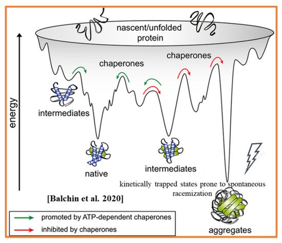
  24. Balchin, D.; Hayer-Hartl, M.; Hartl, F.U. Recent advances in understanding catalysis of protein folding by molecular chaperones. FEBS Lett. 2020, 594, 2770–2781.
  25. Lester, A.W.; Moffat, S.D.; Wiener, J.; Barnes, C.A.; Wolbers, T. The Aging Navigational System. Neuron 2017, 95, 1019–1035.
  26. Yu, S.; Boone, A.P.; He, C.; Davis, R.C.; Hegarty, M.; Chrastil, E.R.; Jacobs, E.G. Age-Related Changes in Spatial Navigation Are Evident by Midlife and Differ by Sex. Psychol. Sci. 2021, 32, 692–704.
  27. Eturgeon, M.; Elustig, C.; Meck, W.H. Cognitive Aging and Time Perception: Roles of Bayesian Optimization and Degeneracy. Front. Aging Neurosci. 2016, 8, 102.
  28. Adamo, D.E.; Briceño, E.M.; Sindone, J.A.; Alexander, N.B.; Moffat, S.D. Age differences in virtual environment and real world path integration. Front. Aging Neurosci. 2012, 4, 26.
  29. Flatt, T.; Schmidt, P.S. Integrating evolutionary and molecular genetics of aging. Biochim. Biophys. Acta (BBA) Gen. Subj. 2009, 1790, 951–962.
  30. Flatt, T.; Partridge, L. Horizons in the evolution of aging. BMC Biol. 2018, 16, 1–13.
  31. Johnson, A.A.; Shokhirev, M.N.; Shoshitaishvili, B. Revamping the evolutionary theories of aging. Ageing Res. Rev. 2019, 55, 100947.
  32. Turan, Z.G.; Parvizi, P.; Dönertaş, H.M.; Tung, J.; Khaitovich, P.; Somel, M. Molecular footprint of Medawar’s mutation accumulation process in mammalian aging. Aging Cell 2019, 18, e12965.
  33. Medawar, P. An Unsolved Problem of Biology; H.K Lewis: London, UK, 1952.
  34. Williams, G.C. Pleiotropy, natural selection, and the evolution of senescence. Sci. Aging Knowl. Environ. 1957, 11, 398–411.
  35. Graham, J.E.; Christian, L.; Kiecolt-Glaser, J.K. Stress, Age, and Immune Function: Toward a Lifespan Approach. J. Behav. Med. 2006, 29, 389–400.
  36. Uhart, M.; Oswald, L.; McCaul, M.E.; Chong, R.; Wand, G.S. Hormonal Responses to Psychological Stress and Family History of Alcoholism. Neuropsychopharmacology 2006, 31, 2255–2263.
  37. Clark, B.L.; Thomas, P.G. A Cell for the Ages: Human γδ T Cells across the Lifespan. Int. J. Mol. Sci. 2020, 21, 8903.
  38. Riboni, F.V.; Belzung, C. Stress and psychiatric disorders: From categorical to dimensional approaches. Curr. Opin. Behav. Sci. 2017, 14, 72–77.
  39. Esch, T.; Stefano, G.B.; Fricchione, G.L.; Benson, H. The role of stress in neurodegenerative diseases and mental disorders. Neuro Endocrinol. Lett. 2002, 23, 199–208.
  40. Dayas, C.V.; Buller, K.M.; Crane, J.; Xu, Y.; Day, T. Stressor categorization: Acute physical and psychological stressors elicit distinctive recruitment patterns in the amygdala and in medullary noradrenergic cell groups. Eur. J. Neurosci. 2001, 14, 1143–1152.
  41. Carter, J.R.; Goldstein, D.S. Sympathoneural and Adrenomedullary Responses to Mental Stress. Compr. Physiol. 2014, 5, 119–146.
  42. Godoy, L.D.; Rossignoli, M.T.; Delfino-Pereira, P.; Garcia-Cairasco, N.; de Lima Umeoka, E.H. A Comprehensive Overview on Stress Neurobiology: Basic Concepts and Clinical Implications. Front. Behav. Neurosci. 2018, 12, 127.
  43. Shonkoff, J.P.; Boyce, W.T.; McEwen, B.S. Neuroscience, Molecular Biology, and the Childhood Roots of Health Disparities: Building a new framework for health promotion and disease prevention. JAMA 2009, 301, 2252–2259.
  44. Miller, G.E.; Chen, E.; Parker, K.J. Psychological stress in childhood and susceptibility to the chronic diseases of aging: Moving toward a model of behavioral and biological mechanisms. Psychol. Bull. 2011, 137, 959–997.
  45. Danese, A.; Caspi, A.; Williams, B.; Ambler, A.; Sugden, K.; Mika, J.; Werts, H.; Freeman, J.; Pariante, C.M.; Moffitt, T.; et al. Biological embedding of stress through inflammation processes in childhood. Mol. Psychiatry 2010, 16, 244–246.
  46. Mifsud, K.R.; Gutièrrez-Mecinas, M.; Trollope, A.F.; Collins, A.; Saunderson, E.A.; Reul, J.M. Epigenetic mechanisms in stress and adaptation. Brain Behav. Immun. 2011, 25, 1305–1315.
  47. Eachus, H.; Cunliffe, V. Biological Embedding of Psychosocial Stress over the Life Course. Epigenetics Aging Longev. 2018, 14, 251–270.
  48. Santos, A.C.L.; Muniz, C.R.; Oliveira, L.T.; Souza, J.T. Contributions of a modified electrodynamics to the molecular biochirality. Chirality 2020, 32, 1186–1190.
  49. Manhas, R.; Rath, P.C. Ribosome, protein synthesis, and aging. In Models, Molecules and Mechanisms in Biogerontology; Springer: Singapore, 2020; pp. 67–87.
  50. McCudden, C.R.; Kraus, V.B. Biochemistry of amino acid racemization and clinical application to musculoskeletal disease. Clin. Biochem. 2006, 39, 1112–1130.
  51. Morrow, S.M.; Bissette, A.; Fletcher, S.P. Transmission of chirality through space and across length scales. Nat. Nanotechnol. 2017, 12, 410–419.
  52. Pályi, G.; Zucchi, C.; Caglioti, L. Advances in BioChirality; Elsevier: Amsterdam, The Netherlands, 1999.
  53. Inaki, M.; Liu, J.; Matsuno, K. Cell chirality: Its origin and roles in left–right asymmetric development. Philos. Trans. R. Soc. B Biol. Sci. 2016, 371, 20150403.
  54. Dyakin, V.V.; Lucas, J.; Dyakina-Fagnano, N.V.; Posner, E.V. The Chain of Chirality Transfer as Determinant of Brain Functional Laterality. Breaking the Chirality Silence: Search for New Generation of Biomarkers; Relevance to Neurodegenerative Diseases, Cognitive Psychology, and Nutrition Science. Neurol. Neurosci. Res. 2017, 1, 2.
  55. Liu, B.; Pappas, C.; Ottelé, J.; Schaeffer, G.; Jurissek, C.; Pieters, P.F.; Altay, M.; Marić, I.; Stuart, M.C.A.; Otto, S. Spontaneous Emergence of Self-Replicating Molecules Containing Nucleobases and Amino Acids. J. Am. Chem. Soc. 2020, 142, 4184–4192.
  56. Plasson, R.; Brandenburg, A. Homochirality and the Need for Energy. Orig. Life Evol. Biosph. 2009, 40, 93–110.
  57. Lambeth, T.R.; Riggs, D.L.; Talbert, L.E.; Tang, J.; Coburn, E.; Kang, A.S.; Noll, J.; Augello, C.; Ford, B.D.; Julian, R.R. Spontaneous Isomerization of Long-Lived Proteins Provides a Molecular Mechanism for the Lysosomal Failure Observed in Alzheimer’s Disease. ACS Central Sci. 2019, 5, 1387–1395.
  58. Hinterholzer, A.; Stanojlovic, V.; Regl, C.; Huber, C.G.; Cabrele, C.; Schubert, M. Detecting aspartate isomerization and backbone cleavage after aspartate in intact proteins by NMR spectroscopy. J. Biomol. NMR 2021, 75, 71–82.
  59. Lehmann, J.; Ye, S. D Amino Acids Highlight the Catalytic Power of the Ribosome. Cell Chem. Biol. 2019, 26, 1639–1641.
  60. Englander, M.T.; Avins, J.L.; Fleisher, R.; Liu, B.; Effraim, P.R.; Wang, J.; Schulten, K.; Leyh, T.S.; Gonzalez, R.L.; Cornish, V.W. The ribosome can discriminate the chirality of amino acids within its peptidyl-transferase center. Proc. Natl. Acad. Sci. USA 2015, 112, 6038–6043.
  61. Huang, P.-Y.; Wang, F.; Narasimhan, K.; Chatman, K.; Aach, J.; Trauger, S.A.; Spoering, R.; Church, G.M. Toward D-Peptide Biosyn-thesis: Elongation Factor P Enables Ribosomal Incorporation f Consecutive D-Amino Acids. bioRxiv 2017, 125930.
  62. Raghunathan, S.; Yadav, K.; Rojisha, V.C.; Jaganade, T.; Prathyusha, V.; Bikkina, S.; Lourderaj, U.; Priyakum, U.D. Transition between - and -stereoisomers without bond breakng. Phys. Chem. Chem. Phys. 2020, 22, 14983–14991.
  63. Grassi, L.; Cabrele, C. Susceptibility of protein therapeutics to spontaneous chemical modifications by oxidation, cyclization, and elimination reactions. Amino Acids 2019, 51, 1409–1431.
  64. Schneiderman, N.; Ironson, G.; Siegel, S.D. Stress and Health: Psychological, Behavioral, and Biological Determinants. Annu. Rev. Clin. Psychol. 2005, 1, 607–628.
  65. Dziechciaż, M.; Filip, R. Biological psychological and social determinants of old age: Bio-psycho-social aspects of human aging. Ann. Agric. Environ. Med. 2014, 21, 835–838.
  66. Kohda, M.; Hotta, T.; Takeyama, T.; Awata, S.; Tanaka, H.; Asai, J.-Y.; Jordan, A.L. If a fish can pass the mark test, what are the implications for consciousness and self-awareness testing in animals? PLoS Biol. 2019, 17, e3000021.
  67. Poplin, L.; Delong, R. Accelerated Aging due to Enzymatic Racemization. Gerontology 1978, 24, 365–368.
  68. Fujii, N.; Ishibashi, Y.; Satoh, K.; Fujino, M.; Harada, K. Simultaneous racemization and isomerization at specific aspartic acid residues in αB-crystallin from the aged human lens. Biochim. Biophys. Acta (BBA) Protein Struct. Mol. Enzym. 1994, 1204, 157–163.
  69. Mori, H.; Ishii, K.; Tomiyama, T.; Furiya, Y.; Sahara, N.; Asano, S.; Endo, N.; Shirasawa, T.; Takio, K. Racemization: Its Biological Significance on Neuropathogenesis of Alzheimer’s Disease. Tohoku J. Exp. Med. 1994, 174, 251–262.
  70. Holloszy, J.O.; Nair, K.S. Muscle Protein Turnover: Methodological Issues and the Effect of Aging. J. Gerontol. Ser. A Boil. Sci. Med. Sci. 1995, 50, 107–112.
  71. Harding, J.J.; Beswick, H.T.; Ajiboye, R.; Huby, R.; Blakytny, R.; Rixon, K.C. Non-enzymic post-translational modification of proteins in aging. A review. Mech. Ageing Dev. 1989, 50, 7–16.
  72. Maddox, J.L. The Encyclopedia of Aging: A Comprehensive Resource in Gerontology and Geriatrics, 3rd ed.; Springer: Berlin/Heidelberg, Germany, 2001; pp. 568, 854–856, 1117.
  73. Ritz-Timme, S.; Collins, M.J. Racemization of aspartic acid in human proteins. Ageing Res. Rev. 2002, 1, 43–59.
  74. Inoue, K.; Hosaka, D.; Mochizuki, N.; Akatsu, H.; Tsutsumiuchi, K.; Hashizume, Y.; Matsukawa, N.; Yamamoto, T.; Toyo’Oka, T. Simultaneous Determination of Post-Translational Racemization and Isomerization of N-Terminal Amyloid-β in Alzheimer’s Brain Tissues by Covalent Chiral Derivatized Ultraperformance Liquid Chromatography Tandem Mass Spectrometry. Anal. Chem. 2013, 86, 797–804.
  75. Morimoto, R.I.; Cuervo, A.M. Protein Homeostasis and Aging: Taking Care of Proteins from the Cradle to the Grave. J. Gerontol. Ser. A Boil. Sci. Med. Sci. 2009, 64, 167–170.
More
Information
Subjects: Biophysics
Contributor MDPI registered users' name will be linked to their SciProfiles pages. To register with us, please refer to https://encyclopedia.pub/register :
View Times: 555
Entry Collection: Neurodegeneration
Revisions: 2 times (View History)
Update Date: 17 Jan 2022
1000/1000
Video Production Service