Submitted Successfully!
To reward your contribution, here is a gift for you: A free trial for our video production service.
Thank you for your contribution! You can also upload a video entry or images related to this topic.
Version Summary Created by Modification Content Size Created at Operation
1 + 2530 word(s) 2530 2022-01-12 04:25:41

Video Upload Options

Do you have a full video?

Confirm

Are you sure to Delete?
Cite
If you have any further questions, please contact Encyclopedia Editorial Office.
Rodrigues, M.A. Zoonotic Visceral Leishmaniasis: Blood Macrophages and Kupffer Cells. Encyclopedia. Available online: https://encyclopedia.pub/entry/18201 (accessed on 05 May 2024).
Rodrigues MA. Zoonotic Visceral Leishmaniasis: Blood Macrophages and Kupffer Cells. Encyclopedia. Available at: https://encyclopedia.pub/entry/18201. Accessed May 05, 2024.
Rodrigues, Maria Armanda. "Zoonotic Visceral Leishmaniasis: Blood Macrophages and Kupffer Cells" Encyclopedia, https://encyclopedia.pub/entry/18201 (accessed May 05, 2024).
Rodrigues, M.A. (2022, January 13). Zoonotic Visceral Leishmaniasis: Blood Macrophages and Kupffer Cells. In Encyclopedia. https://encyclopedia.pub/entry/18201
Rodrigues, Maria Armanda. "Zoonotic Visceral Leishmaniasis: Blood Macrophages and Kupffer Cells." Encyclopedia. Web. 13 January, 2022.
Zoonotic Visceral Leishmaniasis: Blood Macrophages and Kupffer Cells
Edit

Leishmania infantum is a parasite that causes zoonotic visceral leishmaniasis, a disease that affects humans, wild and domestic animals, mainly domestic dogs. Macrophages are cells of the immune system, existing in the peripheral blood and associated with different tissues in the mammal body, having the task to protect against microbiological threats. Interestingly, Leishmania can manipulate the macrophages into a non-active ghost-like state, allowing the parasite to stay in the host. The liver, which is a vital organ and a target for the parasite, has a resident population of macrophages designated as Kupfer cells. Therefore, a better understanding of the immune mechanisms exhibited by the macrophages when facing Leishmania parasite is needed to improve control strategies.

blood macrophages Kupffer cells innate immunity Leishmania infantum zoonotic visceral leishmaniasis Liver

1. Introduction

Monocytes are a pleiotropic leucocyte population derived from the bone marrow that constitutes part of the mononuclear phagocyte system (MPS). These cells circulate in the bloodstream and perform crucial functions as effector cells of the innate immune system. They express several chemokine receptors and adhesion molecules and are easily engaged to sites of infection [1]. Blood circulating monocytes can be activated and terminally differentiated into macrophages (MØs), constituting a specialized cell population able to perform extensive phagocytosis of invading microorganisms and foreign bodies, and to clearance of cell debris, modified or damaged cells, that do not express specific markers of normal cells [2]. Monocytes/macrophages, as well as polymorphonuclear neutrophils (PMNs), express several pattern recognition receptors (PRRs), such as Toll-like receptors (TLR) and nucleotide-binding oligomerization domain-like (NOD) receptors. These innate immune receptors are capable of sensing highly conserved and distinct pathogen-associated molecular patterns (PAMPs) as well as damage-associated molecular patterns (DAMPs), produced by body cells in the event of cellular and/or tissue injury [3][4]. TLRs constitute transmembrane complex proteins that can sense PAMPs [such as lipids, nucleic acids and lipopolysaccharide (LPS)] in the extracellular and/or intracellular space [3]. NODs act exclusively as intracytoplasmic sensors and can recognize different structural core motifs, for example, peptidoglycan, a component of bacterial cell walls that is recognized by NOD1 and NOD2 [5].
Mammals have several specialized MØs populations, usually associated with tissues, that may differ from blood-MØs in lineage origin as well as in immunological behavior. These include the Kupffer cells (KCs) in the liver, red pulp macrophages in the spleen, Langerhans cells in the skin, cardiac-resident MØs, and alveolar MØs in the lung. Tissue MØs act as sentinels, detecting pathogens and cellular injuries, and constitute themselves a different cell subset [6][7]. Only recently has the origin of this tissue-macrophage population, such KCs, been established. They are originated from the fetal yolk-sack, and established during embryonic development, persisting independently of blood monocytes as a predominantly self-renew population [6][8].
KCs constitute the largest population of resident tissue MØs in the mammal body and these cells undertake crucial innate immune functions. KCs are mainly localized in the hepatic sinusoids, where, from this prime location, can proficiently perform immune surveillance on pathogens, particulates and immunoreactive material derived from the gastrointestinal tract and entering the liver via the portal vein or arterial circulation, working as the first line of defense [9]. Although KCs are capable of being immunologically activated, in a healthy liver, KCs exhibit a dominant immuno-tolerant phenotype, primed by the natural liver’s tolerogenic microenvironment. This tolerance is essential to prevent undesired immune responses in the face of incoming immunoreactive materials from the bloodstream into the liver. However, under certain conditions, KCs can be activated and shift from tolerogenic phenotype to a pathologically activated state that may result in hepatocellular injury and damage if not properly controlled [10]. Thus, preserving a balanced functional activity of KCs is critical for the maintenance of a healthy organism.
Therefore, MØs and KCs can be effective phagocytic cells with the ability to inactivate pathogens. On the other hand, these cells play the role o Leishmania host cell, allowing parasite replication and stable dispersion inside the mammal host. Leishmania parasites are highly adapted to the hostile environment inside the phagolysosome, being even able to manipulate MØs immune activation [11].
L. infantum comprises the aetiological agent of zoonotic visceral leishmaniasis (ZVL) and canine leishmaniasis (CanL). In a natural infection, L. infantum promastigotes are inoculated into the mammal dermis by the sandfly during a blood meal. Parasites are then phagocyted by PMNs and blood macrophages [12]Leishmania parasites have evolved several mechanisms to avoid and subvert macrophage activity, evading the fusion of phagosome with the lysosome and differentiating into the intra-macrophagic form, the amastigote [13][14]. Once inside the circulating blood-MØs, parasites are transported into the inner organs, namely to the spleen and liver. In the liver, amastigotes encounter KCs, which can be infected and where the parasite can persist. Thus, MØs play a dual role in Leishmania infection as these cells can promote the destruction of internalized parasites and also provide a safe place for Leishmania replication and dispersion to MPS. MØs are key to disease progression and the success or failure of the infection depends on the interplay between the parasite and the host’s immune response.

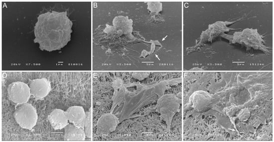
2. L. infantum Can Infect KCs

In blood-MØs and KCs were infected with different L. infantum evolutive morphological forms, mimicking the natural progression of parasite infection. Circulating blood-MØs were isolated and infected by L. infantum promastigotes, the parasite morphological form deposit by sand fly in the dermis of the host. Liver KCs were exposed to L. infantum amastigotes, the parasite form that reaches the internal organs of mammal host. Virulent L. infantum promastigotes were used (Figure 1A) to differentiate amastigotes. In axenic conditions, parasites acquire an amastigote-like morphology (Figure 1B), with an oval shape and a residual flagellum visible in the flagellar pocket (Figure 1C).
Figure 1. Canine blood-MØs and KCs exposed to L. infantum virulent promastigotes or axenic amastigotes. L. infantum virulent promastigotes (yellow) showing elongated flagellum characteristic of L. infantum metacyclic promastigotes (A) and axenic amastigotes (B) with a characteristic oval body (yellow) and residual flagella (C) (green) were observed under scanning electron microscopy (SEM). The acquired images were artificially colored. Blood-MØs (uninfected-D) were exposed to virulent promastigotes for 3 h (E) and 5 h (F). Interaction of promastigotes with the MØ membrane can be observed (white arrows) along with parasite internalization (E,F). Uninfected KCs (G) and KCs exposed to axenic L. infantum amastigotes for 1.5 h (H) and 5 h (I). Internalization of amastigotes by KCs (white arrows) can be observed (I). Blood-MØs exposed to virulent L. infantum promastigotes for 3 h stained with Hemacolor staining kit were observed by optical microscopy (magnification 1000×) (J). Black arrows indicate promastigote internalization. KCs exposed to L. infantum amastigotes for 3 h and stained with DAPI (K) (blue) were observed by fluorescence microscopy. Acquired images show parasites binding to the KCs membrane. N-KCs nucleus.
Canine blood-MØs were exposed to L. infantum virulent promastigotes and parasite internalization was observed (Figure 1D–F,J). After 3 h of incubation, promastigotes were internalized by blood-MØs and more than one parasite per cell could be observed (Figure 1J). In turn, KCs were exposed to amastigotes (Figure 1G–I,K). After 5 h of incubation, L. infantum axenic amastigotes were internalized by KCs (Figure 1I,K), while clearance of extracellular amastigotes was noticed (Figure 1H,I). Amastigote internalization by KCs was observed by fluorescence microscopy (Figure 1K). 

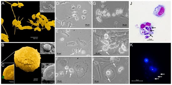
3. L. infantum Infected Blood-MØs Have Increased Lifespan

Blood-MØs viability was assessed to ensure that cells remain functional after infection with L. infantum. Remarkably, the exposition of blood-MØs to parasites resulted in cell’s increasing their lifespan and presented less necrotic/dead cells when compared to non-infected cells. Non-infected blood-MØs viability remained similar between 1.5 h and 5 h of culture, reflecting the natural cell turnover in the population.

4. Infected Blood-MØs Display a Peak of PRR Gene Expression in Response to L. infantum

Innate immune receptors for intracellular and cytoplasmatic PAMPs (NOD1, NOD2 and TLR9) and extracellular PAMPs (TLR2 and TLR4) were accessed. Overall, blood-MØs exposed to L. infantum promastigotes revealed higher levels of induction of gene expression for all tested receptors, as well as a different activation pattern from KCs. After an early (1.5 h and 3 h) exposure to L. infantum promastigotes, blood-MØs showed a significantly higher accumulation of mRNA for all the assessed PRRs followed by a sharp decrease (after 5h) on PRRs gene expression. This might be evidencing a possible inhibition of immune activation of cell sensors exerted by the parasite on infected blood-MØs. Infected blood-MØs exhibited a strong increase in TLR4 and a slight increase in NOD1 expression from 1.5 h to 3 h of exposure to virulent parasites, suggesting that from all the tested PRRs, TLR4 and NOD1 could be engaged in parasite’s antigen recognition. Interestingly, KCs exposed to L. infantum amastigotes revealed a different PRR pattern of induction. Although expressing lower levels of PRRs than infected blood-MØs, parasite exposed KCs showed a strong increase of TLR2 mRNA (after 5 h incubation time) and a moderate rise of NOD1 (3 h to 5 h incubation time) and TLR4 (from 1.5 h to 5 h exposure to parasites) gene expression during the incubation time. Altogether suggesting that TLR2, TLR4, and NOD1 could be involved in parasite antigen’s recognition by KCs.

5. Blood-MØs Phagocyte Parasites While KCs Release Extracellular Traps

Blood-MØs and KCs were analyzed by scanning electron microscopy (SEM) for cellular morphology alterations resulting from cellular immunological activation (Figure 2). After 3 h exposure to virulent L. infantum promastigotes, blood-MØs emitted long and thin pseudopodia to entrap promastigotes, which are compatible with active cellular phagocytosis (Figure 2B,C). Canine KCs, after 5 h of exposure to amastigotes, exhibited morphological alterations compatible with the formation of extracellular traps (ETs), with the complete extrusion of cytoplasmatic cell content and formation of fibers (Figure 2E,F). This constitutes the first evidence of ETs emission by canine KCs activated by L. infantum amastigote exposure.
Figure 2. Blood-MØs phagocyting promastigotes and KCs emitting extracellular traps (ETs) in response to L. infantum. Uninfected blood-MØs (A) and blood-MØs exposed to virulent L. infantum promastigotes for 3 h (B,C) were observed by SEM and images were acquired. White arrows point out fiber-like structures entrapping promastigotes. Uninfected KCs (D) and KCs releasing ETs after 5 h of incubation with L. infantum axenic amastigotes (E,F) can be observed. White arrows indicate a fiber-like structure and cytoplasm extravasation.

6. L. infantum Primes Blood-MØs for M2 Phenotype

Alteration in gene expression of PRRs was accompanied by significant variations on key macrophage cytokine in blood-MØs and KCs exposed to virulent L. infantum parasites. Overall, blood-MØs exhibited higher levels of gene expression of cytokines than KCs, but only for the first 3 h of parasite exposure. Infected blood-MØs exhibited a strong increase in IL-4 and IL-10 as dominantly expressed cytokines after 3 h of contact with parasites. The generation of high levels of IL-4 may be correlated with primming for the M2 macrophage phenotype. Interestingly, KCs exposed to L. infantum amastigotes revealed a different cytokine pattern. Although expressing lower levels of cytokines that infected blood-MØs, KCs strongly generated high levels of IL-10 mRNA and moderate levels of TGF-β. However, parasite-exposed KCs were also able to generate pro-inflammatory IL-12 and TNF-α mRNA during the parasite incubation time. In vivo, this event might lead to the activation of immune response in surrounding cells and recruitment of leukocytes, initiating an effective anti-Leishmania local immune response. 

7. Proposed model

Based on accumulated experimental pieces of evidence, it can be proposed a model of Leishmania-MØs interaction for blood-MØs and KCs (Figure 3). Blood-MØs, are “ready for action” cells by nature, with a myriad of immune tools, and are rapidly activated in the presence of L. infantum, upregulating TLRs and NODs, particularly TLR4 and NOD1, and release NO. However, shortly after parasite phagocytosis and internalization, blood-MØs became anergic and immune-dormant. These M2 primed cells upregulate anti-inflammatory cytokines (IL-4 and IL-10) and support parasite survival, evidencing a long parasite-host adaptation process, since L. infantum can subvert the immune activation state of the blood-MØs. In the liver, KCs are primed by the microenvironment towards immune tolerance and exhibit lower immune activation when encountering L. infantum amastigotes [15]. However, KCs can sense the L. infantum amastigotes by cell’s innate immune receptors, particularly transmembrane TLR2 and TLR4 and intracellular NOD1, generating low amounts of IL-12 and TNF-α, produce NO, and emit METs, favoring the availability of parasite antigens in the liver milieu which can lead to the recruitment of other leukocytes, especially CD8+T cells and natural killer cells that can initiate the formation of a granuloma response. Granuloma is a typical liver structure associated with parasite restriction and local control of the infection. However, the liver is a complex organ and the orchestration of the anti-Leishmania immune response seems derived from a multifaceted interaction between several types of cells. Hepatocytes represent the majority of the liver cells and are normally associated with metabolic functions. Yet, hepatocytes have been recently implicated in the orchestration of the liver’s anti-Leishmania immune response, by playing a potentially key role in the crosstalk between liver cells, as hepatocytes are also able to sense and react to the parasite, upregulating PRRs as well as generating immune mediators [[16][17]], therefore potentially amplifying the immune activation signals emitted by KCs.

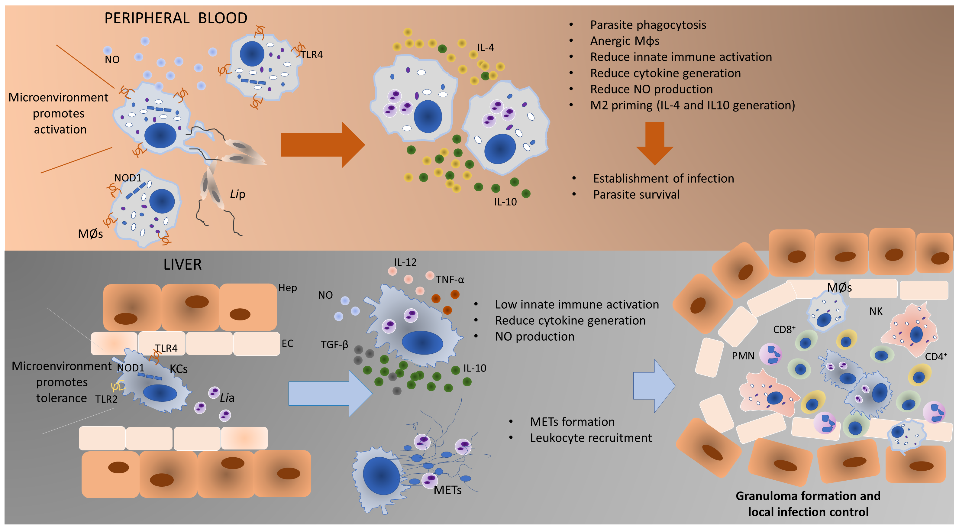
Figure 3. Proposed model of Leishmania-MØs interaction in blood and liver. Blood-MØs are rapidly activated in the presence of L. infantum, upregulating innate sensors (PRRs) and immune mediators (cytokines). However, shortly after parasite internalization blood-MØs became anergic and permissive to parasite survival. In the liver, KCs are primed by the microenvironment towards immune tolerance and exhibit low immune activation towards L. infantum. However, these cells can generate low levels of pro-inflammatory cytokines, release NO and emit METs in the presence of amastigotes, which will recruit other leukocytes and may initiate the constitution of a granuloma. The formation of granuloma is a typical hepatic structure associated with parasite restriction and local control of the infection. MØs-blood macrophages; Lip–L. infantum promastigotes; KCs-Kupffer cells; Lia–L. infantum amastigotes; METs-macrophage extracellular traps; TLR4-Toll-like receptor 4; TLR2- Toll-like receptor 2; NO-nitric oxide; NOD1-nucleotide-binding oligomerization domain-like (NOD) receptor 1; Hep-Hepatocyte; EC-Endothelial Cell; CD8+-cytotoxic T lymphocytes; CD4+-helper T lymphocytes; NK-Natural Killer cells; PMNs-Polymorphonuclear leukocytes.

8. Conclusions

The immune activation potential of two different lineages of MØs in the context of CanL is demonstrated and highlighted the close evolution of Leishmania and dog macrophage. L. infantum can take advantage of the natural predisposition of blood-MØs to be activated and phagocyte pathogens and subvert the cell’s immune activation mechanisms, which allow parasite replication and dissemination, establishing infection in the host and assuring the completion of the parasite life cycle. On the other hand, liver KCs, primed for immune tolerance, are not extensively activated by the presence of this pathogen, allowing L. infantum to establish and replicate. However, KCs can activate other mechanisms, such as NO production and METs release, to ensure parasite antigen exposition, launching a cascade of leukocyte recruitment and activation to locally control the infection. Altogether KCs reveal a different response pattern from blood-MØs when facing L. infantum. In addition, KCs response appears to be more efficient in controlling this parasite, thus contributing to the ability of the liver to naturally control parasite dissemination. Understanding the mechanism of liver resistance to L. infantum infection may lead to a new paradigm in CanL epidemiology regarding the liver as a possible parasite-reservoir organ.

References

  1. Van Furth, R.; Beekhuizen, H. Monocytes. In Encyclopedia of Immunology; Elsevier: Amsterdam, The Netherlands, 1998; pp. 1750–1754.
  2. Mosser, D.M.; Edwards, J.P. Exploring the full spectrum of macrophage activation. Nat. Rev. Immunol. 2008, 8, 958–969.
  3. Akira, S.; Uematsu, S.; Takeuchi, O. Pathogen recognition and innate immunity. Cell 2006, 124, 783–801.
  4. Zhou, L.; Cao, X.; Fang, J.; Li, Y.; Fan, M. Macrophages polarization is mediated by the combination of PRR ligands and distinct inflammatory cytokines. Int. J. Clin. Exp. Pathol. 2015, 8, 10964–10974.
  5. Shaw, M.H.; Reimer, T.; Kim, Y.G.; Nuñez, G. NOD-like receptors (NLRs): Bona fide intracellular microbial sensors. Curr. Opin. Immunol. 2008, 20, 377–382.
  6. Yona, S.; Kim, K.W.; Wolf, Y.; Mildner, A.; Varol, D.; Breker, M.; Strauss-Ayali, D.; Viukov, S.; Guilliams, M.; Misharin, A.; et al. Fate Mapping Reveals Origins and Dynamics of Monocytes and Tissue Macrophages under Homeostasis. Immunity 2013, 38, 79–91.
  7. Ginhoux, F.; Guilliams, M. Tissue-Resident Macrophage Ontogeny and Homeostasis. Immunity 2016, 44, 439–449.
  8. Epelman, S.; Lavine, K.J.; Randolph, G.J. Origin and Functions of Tissue Macrophages. Immunity 2014, 41, 21–35.
  9. Jenne, C.N.; Kubes, P. Immune surveillance by the liver. Nat. Immunol. 2013, 14, 996–1006.
  10. Dixon, L.J.; Barnes, M.; Tang, H.; Pritchard, M.T.; Nagy, L.E. Kupffer cells in the liver. Compr. Physiol. 2013, 3, 785–797.
  11. Tomiotto-Pellissier, F.; Bortoleti, B.T.d.; Assolini, J.P.; Gonçalves, M.D.; Carloto, A.C.M.; Miranda-Sapla, M.M.; Conchon-Costa, I.; Bordignon, J.; Pavanelli, W.R. Macrophage Polarization in Leishmaniasis: Broadening Horizons. Front. Immunol. 2018, 9, 2529.
  12. Pereira, M.A.; Alexandre-Pires, G.; Câmara, M.; Santos, M.; Martins, C.; Rodrigues, A.; Adriana, J.; Passero, L.F.D.; da Fonseca, I.P.; Santos-Gomes, G. Canine neutrophils cooperate with macrophages in the early stages of Leishmania infantum in vitro infection. Parasite Immunol. 2019, 41, e12617.
  13. Olivier, M.; Gregory, D.J.; Forget, G. Subversion mechanisms by which Leishmania parasites can escape the host immune response: A signaling point of view. Clin. Microbiol. Rev. 2005, 18, 293–305.
  14. Bogdan, C. Macrophages as host, effector and immunoregulatory cells in leishmaniasis: Impact of tissue micro-environment and metabolism. Cytokine X 2020, 2, 100041.
  15. Rodrigues A., Santos-Mateus D., Alexandre-Pires G., Valério-Bolas A., Rafael-Fernandes M., Pereira MA., Ligeiro D., Jesus J., Alves-Azevedo R., Lopes-Ventura S., Santos M., Tomás A., Pereira da Fonseca I., Santos-Gomes G.; Leishmania infantum exerts immunomodulation in canine Kupffer cells reverted by meglumine antimoniate. Comparative Immunology, Microbiology and Infectious Diseases 2017, 55, 42-52, https://doi.org/10.1016/j.cimid.2017.09.004.
  16. Rodrigues, A., Alexandre-Pires, G., Valério-Bolas, A., Santos-Mateus, D., Rafael-Fernandes, M., Pereira, M., Ligeiro, D., Nunes, T., Alves-Azevedo R., Lopes-Ventura S., Santos M., Tomás A., Pereira da Fonseca I., Santos-Gomes, G.; Dog hepatocytes are key effector cells in the liver innate immune response to Leishmania infantum.. Parasitology 2019, 146, 753-764, doi:10.1017/S0031182018002068.
  17. Armanda V. Rodrigues; Graça Alexandre-Pires; Ana Valério-Bolas; David Santos-Mateus; Mariana Rafael-Fernandes; Maria A. Pereira; Dário Ligeiro; Telmo Nunes; Raquel Alves-Azevedo; Marcos Santos; et al.Isabel Pereira Da FonsecaGabriela Santos-Gomes 3D-Hepatocyte Culture Applied to Parasitology: Immune Activation of Canine Hepatic Spheroids Exposed to Leishmania infantum. Biomedicines 2020, 8, 628, 10.3390/biomedicines8120628.
More
Information
Contributor MDPI registered users' name will be linked to their SciProfiles pages. To register with us, please refer to https://encyclopedia.pub/register :
View Times: 418
Revision: 1 time (View History)
Update Date: 13 Jan 2022
1000/1000