Submitted Successfully!
To reward your contribution, here is a gift for you: A free trial for our video production service.
Thank you for your contribution! You can also upload a video entry or images related to this topic.
Version Summary Created by Modification Content Size Created at Operation
1 + 2656 word(s) 2656 2021-12-22 11:38:39

Video Upload Options

Do you have a full video?

Confirm

Are you sure to Delete?
Cite
If you have any further questions, please contact Encyclopedia Editorial Office.
Huang, N. Cell Therapies for Treatment of Volumetric Muscle Loss. Encyclopedia. Available online: https://encyclopedia.pub/entry/17946 (accessed on 03 September 2024).
Huang N. Cell Therapies for Treatment of Volumetric Muscle Loss. Encyclopedia. Available at: https://encyclopedia.pub/entry/17946. Accessed September 03, 2024.
Huang, Ngan. "Cell Therapies for Treatment of Volumetric Muscle Loss" Encyclopedia, https://encyclopedia.pub/entry/17946 (accessed September 03, 2024).
Huang, N. (2022, January 10). Cell Therapies for Treatment of Volumetric Muscle Loss. In Encyclopedia. https://encyclopedia.pub/entry/17946
Huang, Ngan. "Cell Therapies for Treatment of Volumetric Muscle Loss." Encyclopedia. Web. 10 January, 2022.
Cell Therapies for Treatment of Volumetric Muscle Loss
Edit

Volumetric muscle loss (VML) is the traumatic, surgical or degenerative loss of a substantial portion of bulk skeletal muscle in a manner that overwhelms the endogenous repair capacity of the muscle and results in impaired scar tissue formation. Cell-based therapies have emerged as a promising approach to promote skeletal muscle regeneration following injury and/or disease. Stem cell populations, such as muscle stem cells, mesenchymal stem cells and induced pluripotent stem cells (iPSCs), have shown a promising capacity for muscle differentiation. Support cells, such as endothelial cells, nerve cells or immune cells, play a pivotal role in providing paracrine signaling cues for myogenesis, along with modulating the processes of inflammation, angiogenesis and innervation. 

tissue engineering cell transplantation stem cells satellite cells induced pluripotent stem cells volumetric muscle loss

1. Introduction

Volumetric muscle loss (VML) is the traumatic, surgical or degenerative loss of a substantial portion of bulk skeletal muscle in a manner that overwhelms the endogenous repair capacity of the muscle and results in impaired scar tissue formation. VML is associated with chronic functional disability in many military and civilian populations following battlefield injuries, car accidents, tumor ablation or degenerative diseases [1][2]. VML lacks a standard treatment to replace the lost tissue with contractile muscle or restore muscle strength. Current surgical approaches, such as autologous free flap grafting, scar tissue debridement or minced skeletal tissue transfer, are utilized to reconstruct the tissue defects [3][4]. However, these techniques are limited by tissue availability, the need for highly skilled surgeons, significant donor site morbidity and functional deficiencies. Furthermore, the efficacy of these techniques for the repair of severe injuries, such as VML, has not been demonstrated clinically.
Skeletal muscle consists of bundles of oriented multi-nucleated muscle fibers (myofibers) that are surrounded by connective tissue, along with branches of blood vessels and peripheral nerves that supply blood flow and electrical signals to the muscle. Thus, a variety of cell types work in concert to form the skeletal muscle structure (Figure 1).
Figure 1. Schematic of skeletal muscle structure and different cell types in skeletal muscle.

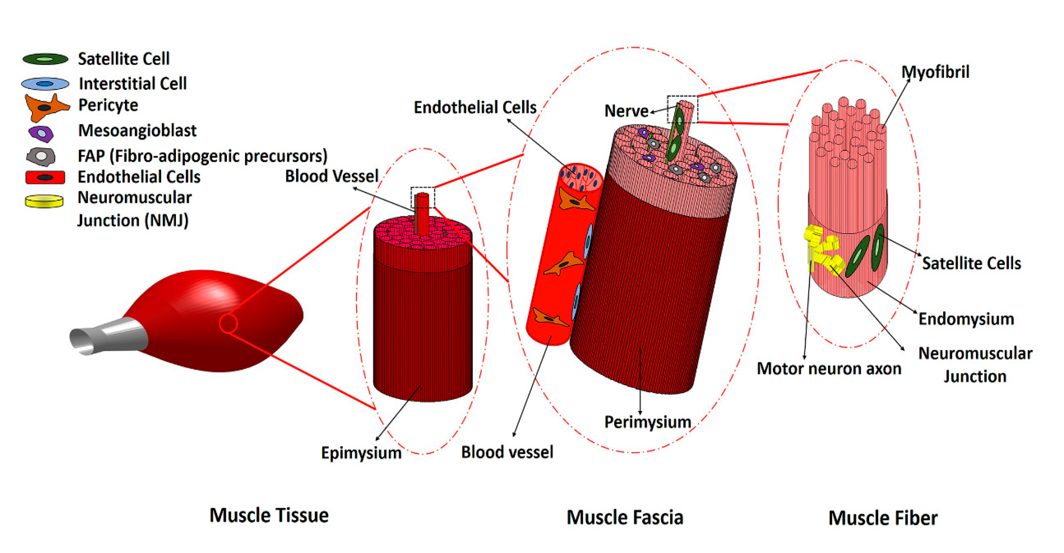
2. Main Myogenic Cell Sources

A variety of cells reside within skeletal muscle, including muscle satellite cells, pericytes, vascular lineages, interstitial stem cells and fibro/adipogenic progenitors (FAPs). Among these cell types, satellite cells are primarily responsible for natural skeletal muscle repair and regeneration. Satellite cells are muscle stem cells (MuSCs) that reside beneath the basal lamina surrounding each myofiber. These cells are a heterogenous population, but commonly express paired box protein-7 (Pax7), a transcription factor that plays role in myogenesis and regulates the proliferation of MuSCs. In response to myofiber injury, quiescent satellite cells activate, proliferate, and give rise to myogenic progenitor cells (MPCs). MPCs differentiate into myoblasts and can fuse to form multinucleated myotubes, which mature into newly formed myofibers.
Although MuSCs are considered promising cell sources for VML treatment, some of the limitations associated with satellite cells include their low abundance within muscle, challenges in isolating and purifying these cells, limited self-renewal and differentiation potential in vitro, and low engraftment post-transplantation [5][6]. The in vitro expansion of satellite cells can result in the loss of their innate myogenic behavior, but multiple research studies have demonstrated that the regenerative capacity of satellite cells can be regulated by biochemical and biophysical microenvironmental cues, such as peptide functionalization, substrate stiffness and three-dimensionality [7][8][9][10]. For example, Gilbert et al. showed that hydrogels of muscle-like stiffness could recapitulate the rigidity features of the stem cell niche. MuSCs cultured on these 12 kPa stiffness hydrogels could preserve self-renewal characteristics in vitro, in contrast to commonly used rigid plastic culture dishes [10]. Additionally, Pruller et al. showed that murine MuSCs embedded in collagen I, polyethylene glycol (PEG)-fibrinogen and three-dimensional (3D) fibrin scaffolds did not show any myogenic differentiation [9]. However, placing freshly isolated myofibers within 3D scaffolds significantly improved the myogenic behavior of satellite cells on the myofibers, in part because the cells were still in their native niche [9].
Given the limitations of MuSCs, mesenchymal stem cells (MSCs) are an alternative cell source. The intramuscular injection of MSCs has accelerated muscle repair in injured skeletal muscle in animal models [11]. MSCs are a class of non-hematopoietic multipotent adult stem cells which can differentiate into many cell types, including osteoblasts, adipocytes, chondrocytes and myoblasts. They are present in a variety of adult tissues, including bone marrow, birth-derived tissues and dental pulp, however bone marrow and adipose tissue are the two main sources of MSCs. Compared to satellite cells, MSCs have greater abundance, and can be harvested by minimally invasive processes like bone marrow extraction or lipoaspiration [12]. MSCs are immunologically advantageous because of their low expression level of major histocompatibility complex class I (MHC I) and II (MHC II) proteins, which makes them attractive for transplantation [13]. Although MSCs can be a suitable cell source for clinical treatment of VML in future, many studies have shown the low engraftment of MSCs and a lack of tissue-specific differentiation. It is not clear that MSCs are beneficial in muscle regeneration just by secreting immunomodulatory and paracrine signals, or by myogenic differentiation. More research studies should be conducted to optimize MSC isolation and the myogenic differentiation process. The identification of suitable microenvironmental factors that support the myogenic phenotype will be important for harnessing the therapeutic potential of MSCs, as is the understanding of how MSCs participate in muscle regeneration either by paracrine signals or myogenic differentiation.
Induced pluripotent stem cells (iPSCs) represent another attractive myogenic cell source for skeletal muscle regeneration, due to their unlimited self-renewal capacity and capacity to differentiate into myogenic cells. The iPSCs can be directly reprogrammed into skeletal muscle cells via the overexpression of myogenic transcription factors, such as Pax7 or MyoD, or by small molecules [14][15]. Genetic reprogramming is more efficient than small molecule-based reprogramming, and can generate 3D contractile skeletal muscle that fuses with the existing damaged tissues [16]. However, the risks of genetic instability and tumorgenicity make reprogramming unreliable for clinical studies and VML treatment. Besides these main cell types, other potential cell sources for skeletal muscle regeneration are summarized in Table 1, and have been broadly discussed elsewhere [17]. Few studies have specifically investigated many of these myogenic cell sources, such as pericytes or mesoangioblasts, for VML repair. The critical factors for choosing a suitable myogenic cell source for treatment of VML include the potential for myogenic differentiation, availability, ease of isolation and sorting, as well as ease of in vitro expansion and immunogenicity. Adipose-derived MSCs (ASCs) are abundant and easy to isolate from adipose tissue with low immunogenicity. However, it is not clear that they directly differentiate into muscle cells, or whether their impact is more immunomodulatory using paracrine signals. In Table 1, different myogenic cell sources are compared, and their pros and cons are outlined.
Table 1. Cell Sources for Regeneration of Skeletal Muscle.

Cell Types

Markers

Location

Advantages

Disadvantages

Reference

MuSCs

Pax7+, CD56+,

MyoD+

Under basal

lamina of

muscle fibers.

Critical to native skeletal muscle regeneration.

High myogenic potential.

Isolation is invasive and low yield.

Loss of self-renewal potential during in vitro expansion.

Loss of differentiation potential after in vivo transplantation.

[6][18][19]

Mesenchymal stem cells (MSCs)

CD90+, CD44+,

CD29+, CD105+, CD13+, CD73+, CD166+, CD45, CD34, CD14

Adipose tissue, bone marrow (BM), umbilical cord (UC).

Abundance of adipose tissue.

Ease of isolation from adipose tissue.

Low expression of MHC-I and MHC-II

Immunomodulatory effect.

Invasive isolation for BM-MSCs.

Poor myogenic differentiation capacity.

[6][18]

Myo-endothelial cells

CD34+, CD144+, CD56+, CD31+, CD45

Periphery of myofibers close to blood vessels.

Have both angiogenic and myogenic capacity.

Laborious isolation and purification process.

Limited literature on their role in skeletal muscle regeneration.

[20]

Mesoangioblasts

CD34+, Sca-1+, CD31+, c-Kit+, CD45

Walls of microvessels.

High proliferative capacity in vitro.

Multipotent cells with potential to differentiate into skeletal muscle

Invasive isolation procedure.

Lack of studies for VML treatment.

[21]

Pericytes

CD146+, NG2+, ALP+, PDGFR-β+

Periphery of

capillaries and microvessels.

Pericyte myogenesis naturally occurs during development and regeneration of muscle.

High muscle differentiation potential.

Lack of MHCII expression.

Limited literature on their potential in skeletal muscle regeneration and VML.

[6][18]

CD133+ progenitor cells

CD133+, CD34+/−, CD90+/−, CD146+

Periphery of myofibers close to

blood vessels.

Availability and ease of purification from peripheral blood

Myogenic and angiogenic capacity.

Reduction of myogenic potential following in vitro culture.

[18][22]

Induced pluripotent stem cells (iPSCs)

Oct4+, Sox2+,

KLF4+, and c-Myc+

All tissues, mainly skin.

Unlimited self-renewal in vitro.

Patient-derived autologous cells.

Myogenic differentiation capacity.

Risk of tumorigenicity and genetic instability.

[6][18]

Embryonic stem cells (ESCs)

Oct4+, Sox2+,

KLF4+, and c-Myc+

Inner cell

mass of

blastocyst.

Unlimited self-renewal in vitro.

Myogenic differentiation capacity

Ethical concerns

Inefficient isolation process.

Risk of tumorigenicity.

Risk associated with immune response.

[6]

Muscle side population cells (SPs)

CD45, c-Kit, Sca1+, ABCG2+, Pax7, Myf5, Desmin

Interstitial space of skeletal muscle.

Myogenic differentiation capacity in vivo.

Low availability

Lack of specific phenotypic markers.

Poor myogenic differentiation in vitro.

Limited literature on their potential for skeletal muscle regeneration and VML.

[19][23]

Abbreviations: Stem cells antigen -1 (Sca-1), Alkaline phosphatase (ALP), Platelet-derived growth factor receptor beta (PDGFR-β), Neural/glial antigen 2 (NG 2), ATP binding cassette subfamily G member 2 (ABCG-2), Octamer-binding transcription factor 4 (Oct-4), SRY (sex determining region Y)-box 2 (Sox-2), Kruppel-like factor 4 (KLF4), Myogenic factor 5 (Myf5).

3. Non-Myogenic Cells

In addition to resident MuSCs, the normal functioning and regeneration of skeletal muscle tissue are supported by fibroblasts, endothelial cells, neural cells, fibro/adipogenic progenitor (FAP) cells and infiltrating immune cells. FAPs are muscle-resident progenitor cells of mesenchymal origin, expressing stem cells antigen-1 (Sca-1) and platelet-derived growth factor receptor beta (PDGFR-β) surface markers, which can differentiate into fibroblasts and adipocytes. Following muscle injury, FAPs proliferate and enhance myogenic differentiation by generating pro-differentiation signals for MPCs [24]. The intercellular interactions and paracrine effects of the above-mentioned support cells on MuSCs are important for the regeneration of damaged nervous and vascular tissues that are often associated with VML [25][26]. For example, pericytes and MSCs regulate myogenesis through paracrine effects on other cell types, such as macrophages. Chronic inflammation and the delayed transition of inflammatory to anti-inflammatory phase in VML impairs the hemostasis of many intracellular interactions, and results in fibrotic muscle degeneration. Persistent inflammatory signals such as TGF-β prevent FAPs, apoptosis and lead to their pathological accumulation and differentiation into fibroblasts [27]. MSCs derived from bone marrow or adipose tissue can ameliorate the local immunological response by suppressing inflammatory cytokines and modulating local immune cells in the injured muscle, as discussed in [28].
Multi-cellular culture systems have been developed to study the intercellular and paracrine interactions that are necessary for muscle formation and function. For example, Ostrovidov et al. demonstrated that the co-culture of a PC12 neural cell line with a C2C12 myoblast cell line could induce C2C12 myoblast differentiation and myotube formation [26]. Neural cell sources, such as human neural stem cells and iPSC-derived neurons, in co-culture with skeletal muscle cells could form functional neuromuscular junctions (NMJ) and have improved muscle differentiation and myotube formation [29]. Laternser et al. demonstrated that the interaction between tendon and muscle cells affects the functionality of the regenerated muscle. 3D muscle and tendon tissues were developed by printing human myoblasts and rat tenocytes within bioink layers. The printed myofibers were functional and could contract following electrical stimulation. The increased expression of muscle and tendon marker genes indicated the good differentiation of cells [30]. Additionally, endothelial cells modulate skeletal MuSCs differentiation and proliferation by secreting growth factors such as vascular endothelial growth factor (VEGF), insulin growth factor (IGF-1) and hepatocyte growth factor (HGF) [31].

4. Preclinical VML Treatment Studies

4.1. Animal Models of VML

Approximately 90% of preclinical models of VML have been conducted in mice and rats [32]. However, a standard VML model with respect to muscle anatomical location and defect size has not been defined. The latissimus dorsi (LD), tibialis anterior (TA), quadriceps and abdominal wall muscles are the most frequently ablated muscles in experimentally induced VML [32]. Although in most VML models more than 20% of the muscle mass is ablated, Anderson et al. reported that 15% muscle ablation was the critical threshold for irreversible muscle loss. The critical threshold was characterized by persistent fibrotic and inflammatory response, as well as incomplete innervation and partial myofiber regeneration [33]. Full-thickness injuries of 2, 3 and 4 mm diameters were created in the quadricep muscles of mice, which resulted in 5%, 15% and 30% muscle mass defects, respectively. In addition to the size of defect, other factors could mediate the healing outcome of the VML lesion; including whether the defect is partial or full-thickness, how the suture for wound closure was applied (e.g., bridging vs. non-bridging lesion) and the proportion of defect size to animal weight. In addition, myofiber density and the amount of satellite cells in the muscle tissue varies between male and female animals, and across different ages. Biomechanical loading is also different in each muscle tissue. Considering these parameters, VML rodent models are beneficial for examining therapies or understanding the pathophysiology of VML and skeletal muscle regeneration. However, the treatment results of rodent preclinical studies should not be extrapolated for human VML treatments without considering factors such as mechanical loading. In other words, the current rodent VML models fit better for small non-appendicular muscles like facial muscles than large, appendicular muscles.

4.2. Cell-Seeded Scaffolds for Preclinical Treatment of VML

The direct transplantation of MuSCs and MPCs into damaged tissue results in poor cell survival and limited engraftment due to a lack of sufficient cell support and the harsh injured tissue microenvironment [34]. Scaffolds support cell attachment, survival and differentiation, and also facilitate topographical, biomechanical and biochemical cues to promote myogenesis [32][35]. The transplantation of muscle stem cells seeded in decellularized bladder matrices into rat TA muscle with VML injury showed that muscle peak isometric torque was already enhanced by the use of scaffolds alone, but the inclusion of cells with the scaffolds doubled the functional recovery of the muscle [36]. Aligned nanofibrillar collagen scaffolds direct the alignment of murine myoblasts, and promote myotube fusion and contractility in the engineered muscle tissue [37]. Scaffolds, in addition to physically filling in the lost muscle volume and augmenting force transmission across the defect, can be used as combined delivery vehicles for cells and growth factors. Macroporous alginate hydrogels simultaneously delivered murine myoblasts along with a VEGF/IGF-1 cocktail. The results showed a significant increase in the muscle contractile force and a decrease in fibrosis [38]. A more extensive discussion of scaffold biomaterials for skeletal muscle regeneration in VML is found in [18][39].

4.3. MuSC-Based Therapies for Preclinical Treatment of VML

MuSCs derived from newborn mice and embedded in fibrin hydrogel were able to engraft and differentiate into new myofibers in NOD SCID mice with VML. This led to a 30% increase in muscle mass and a 50% reduction in fibrosis area, compared to untreated muscles. The detection of LacZ+/Pax7+ in the cross section of engrafted muscle demonstrated that the transplanted MuSCs, labeled with LacZ, could restore satellite cells. In addition, LacZ+ donor-derived cells contributed to an average of 26% of the new vessels formed in engrafted regions [40]. Although this study showed evidence of neotissue formation in a VML animal model using histology and immunohistochemical analysis, no functional measurements were taken to compare recovered muscle with uninjured tissue, such as the level of nerve and force restoration, muscle strength deficits, and measurements of peak isometric tetanic force or torque.

4.4. MSC-Based Therapies for Preclinical Treatment of VML

Besides muscle stem and progenitor cells, MSCs have been tested in rodent VML models [41][42]. Recent studies suggest that the pro-regenerative outcomes of these cells are not due mainly to the direct contribution of these cells to de novo myofiber formation, but instead to their paracrine effects, such as immunomodulation, inducing vascularization, mediating with fibrotic response and signaling to endogenous cells. In one study, bone marrow-derived MSCs (BM-MSCs) encapsulated in plasma-rich platelet-derived fibrin microbeads could accelerate muscle regeneration in a rat VML model of the ablated biceps femoris muscle. At 180 days following implantation, animals that did not have microbeads and MSCs showed incomplete repair with scar formation, whereas the presence of microbeads decreased collagen deposition and caused the formation of aligned myofibers. Based on histomorphometric analysis at 30, 60 and 180 days following implantation, MSCs encapsulated in fibrin showed immunomodulatory effects, and sped up the healing process compared to fibrin alone [43].  

References

  1. Corona, B.T.; Rivera, J.C.; Owens, J.G.; Wenke, J.C.; Rathbone, C.R. Volumetric muscle loss leads to permanent disability following extremity trauma. J. Rehabil. Res. Dev. 2015, 52, 785–792.
  2. Garg, K.; Ward, C.L.; Hurtgen, B.J.; Wilken, J.M.; Stinner, D.J.; Wenke, J.C.; Owens, J.G.; Corona, B.T. Volumetric muscle loss: Persistent functional deficits beyond frank loss of tissue. J. Orthop. Res. 2014, 33, 40–46.
  3. Corona, B.T.; Wenke, J.C.; Ward, C.L. Pathophysiology of volumetric muscle loss injury. Cells Tissues Organs 2016, 202, 180–188.
  4. Lin, C.-H.; Lin, Y.-T.; Yeh, J.-T.; Chen, C.-T. Free Functioning muscle transfer for lower extremity posttraumatic composite structure and functional defect. Plast. Reconstr. Surg. 2007, 119, 2118–2126.
  5. Ding, S.; Wang, F.; Liu, Y.; Li, S.; Zhou, G.; Hu, P. Characterization and isolation of highly purified porcine satellite cells. Cell Death Discov. 2017, 3, 17003.
  6. Pantelic, M.N.; Larkin, L.M.; Pantellic, M.N. Stem cells for skeletal muscle tissue engineering. Tissue Eng. Part B Rev. 2018, 24, 373–391.
  7. Garcia, J.M.S.; Panitch, A.; Calve, S. Functionalization of hyaluronic acid hydrogels with ECM-derived peptides to control myoblast behavior. Acta Biomater. 2019, 84, 169–179.
  8. Patel, K.H.; Dunn, A.J.; Talovic, M.; Haas, G.J.; Marcinczyk, M.; Elmashhady, H.; Kalaf, E.A.G.; Sell, S.A.; Garg, K.; Growney, E. Aligned nanofibers of decellularized muscle ECM support myogenic activity in primary satellite cells in vitro. Biomed. Mater. 2019, 14, 035010.
  9. Prueller, J.; Mannhardt, I.; Eschenhagen, T.; Zammit, P.S.; Figeac, N. Satellite cells delivered in their niche efficiently generate functional myotubes in three-dimensional cell culture. PLoS ONE 2018, 13, e0202574.
  10. Gilbert, P.M.; Havenstrite, K.L.; Magnusson, K.E.G.; Sacco, A.; Leonardi, N.A.; Kraft, P.; Nguyen, N.K.; Thrun, S.; Lutolf, M.P.; Blau, H.M. Substrate elasticity regulates skeletal muscle stem sell self-renewal in culture. Science 2010, 329, 1078–1081.
  11. Mori, R.; Kamei, N.; Okawa, S.; Nakabayashi, A.; Yokota, K.; Higashi, Y.; Ochi, M. Promotion of skeletal muscle repair in a rat skeletal muscle injury model by local injection of human adipose tissue-derived regenerative cells. J. Tissue Eng. Regen. Med. 2012, 9, 1150–1160.
  12. Berebichez-Fridman, R.; Montero-Olvera, P.R. Sources and Clinical Applications of Mesenchymal Stem Cells. Sultan Qaboos Univ. Med. J. 2018, 18, 264–277.
  13. Jacobs, S.A.; Roobrouck, V.D.; Verfaillie, C.M.; Van Gool, S.W. Immunological characteristics of human mesenchymal stem cells and multipotent adult progenitor cells. Immunol. Cell Biol. 2013, 91, 32–39.
  14. Kodaka, Y.; Rabu, G.; Asakura, A. Skeletal Muscle Cell Induction from Pluripotent Stem Cells. Stem Cells Int. 2017, 2017, 1–16.
  15. del Carmen Ortuño-Costela, M.; García-López, M.; Cerrada, V.; Gallardo, M.E. iPSC s: A powerful tool for skeletal muscle tissue engineering. J. Cell. Mol. Med. 2019, 23, 3784–3794.
  16. Rao, L.; Qian, Y.; Khodabukus, A.; Ribar, T.; Bursac, N. Engineering human pluripotent stem cells into a functional skeletal muscle tissue. Nat. Commun. 2018, 9, 126.
  17. De Micheli, A.J.; Laurilliard, E.J.; Heinke, C.L.; Ravichandran, H.; Fraczek, P.; Soueid-Baumgarten, S.; De Vlaminck, I.; Elemento, O.; Cosgrove, B.D. Single-Cell Analysis of the Muscle Stem Cell Hierarchy Identifies Heterotypic Communication Signals Involved in Skeletal Muscle Regeneration. Cell Rep. 2020, 30, 3583–3595.e5.
  18. Dunn, A.; Talovic, M.; Patel, K.; Patel, A.; Marcinczyk, M.; Garg, K. Biomaterial and stem cell-based strategies for skeletal muscle regeneration. J. Orthop. Res. 2019, 37, 1246–1262.
  19. Yin, H.; Price, F.; Rudnicki, M.A. Satellite Cells and the Muscle Stem Cell Niche. Physiol. Rev. 2013, 93, 23–67.
  20. Zheng, B.; Cao, B.; Crisan, M.; Sun, B.; Li, G.; Logar, A.; Yap, S.; Pollett, J.B.; Drowley, L.; Cassino, T.; et al. Prospective identification of myogenic endothelial cells in human skeletal muscle. Nat. Biotechnol. 2007, 25, 1025–1034.
  21. Sirabella, D.; De Angelis, L.; Berghella, L. Sources for skeletal muscle repair: From satellite cells to reprogramming. J. Cachexia Sarcopenia Muscle 2013, 4, 125–136.
  22. Torrente, Y.; Belicchi, M.; Marchesi, C.; D’Antona, G.; Cogiamanian, F.; Pisati, F.; Gavina, M.; Giordano, R.; Tonlorenzi, R.; Fagiolari, G.; et al. Autologous transplantation of muscle-derived CD133+ stem cells in Duchenne muscle patients. Cell Transplant. 2007, 16, 563–577.
  23. Asakura, A.; Seale, P.; Girgis-Gabardo, A.; Rudnicki, M.A. Myogenic specification of side population cells in skeletal muscle. J. Cell Boil. 2002, 159, 123–134.
  24. Joe, A.W.B.; Yi, L.; Natarajan, A.; Le Grand, F.; So, L.; Wang, J.; Rudnicki, M.A.; Rossi, F.M. Muscle injury activates resident fibro/adipogenic progenitors that facilitate myogenesis. Nature 2010, 12, 153–163.
  25. Wosczyna, M.N.; Rando, T.A. A Muscle Stem Cell Support Group: Coordinated Cellular Responses in Muscle Regeneration. Dev. Cell 2018, 46, 135–143.
  26. Ostrovidov, S.; Ahadian, S.; Ramon-Azcon, J.; Hosseini, V.; Fujie, T.; Parthiban, S.P.; Shiku, H.; Matsue, T.; Kaji, H.; Ramalingam, M. Three-dimensional co-culture of C2C12/PC12 cells improves skeletal muscle tissue formation and function. J. Tissue Eng. Regen. Med. 2017, 11, 582–595.
  27. Biferali, B.; Proietti, D.; Mozzetta, C.; Madaro, L. Fibro–Adipogenic Progenitors Cross-Talk in Skeletal Muscle: The Social Network. Front. Physiol. 2019, 10, 10.
  28. Gao, F.; Chiu, S.M.; Motan, D.A.L.; Zhang, Z.; Chen, L.; Ji, H.-L.; Tse, H.-F.; Fu, Q.-L.; Lian, Q. Mesenchymal stem cells and immunomodulation: Current status and future prospects. Cell Death Dis. 2016, 7, 2062.
  29. Kim, J.H.; Kim, I.; Seol, Y.-J.; Ko, I.K.; Yoo, J.J.; Atala, A.; Lee, S.J. Neural cell integration into 3D bioprinted skeletal muscle constructs accelerates restoration of muscle function. Nat. Commun. 2020, 11, 1–12.
  30. Laternser, S.; Keller, H.; Leupin, O.; Rausch, M.; Graf-Hausner, U.; Rimann, M. A Novel Microplate 3D Bioprinting Platform for the Engineering of Muscle and Tendon Tissues. SLAS Technol. Transl. Life Sci. Innov. 2018, 23, 599–613.
  31. Latroche, C.; Weiss-Gayet, M.; Muller, L.; Gitiaux, C.; Leblanc, P.; Liot, S.; Ben-Larbi, S.; Abou-Khalil, R.; Verger, N.; Bardot, P.; et al. Coupling between Myogenesis and Angiogenesis during Skeletal Muscle Regeneration is Stimulated by Restorative Macrophages. Stem Cell Rep. 2017, 9, 2018–2033.
  32. Sarrafian, T.L.; Bodine, S.C.; Murphy, B.; Grayson, J.K.; Stover, S.M. Extracellular matrix scaffolds for treatment of large volume muscle injuries: A review. Veter. Surg. 2018, 47, 524–535.
  33. Anderson, S.E.; Han, W.M.; Srinivasa, V.; Mohiuddin, M.; Ruehle, M.A.; Moon, J.Y.; Shin, E.; Emeterio, C.L.S.; Ogle, M.E.; Botchwey, E.A.; et al. Determination of a Critical Size Threshold for Volumetric Muscle Loss in the Mouse Quadriceps. Tissue Eng. Part C Methods 2019, 25, 59–70.
  34. Skuk, D.; Caron, N.J.; Goulet, M.; Roy, B.; Tremblay, J.P. Resetting the Problem of Cell Death Following Muscle-Derived Cell Transplantation: Detection, Dynamics and Mechanisms. J. Neuropathol. Exp. Neurol. 2003, 62, 951–967.
  35. Wolf, M.T.; Dearth, C.L.; Sonnenberg, S.B.; Loboa, E.G.; Badylak, S.F. Naturally derived and synthetic scaffolds for skeletal muscle reconstruction. Adv. Drug Deliv. Rev. 2015, 84, 208–221.
  36. Corona, B.T.; Ward, C.L.; Baker, H.B.; Walters, J.T.R.; Christ, G.J. Implantation ofIn VitroTissue Engineered Muscle Repair Constructs and Bladder Acellular Matrices Partially RestoreIn VivoSkeletal Muscle Function in a Rat Model of Volumetric Muscle Loss Injury. Tissue Eng. Part A 2013, 20, 705–715.
  37. Nakayama, K.H.; Quarta, M.; Paine, P.; Alcazar, C.; Karakikes, I.; Garcia, V.; Abilez, O.; Calvo, N.S.; Simmons, C.S.; Rando, T.A.; et al. Treatment of volumetric muscle loss in mice using nanofibrillar scaffolds enhances vascular organization and integration. Commun. Biol. 2019, 2, 170.
  38. Borselli, C.; Cezar, C.A.; Shvartsman, D.; VanDenburgh, H.H.; Mooney, D.J. The role of multifunctional delivery scaffold in the ability of cultured myoblasts to promote muscle regeneration. Biomaterials 2011, 32, 8905–8914.
  39. Grasman, J.; Zayas, M.J.; Page, R.L.; Pins, G.D. Biomimetic scaffolds for regeneration of volumetric muscle loss in skeletal muscle injuries. Acta Biomater. 2015, 25, 2–15.
  40. Matthias, N.; Hunt, S.D.; Wu, J.; Lo, J.; Callahan, L.A.S.; Li, Y.; Huard, J.; Darabi, R. Volumetric muscle loss injury repair using in situ fibrin gel cast seeded with muscle-derived stem cells (MDSCs). Stem Cell Res. 2018, 27, 65–73.
  41. Kesireddy, V. Evaluation of adipose-derived stem cells for tissue-engineered muscle repair construct-mediated repair of a murine model of volumetric muscle loss injury. Int. J. Nanomed. 2016, 11, 1461–1473.
  42. Qiu, X.; Liu, S.; Zhang, H.; Zhu, B.; Su, Y.; Zheng, C.; Tian, R.; Wang, M.; Kuang, H.; Zhao, X.; et al. Mesenchymal stem cells and extracellular matrix scaffold promote muscle regeneration by synergistically regulating macrophage polarization toward the M2 phenotype. Stem Cell Res. Ther. 2018, 9, 88.
  43. Lalegül-Ülker, Ö.; Şeker, Ş.; Elçin, A.E.; Elçin, Y.M. Encapsulation of bone marrow-MSCs in PRP-derived fibrin microbeads and preliminary evaluation in a volumetric muscle loss injury rat model: Modular muscle tissue engineering. Artif. Cells Nanomed. Biotechnol. 2018, 47, 10–21.
More
Information
Contributor MDPI registered users' name will be linked to their SciProfiles pages. To register with us, please refer to https://encyclopedia.pub/register :
View Times: 348
Revision: 1 time (View History)
Update Date: 10 Jan 2022
1000/1000
ScholarVision Creations