Submitted Successfully!
To reward your contribution, here is a gift for you: A free trial for our video production service.
Thank you for your contribution! You can also upload a video entry or images related to this topic.
Version Summary Created by Modification Content Size Created at Operation
1 + 3857 word(s) 3857 2022-01-06 04:48:38 |
2 update references and layout Meta information modification 3857 2022-01-10 03:32:19 |

Video Upload Options

Do you have a full video?

Confirm

Are you sure to Delete?
Cite
If you have any further questions, please contact Encyclopedia Editorial Office.
Jerzykiewicz, J. Polyethyleneimine-Based Lipopolyplexes. Encyclopedia. Available online: https://encyclopedia.pub/entry/17926 (accessed on 30 June 2024).
Jerzykiewicz J. Polyethyleneimine-Based Lipopolyplexes. Encyclopedia. Available at: https://encyclopedia.pub/entry/17926. Accessed June 30, 2024.
Jerzykiewicz, Julia. "Polyethyleneimine-Based Lipopolyplexes" Encyclopedia, https://encyclopedia.pub/entry/17926 (accessed June 30, 2024).
Jerzykiewicz, J. (2022, January 09). Polyethyleneimine-Based Lipopolyplexes. In Encyclopedia. https://encyclopedia.pub/entry/17926
Jerzykiewicz, Julia. "Polyethyleneimine-Based Lipopolyplexes." Encyclopedia. Web. 09 January, 2022.
Polyethyleneimine-Based Lipopolyplexes
Edit

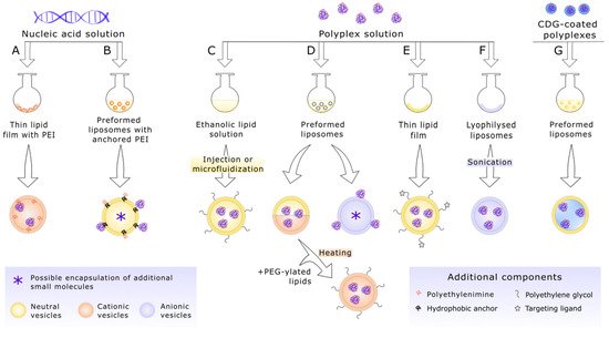
Lipopolyplexes based on poliethylenimine are an interesting platform for future anti-cancer gene therapies. The carrier consists of nucleic acids condensed with poliethylenimine chains and enclosed in lipid vesicles. Lipopolyplexes could be very versatile, what enables tailoring the carrier for specific thereapeutic needs, however the preparation process is a multistage and fairly sensitive one, which additionally requires a specific balance to be maintained between its stability in the body, which would allow the appropriate dose of the preparation to reach the target site, and the ability to release nucleic acid at the right place and time.

lipopolyplexes polyethyleneimine nucleic acids lipids liposomes non-viral vectors gene therapy cancer

1. Polyethyleneimine-Based Lipopolyplexes as Nucleic Acid Carriers

Recent years have witnessed rapidly growing interest in application of gene therapies for cancer treatment. However, this strategy requires nucleic acid carriers that are both effective and safe. In this context, non-viral vectors have advantages over their viral counterparts. In particular, lipopolyplexes – nanocomplexes consisting of nucleic acids condensed with polyvalent molecules and enclosed in lipid vesicles – currently offer great promise. Lipopolyplexes combine polyplexes and lipid vesicles, drawing benefits from both systems. A wide range of available lipids capable of spontaneous formation of a stable bilayer and the ease of its modification permit the preparation of a carrier tailored for individual therapeutic needs. On the other hand polyethylenimine (PEI) – a relatively easy to manufacture (and inexpensive) polycation, used to introduce nucleic acids into eukaryotic cells in vitro since 1995. [1] The structure of lipopolyplexes based on polyethyleneimine varies depending on formulation; exemplary components and the schematic layout of such carriers are shown in Figure 1.
Figure 1. Structure and components of various polyethyleneimine-based lipopolyplex carriers for gene delivery. A’—Reversed lipopolyplexes with polyethyleneimine bound covalently to hydrophobic anchor (e.g., triamcinolone acetonide) embedded in a neutral lipid bilayer; A”—reversed lipopolyplexes composed of anionic lipids that enable docking of positively charged polyplexes based on polyethyleneimine; B—“classical” lipopolyplexes with the addition of cationic lipids; C—lipopolyplex with targeting ligands (e.g., antibodies, transferrin, aptamers) conjugated to the surface via polyethylene glycol (PEG) modified lipids. bPEI—branched polyethyleneimine; DMPG—1.2-dimyristoyl-sn-glycero-3-phospho-(1′-rac-glycerol) (sodium salt), negatively charged; DOPE—1.2-dioleoyl-sn-glycero-3-phosphoethanolamine; DOTAP—1.2-dioleoyl-3-trimethylammonium-propane (chloride salt), positively charged; DPPC—1.2-dipalmitoyl-sn-glycero-3-phosphocholine; DSPE-PEG—1.2-distearoyl-sn-glycero-3-phosphorylethanolamine conjugated with polyethylene glycol chains; lPEI—linear polyethyleneimine.
The use of lipopolyplexes usually allows for much more effective transfection, both in vitro and in vivo, compared to polyplexes alone [2][3]. More specifically, lipopolyplexes present satisfactory pharmacokinetics when administered intravenously [4][5]. In the case of carriers without any specific targeting ligands on their surface, e.g., those coated with polyethylene glycol (see below), which makes them stealth carriers and thus more stable in the bloodstream [6][7], the main parameter limiting penetration of tissues is size. In anti-cancer therapies, this allows the use of a passive targeting strategy based on the occurrence of an enhanced permeability and retention effect (EPR), which refers to abnormal structure of endothelium of blood vessels and lack of lymphatics surrounding the tumor [5][8][9]. Moreover, the modular architecture of lipopolyplexes enables further modifications of their surface, including addition of targeting molecules, such as antibodies and their fragments, peptides, and proteins, aptamers, or sugar moieties, not interfering with either the structure of the polyplexes located inside of the vesicles or the mechanical properties of the lipid shell [10][11]. Receptors overexpressed at the surface of cells exhibiting compromised mechanisms of cell proliferation and/or survival regulation have become one of the most common targets, for instance, folate receptor α (FRα) or transferrin receptor 1 (TfR1) [11][12][13]. Exposure of their agonists on the surface of genetic drug carriers often leads to improvement of their internalization kinetics [11]. Such active targeting also reduces cytotoxicity to normal cells, while maintaining efficient transfection of pathological ones [5][14]. For example, cruciality of targeting in some cases was demonstrated on patient-derived glioblastoma stem-like cells, where non-functionalized lipopolyplexes were internalized to a negligible degree. In contrast, vesicles exposing on the surface fibronectin-mimetic peptide that specifically targets the α5β1 integrin (protein overexpressed in glioblastomas) presented satisfactory transfection efficiency in vitro [15]. Nevertheless, it should be taken into consideration that some ligands are specific only for cancer cells of a given phenotype (e.g., epidermal growth factor variant III, expressed in patients with glioblastoma multiforme and incapable of binding any known ligand [16]). However, they may vary between different types of tumor (e.g., heterogeneity of breast cancers, markers of which differ both among patients and within each individual tumor in one body [17]), but also changes related to the tumor progression. An exemplary case is metastatic tumors, as evolution of their genotypes occurs independently of the primary tumor and provides, inter alia, organ-specific adaptation to a new microenvironment. For instance, upregulation of L1 cell adhesion molecule (L1CAM) initiates outgrowths of colorectal cancer into perivascular sites [18]. All of the above might result in making some groups of cancer cells non-recognizable to a targeted carrier [19][20].

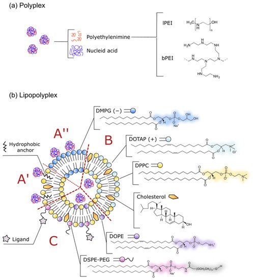
2. Composition of an Effective Lipopolyplex Based on Polyethyleneimine

First of all, a crucial factor to take into consideration is the N/P ratio, which corresponds to the molar ratio of, respectively, nitrogen atoms within positively charged imine groups of a polyethyleneimine to phosphorous atoms comprising anionic phosphate groups in a nucleic acid backbone. This impacts both the size of the resulting polyplexes and their net charge. Excess of PEI not only seems to prevent aggregation of such cationic polyplexes but might also improve transfection efficiency [21][22]. However, the optimal N/P ratio for efficient transfection is unique to each formulation, as it depends on various factors, such as size and topology of both of the polymers, ionic strength of the environment, and even the cell line to be transfected, some of which are discussed below [23][24].
Due to the electrostatic nature of the interactions between PEI and nucleic acids, their association, to a large extent, depends on the factors influencing the ionization of both molecules. For instance, it has been demonstrated that, in the case of 2.5 kDa linear polyethyleneimine (lPEI), both neutral pH and high ionic strength could lead to aggregation of its chains and precipitation, thus reducing the possibility of interaction with polyanionic molecules [25].
Another crucial interplay could be observed between polyplex formation and the molecular weight (MW) of the polyethyleneimine used. Hence, those with lower MW tend to exhibit poorer condensation of DNA and RNA, which can translate into diminished transfection using such complexes alone [21][26][27]. However, this tendency is reduced by enclosing polyplexes in liposomes, due to the lipid envelope properties [2][28]. Thus, the lipid composition seems to be, to a large extent, responsible for a carrier’s transfection efficiency [29]. It is even considered that, while, in the case of lipopolyplexes, PEI is mainly responsible for intracellular protection and distribution of nucleic acids, the lipid envelope plays an analogous role at the extracellular level. As a result, shorter chains of PEI are predominantly used for lipopolyplex formulations intended for in vivo administration, especially given their low cytotoxicity (the higher the MW, the more cytotoxic effect that polyplexes have) [26][30], whilst providing effective protection against nucleases [31][32].
It also seems that the topology of polyethyleneimine molecules is of considerable significance for the effective formation of polyplexes, despite the fact that currently no clear trend in this matter could be delineated. Namely, there are studies recognizing the superiority of linear PEI as a carrier of nucleic acids [21][33], as well as those favoring branched chains [34][35]. Most likely, it further depends on what size and kind of nucleic acid is condensed with a given PEI. For example, branched chains with high MW seem to work better with oligonucleotides, as opposed to 25 kDa lPEI, which is considered the gold standard when transfecting cells with plasmid DNA (pDNA). Interestingly, plasmid linearization hampers the complexation process [36][37][38]. Moreover, Kwok and Hart observed that pDNA complexes are more stable than those with siRNA, and the branched form of PEI is more effective in the case of RNA [39].
Next, the composition of the lipid envelope needs to be selected to a large extent with the aim of increasing, inter alia, the biocompatibility and stability of the carrier, hence the widespread use of glycerophospholipids with dominance of phosphatidylcholines and phosphatidylethanolamines with saturated acyl chains in their structure, which are able to spontaneously form a stable bilayer with low permeability at physiological temperature. In the case of lipopolyplexes, the most commonly used are: stability-enhancing cholesterol, phospholipids—anionic and zwitterionic ones and synthetic cationic lipids, examples and the structure of which are shown in Figure 1 [40][41][42][43][44][45][46][47]. Among these, particularly noteworthy is 1.2-dioleoyl-sn-glycero-3-phosphoethanolamine (DOPE), utilized both in cationic and neutral lipid shells. In the acidic environment of late endosomes, it is able to destabilize the membranes of these organelle and, thereby, partake in the release of the vector into the cytosol, helping to avoid its lysosomal degradation [48]. The displacement of cationic lipids, such as 1.2-dioleoyl-3-trimethylammonium-propane (DOTAP) or alkaline amino acid cholesterol derivatives (e.g., histidylated cholesterol), may be sometimes beneficial, because they exhibit more toxicity as they do not occur naturally in the body and could trigger an adverse interaction with serum and cell components due to the positive charge. However, various studies are still based on such positively charged carriers, due to their high transfection efficiency, as they are capable of electrostatic interaction with biological membranes, thus facilitating both the uptake of the vesicle and its subsequent release from the endosome [49][50][51][52]. It is further debated whether the addition of cationic lipids leads to their interaction with nucleic acids, resulting in further condensation [53][2][47][54].
Finally, the process of lipopolyplex formation itself seems to be equally important as the components; exemplary, commonly employed methods are presented in Figure 2. One of these is dry lipid film rehydration with a polyplex suspension, employed mainly when dealing with oligonucleotides. The utility of this technique, especially for more complex lipid formulations (e.g., immunolipopolyplexes), was demonstrated independently by Meissner et al. [55] and Ko et al. [46] and is still used in most recent studies concerning functionalized lipopolyplexes [15]. Meanwhile, Heyes et al. [43] proposed spontaneous vesicle formation, mixing the preformed polyplexes with an ethanolic lipid solution, which also turns out to be an efficient method of preparing PEGylated carriers for plasmid DNA. Penacho et al. [56] also stated that this method enables more robust transfection than with the hydration-extrusion method, when used to form lipopolyplexes with cationic lipids. Currently, some modification of this strategy comprising of the use of microfluidic technology or the dissolution of polyplexes in high-density polymers, such as poloxamer, have been demonstrated to increase the encapsulation efficiency [57].
Nevertheless, the technique that dominated the majority of published works relies on incubating liposome suspensions with already preformed polyplexes containing small RNA molecules, DNA oligonucleotides or plasmid DNA [2][41][5][9][42][45][47][50][52][58][59]. Presumably, the efficiency of such methods depends on exposure of vesicles to the fusogenic abilities of free polyethyleneimine chains and, in some cases, can be further enhanced by mechanical disruption of the lipid bilayer, e.g., via sonication. When using cationic liposomes, this strategy was demonstrated to be the most efficient one in comparison to mixing the cationic polymer with lipoplexes or with lipids and nucleic acid solutions simultaneously [60]. High transfection using lipopolyplexes obtained by this strategy was reported by Garcìa et al. [54], who carried out comparative tests on cationic liposomes encapsulating polyplexes. Interestingly, in this case, the order in which polymers were mixed together was also crucial; namely, addition of the branched PEI suspension to the DNA solution was found to be much more effective in terms of transfection than the other way around. This phenomenon was also reflected in the study of polyplexes alone conducted in vitro by Cho et al. [61]. The authors suggested that larger aggregates (about 200 nm), formed when PEI solution was instilled into DNA solution, are able to better overcome the barrier of lipid membranes. However, it is unknown whether, in the case of the experiments of Garcia et al. [54], this phenomenon might have translated into more effective internalization of polyplexes into liposomes. Even so, attention should be drawn to the possible dependence of the encapsulation efficiency using the presented method on the composition of the liposomes. Electron microscope observations confirmed that, in the case of an uncharged shell made of 1.2-dipalmitoyl-sn-glycero-3-phosphocholine (DPPC), this technique actually allows the encapsulation of polyplexes inside the vesicles [5]. It also seems that the efficiency of this process could be improved with the use of appropriate lipid anchors (e.g., by coating polyplexes with dodecyl-glucopyranoside derivative (CDG)) or by taking advantage of the electrostatic interactions between positively charged polyplexes and anionic lipids [62][42][47].
Even so, in most circumstances, there is still no clear evidence for complete internalization of complexes when incubating them with preformed liposomes. Thus, bearing in mind that Garcia et al. [54] only evaluated the effectiveness of the carrier based on the expression of the reporter gene, without explicitly estimating encapsulation efficiency of the polyplexes, it might be possible that the latter just associate with the carrier’s surface rather than migrate to the inner hydrophilic compartment of vesicles. Moreover, the presence of additional molecules on the surface, such as hydrophilic PEG, may constitute an additional barrier preventing the penetration of the complexes into the liposomes and/or their adsorption on the surface of such carriers. Hence, in some protocols, PEGylated lipids were added after the incubation of vesicles with polyplexes and subsequently heated to obtain surface-modified lipopolyplexes [50][52].
Despite the fact that most studies are aimed at polyplex encapsulation into liposomes, it does not seem to be necessary to obtain high transfection with simultaneous retained integrity of the therapeutic nucleic acids. Therefore, even just the conjugation of complexes of PEI and DNA or RNA to the surface of the vesicles seems to comply with these conditions [63][64]. Reverse lipopolyplexes, briefly mentioned earlier and illustrated in Figure 1, exactly fulfill the idea of transporting polyplexes as the external layer of the liposomal carrier. Exposure of polyethyleneimine helps to circumvent some limitations of lipid carriers, namely short circulation times (especially considering lipoplexes), simultaneously increasing endosomal release due to its increased efficiency as a proton sponge [38][49]. The standard procedure utilizes either non-covalent adsorption of polyethyleneimine to negatively charged lipids and surfactants constituting the preformed vesicles [65][66] or covalent modification of PEI with some hydrophobic anchor that enables close interaction with lipid bilayers. The reverse phase evaporation method is commonly used in such cases [67]. However, the protocol based on electrostatic interaction between PEI and the liposomal surface is heavily pH-dependent. As Sabín et al. [65] demonstrated, in the case of vesicles composed of zwitterionic lipids, association of PEI occurs only in a pH range providing opposite charges for both particles (e.g., for 1.2-dioleoyl-sn-glycero-3-phosphocholine (DOPC) liposomes, the optimal pH is in a range from 6 to 10). Thus, use of anchors, some of which additionally could facilitate nuclear transport (e.g., triamcinolone acetonide that acts as a nuclear localization signal) [67], might form more stable carriers.
Figure 2. Possible procedures used in lipopolyplex preparation. A—Hydration of thin lipid film prepared from cationic lipids and polyethyleneimine (PEI) with nucleic acid solution [45]; B—reverse lipopolyplexes obtained through complexation of nucleic acid with PEI grafted to neutral lipid vesicles via hydrophobic anchor [67]; C—injection of polyplex solution into ethanolic solution of neutral lipids is a feasible way to obtain PEGylated lipopolyplexes [43][68]; D—incubation of polyplexes with preformed liposomes can result in either reverse lipopolyplexes (when anionic vesicles are used) [65][66] or “classic” lipopolyplexes (neutral or cationic vesicles) [2][41][5][9][42][45][47][50][52][58][59]; for preparation of cationic, stealth lipopolyplexes, PEGylated lipids could be incorporated in preformed carriers via mixture heating [50][52]; E—hydration of thin lipid film enables preparation of targeted lipopolyplexes [55][46]; F—hydration of lyophilized anionic liposomes might help polyplex internalization [42]; G—coating polyplexes with multicarboxyl dodecyl glucopyranoside anchors them to neutral lipid vesicles [62]. The presented approaches could be combined with each other and further modified, which enables a broad array of various lipopolyplexes to be generated.

3. Selected In Vivo Studies on Anticancer PEI-Based Lipopolyplexes

Recent preclinically in vivo tested therapies regarding lipopolyplexes focus mainly on silencing overexpressed and abnormal genes using miRNA, siRNA, and antisense oligonucleotides (ODN) [55][5][9][50][52][59] or the activation of suppressors using saRNA [42]. It is worth noting that these carriers, regardless of the type of nucleic acid, are in prevalence based on low-molecular, thus less toxic polyethyleneimines (2–10 kDa) and have a lipid shell composed of various phosphatidylethanolamines (PE) and phosphatidylcholines (PC). Another remarkable fact is feasibility of most of presented lipopolyplexes to intravenous administration (usually through tail vein in animal models), as it makes them adequate for fighting not only primary tumor but also metastasis.
Linder et al. [9] prepared vesicles with a diameter of around 300 nm and zeta potential close to zero, loaded with polyplexes based on Stat3-siRNA. In pathological conditions this protein is responsible for the activation of various genes that promote cancer, including those related to cell migration [69]. The group demonstrated extension of the life span of mice bearing glioblastoma, despite the fact that this locally administered formulation reached only a fraction of tumor cells. Ewe et al. [5], using the same phenomenon that is RNA interference but a different molecular target (survivin, an apoptosis inhibitor [70]), reduced PC-3 (prostate cancer) cell proliferation in a murine xenograft model, while observing a lack of immunostimulation upon systemic administration of DPPC-based lipopolyplexes. However, the excellent biodistribution of these non-PEGylated vesicles might result in part from intraperitoneal injection, rather than the more clinically relevant intravenous one.
In turn, the team [55] developed an antisense therapy based on vesicles decorated with the anti-CD20 antibody covalently bound to PEGylated lipids. Thanks to the active targeting strategy, this carrier could be utilized against both acute lymphoblastic leukemia and lymphomas, where CD20 is overexpressed on the surface of abnormal white blood cells. The approach chosen here was to decrease the expression of the anti-apoptotic BCL2 gene at the transcription level, which, in consequence, led to a reduction in the level of the cell survival-promoting protein, both in vitro and in NOD/SCID mice bearing xenograft tumors of Daudi human Burkitt’s lymphoma, after intravenous injection. Interestingly, the formulation stability was preserved for at least a year either in suspension or as freeze-dried powder.
An alternative approach was presented by Wang et al. [42], who utilized saRNA to stimulate the expression of the regulatory factor p21. This protein corresponds, inter alia, to cell cycle arrest in cells with damaged genetic material. Thanks to the additional coating with hyaluronic acid (HA), the anionic lipopolyplexes injected intratumorally were able to yield high transfection of colorectal cancer cells with abnormal amounts of CD44 receptor on their surface, since HA promotes adhesion and receptor-mediated endocytosis into such cells. This example represents another feasible strategy to limit cancerous cell division, leading, in consequence, to inhibition of tumor growth in an orthotropic xenograft model.
Xue et al. [62] designed a carrier that structurally mimics lentiviral particles, where a polyplex core is fused to the neutral vesicles using the aforementioned CDG derivative. Although, originally, the outer envelope consisted only of egg lecithin and cholesterol, it was demonstrated that it is possible to incorporate a variety of functional components in it, such as PEGylated lipids. Upon intravenous administration, the nanoparticles seemed to successfully deliver siRNA to the U87-MG glioblastoma cells implanted under the skin of nude mice, silencing the expression of vascular endothelial growth factor (VEGF), and thus reducing capillary density at the tumor site more efficiently than just polyplexes.
A slightly different approach was chosen by Jilek et al. [50] and Petrek et al. [52], who attempted enclosing small, non-coding RNA complexed with branched polyethyleneimine in a PEGylated shell containing cationic lipids. Both studies utilized recombinant miRNA molecules obtained via bacterial fermentation (BERA), that were based on a hybrid tRNA/pre-miRNA scaffold. The first of the mentioned approaches [50] introduced miRNA let-7c to mice bearing orthotopic hepatocellular carcinoma. This molecule, capable of inhibiting expression of the Bcl-xL protein, brings about apoptosis of the mentioned tumor cells in vivo, thus prolonging the life span of the tested animals. Meanwhile, the second team [52] applied an analogous strategy to the double hybrid let-7c/miR-124, controlling the expression of genes of the RAS, VAMP3, and CDK6 families, which prolonged the survival of non-small cell lung cancer xenograft in a murine model. Interestingly, in both studies, no significant changes of the markers (such as alanine aminotransferase, aspartate aminotransferase, albumin, creatinine, blood urea nitrogen, and total bilirubin) in the blood of the animals were detected, which was considered to be a hallmark of low toxicity of the preparations.
The complexity of pathological processes occurring in the cell during neoplasm also prompts the development of complex preparations with more than one molecular target. A remarkable example is the nanoparticles recently developed by Wang et al. [71] that carry simultaneously a plasmid containing the PTEN gene, an important suppressor of the cell cycle, and epigallocatechin gallate (EGCG), a flavonoid exhibiting strong antioxidant activity. The vesicles, containing the flavonoid in the hydrophilic core, additionally had 1.2-distearoyl-sn-glycero-3-phosphoethanolamine (DSPE) incorporated in the envelope conjugated with PEI chains projecting outwards, which enabled condensation of DNA on the carrier’s surface. This formulation takes advantage of EGCG’s ability to scavenge excess free radicals present in the tumor microenvironment, which may inactivate tyrosine phosphatases, such as PTEN. However, this flavonoid not only restores the functionality of the enzyme but also regulates other signaling proteins related to, for example, the apoptotic pathway [64]. Such multifunctional nanocarriers enabled the proliferation of PC-3 neoplastic cells to be limited in vitro, but also led to significant inhibition of tumor growth in vivo [71].
Another appealing example of bifunctional reverse lipopolyplexes was proposed by Mendes et al. [72]. This team developed bPEI-modified liposomes of egg PE and cholesterol that carry siRNA targeting multidrug resistance gene MDR1 and, subsequently, encapsulate chemotherapeutic drug—paclitaxel (PTX). The formulation takes advantage of synergy between PTX and siRNA-MDR1, as the latter downregulated proteins associated with drug resistance (such as P-glycoprotein), thus enhancing efficacy of PTX, a classic cancer drug that prevents microtubule disassembly. Moreover, such lipopolyplexes were able to promote in vivo tumor growth inhibition in an MDR xenograft ovarian tumor model (A2780-ADR cell line). This gives an exciting possibility for future combined therapies against drug-resistant cancers.

4. Summary

The preparation of polyethylenimine-based lipopolyplexes is a multistage and fairly sensitive process, which additionally requires a specific balance to be maintained between its stability in the body, which would allow the appropriate dose of the preparation to reach the target site, and the ability to release nucleic acid at the right place and time. However, as the above examples of research show, such a carrier is a promising platform for future anti-cancer gene therapies, especially due to its versatility. The possibility to select from a broad array of components (DNA in the form of plasmid or oligonucleotides, small RNAs, polyethylenimines of various length and topology, lipids and/or additional targeting ligands) and exchange or modify them without affecting the overall carrier’s functionality makes it possible to tailor a lipopolyplex suitable for a given application. Moreover, lipopolyplexes’ performance in vivo is superior to bare polyplexes or nucleic acids, and even though no formulation has yet reached the transfection effectiveness of viral carriers, they are able to safely deliver therapeutic nucleic acids, notwithstanding the manner in which these molecules are associated with the carrier (i.e. inside a lipid vesicle or on its surface). Finally, the repeatedly emphasized modularity and flexibility of lipopolyplexes allow considerable room for improvement of these carriers in the coming years.

References

  1. Ewe, A.; Schaper, A.; Barnert, S.; Schubert, R.; Temme, A.; Bakowsky, U.; Aigner, A. Storage stability of optimal liposome-polyethylenimine complexes (lipopolyplexes) for DNA or siRNA delivery. Acta Biomater. 2014, 10, 2663–2673.
  2. Hanzlíková, M.; Soininen, P.; Lampela, P.; Männistö, P.T.; Raasmaja, A. The role of PEI structure and size in the PEI/liposome-mediated synergism of gene transfection. Plasmid 2009, 61, 15–21.
  3. Sercombe, L.; Veerati, T.; Moheimani, F.; Wu, S.Y.; Sood, A.K.; Hua, S. Advances and challenges of liposome assisted drug delivery. Front. Pharmacol. 2015, 6, 286.
  4. Ewe, A.; Panchal, O.; Pinnapireddy, S.R.; Bakowsky, U.; Przybylski, S.; Temme, A.; Aigner, A. Liposome-polyethylenimine complexes (DPPC-PEI lipopolyplexes) for therapeutic siRNA delivery in vivo. Nanomed. Nanotechnol. Biol. Med. 2017, 13, 209–218.
  5. Immordino, M.L.; Dosio, F.; Cattel, L. Stealth liposomes: Review of the basic science, rationale, and clinical applications, existing and potential. Int. J. Nanomed. 2006, 1, 297–315.
  6. Needham, D.; McIntosh, T.J.; Lasic, D.D. Repulsive interactions and mechanical stability of polymer-grafted lipid membranes. BBA Biomembr. 1992, 1108, 40–48.
  7. Maeda, H. The enhanced permeability and retention (EPR) effect in tumor vasculature: The key role of tumor-selective macromolecular drug targeting. Adv. Enzym. Regul. 2001, 41, 189–207.
  8. Linder, B.; Weirauch, U.; Ewe, A.; Uhmann, A.; Seifert, V.; Mittelbronn, M.; Harter, P.N.; Aigner, A.; Kögel, D. Therapeutic targeting of stat3 using lipopolyplex nanoparticle-formulated sirna in a syngeneic orthotopic mouse glioma model. Cancers 2019, 11, 333.
  9. Toporkiewicz, M.; Meissner, J.; Matusewicz, L.; Czogalla, A.; Sikorski, A.F. Toward a magic or imaginary bullet? Ligands for drug targeting to cancer cells: Principles, hopes, and challenges. Int. J. Nanomed. 2015, 10, 1399–1414.
  10. Yoo, J.; Park, C.; Yi, G.; Lee, D.; Koo, H. Active targeting strategies using biological ligands for nanoparticle drug delivery systems. Cancers 2019, 11, 640.
  11. Cheung, A.; Bax, H.J.; Josephs, D.H.; Ilieva, K.M.; Pellizzari, G.; Opzoomer, J.; Bloomfield, J.; Fittall, M.; Grigoriadis, A.; Figini, M.; et al. Targeting folate receptor alpha for cancer treatment. Oncotarget 2016, 7, 52553–52574.
  12. Shen, Y.; Li, X.; Dong, D.; Zhang, B.; Xue, Y.; Shang, P. Transferrin receptor 1 in cancer: A new sight for cancer therapy. Am. J. Cancer Res. 2018, 8, 916–931.
  13. Bofinger, R.; Zaw-Thin, M.; Mitchell, N.J.; Patrick, P.S.; Stowe, C.; Gomez-Ramirez, A.; Hailes, H.C.; Kalber, T.L.; Tabor, A.B. Development of lipopolyplexes for gene delivery: A comparison of the effects of differing modes of targeting peptide display on the structure and transfection activities of lipopolyplexes. J. Pept. Sci. 2018, 24, e3131.
  14. Shabana, A.M.; Xu, B.; Schneiderman, Z.; Ma, J.; Chen, C.C.; Kokkoli, E. Targeted liposomes encapsulating mir-603 complexes enhance radiation sensitivity of patient-derived glioblastoma stem-like cells. Pharmaceutics 2021, 13, 1115.
  15. Gan, H.K.; Cvrljevic, A.N.; Johns, T.G. The epidermal growth factor receptor variant III (EGFRvIII): Where wild things are altered. FEBS J. 2013, 280, 5350–5370.
  16. Riggio, A.I.; Varley, K.E.; Welm, A.L. The lingering mysteries of metastatic recurrence in breast cancer. Br. J. Cancer 2020, 124, 13–26.
  17. Ganesh, K.; Basnet, H.; Kaygusuz, Y.; Laughney, A.M.; He, L.; Sharma, R.; O’Rourke, K.P.; Reuter, V.P.; Huang, Y.-H.; Turkekul, M.; et al. L1CAM defines the regenerative origin of metastasis-initiating cells in colorectal cancer. Nat. Cancer 2020, 1, 28–45.
  18. Gao, D.; Mittal, V.; Ban, Y.; Lourenco, A.R.; Yomtoubian, S.; Lee, S. Metastatic tumor cells—Genotypes and phenotypes. Front. Biol. 2018, 13, 277–286.
  19. Fares, J.; Fares, M.Y.; Khachfe, H.H.; Salhab, H.A.; Fares, Y. Molecular principles of metastasis: A hallmark of cancer revisited. Signal Transduct. Target. Ther. 2020, 5, 28.
  20. Dai, Z.; Wu, C. How does DNA complex with polyethylenimine with different chain lengths and topologies in their aqueous solution mixtures? Macromolecules 2012, 45, 4346–4353.
  21. Choosakoonkriang, S.; Lobo, B.A.; Koe, G.S.; Koe, J.G.; Middaugh, C.R. Biophysical characterization of PEI/DNA complexes. J. Pharm. Sci. 2003, 92, 1710–1722.
  22. Chytła, A.; Gajdzik-Nowak, W.; Biernatowska, A.; Sikorski, A.F.; Czogalla, A. High-level expression of palmitoylated MPP1 recombinant protein in mammalian cells. Membranes 2021, 11, 715.
  23. Bono, N.; Ponti, F.; Mantovani, D.; Candiani, G. Non-viral in vitro gene delivery: It is now time to set the bar! Pharmaceutics 2020, 12, 183.
  24. Curtis, K.A.; Miller, D.; Millard, P.; Basu, S.; Horkay, F.; Chandran, P.L. Unusual salt and pH induced changes in polyethylenimine solutions. PLoS ONE 2016, 11, e0158147.
  25. Godbey, W.T.; Wu, K.K.; Mikos, A.G. Size matters: Molecular weight affects the efficiency of poly(ethylenimine) as a gene delivery vehicle. J. Biomed. Mater. Res. 1999, 45, 268–275.
  26. Wagner, M.; Rinkenauer, A.C.; Schallon, A.; Schubert, U.S. Opposites attract: Influence of the molar mass of branched poly(ethylene imine) on biophysical characteristics of siRNA-based polyplexese. RSC Adv. 2013, 3, 12774–12785.
  27. Lampela, P.; Soininen, P.; Urtti, A.; Männistö, P.T.; Raasmaja, A. Synergism in gene delivery by small PEIs and three different nonviral vectors. Int. J. Pharm. 2004, 270, 175–184.
  28. Lampela, P.; Elomaa, M.; Ruponen, M.; Urtti, A.; Männistö, P.T.; Raasmaja, A. Different synergistic roles of small polyethylenimine and Dosper in gene delivery. J. Control. Release 2003, 88, 173–183.
  29. Kunath, K.; von Harpe, A.; Fischer, D.; Petersen, H.; Bickel, U.; Voigt, K.; Kissel, T. Low-molecular-weight polyethylenimine as a non-viral vector for DNA delivery: Comparison of physicochemical properties, transfection efficiency and in vivo distribution with high-molecular-weight polyethylenimine. J. Control. Release 2003, 89, 113–125.
  30. Xun, M.M.; Xiao, Y.P.; Zhang, J.; Liu, Y.H.; Peng, Q.; Guo, Q.; Wu, W.-X.; Xu, Y.; Yu, X.-Q. Low molecular weight PEI-based polycationic gene vectors via Michael addition polymerization with improved serum-tolerance. Polymer 2015, 65, 45–54.
  31. Eigner, A.; Fischer, D.; Merdan, T.; Brus, C.; Kissel, T.; Czubayko, F. Delivery of unmodified bioactive ribozymes by an RNA stabilizing polyethylenimine LMW PEI efficiently down regulates gene expression. Gene Ther. 2002, 9, 1700–1707.
  32. Itaka, K.; Harada, A.; Yamasaki, Y.; Nakamura, K.; Kawaguchi, H.; Kataoka, K. In situ single cell observation by fluorescence resonance energy transfer reveals fast intra-cytoplasmic delivery and easy release of plasmid DNA complexed with linear polyethylenimine. J. Gene Med. 2004, 6, 76–84.
  33. Dai, Z.; Gjetting, T.; Mattebjerg, M.A.; Wu, C.; Andresen, T.L. Elucidating the interplay between DNA-condensing and free polycations in gene transfection through a mechanistic study of linear and branched PEI. Biomaterials 2011, 32, 8626–8634.
  34. Dunlap, D.D.; Maggi, A.; Soria, M.R.; Monaco, L. Nanoscopic structure of DNA condensed for gene delivery. Nucleic Acids Res. 1997, 25, 3095–3101.
  35. Reisinger, H.; Steinfellner, W.; Katinger, H.; Kunert, R. Serum-free transfection of CHO cells with chemically defined transfection systems and investigation of their potential for transient and stable transfection. Cytotechnology 2009, 60, 115–123.
  36. Sundaram, S.; Lee, L.K.; Roth, C.M. Interplay of polyethyleneimine molecular weight and oligonucleotide backbone chemistry in the dynamics of antisense activity. Nucleic Acids Res. 2007, 35, 4396–4408.
  37. Lehner, R.; Wang, X.; Hunziker, P. Plasmid linearization changes shape and efficiency of transfection complexes. Eur. J. Nanomed. 2013, 5, 205–212.
  38. Kwok, A.; Hart, S.L. Comparative structural and functional studies of nanoparticle formulations for DNA and siRNA delivery. Nanomed. Nanotechnol. Biol. Med. 2011, 7, 210–219.
  39. Juszkiewicz, K.; Sikorski, A.F.; Czogalla, A. Building blocks to design liposomal delivery systems. Int. J. Mol. Sci. 2020, 21, 9559.
  40. Pinnapireddy, S.R.; Duse, L.; Strehlow, B.; Schäfer, J.; Bakowsky, U. Composite liposome-PEI/nucleic acid lipopolyplexes for safe and efficient gene delivery and gene knockdown. Colloids Surf. B Biointerfaces 2017, 158, 93–101.
  41. Wang, L.L.; Feng, C.L.; Zheng, W.S.; Huang, S.; Zhang, W.X.; Wu, H.N.; Zhan, Y.; Han, Y.-X.; Wu, S.; Jiang, J.-D. Tumor-selective lipopolyplex encapsulated small active RNA hampers colorectal cancer growth in vitro and in orthotopic murine. Biomaterials 2017, 141, 13–28.
  42. Heyes, J.; Palmer, L.; Chan, K.; Giesbrecht, C.; Jeffs, L.; MacLachlan, I. Lipid encapsulation enables the effective systemic delivery of polyplex plasmid DNA. Mol. Ther. 2007, 15, 713–720.
  43. Huang, X.; Schwind, S.; Yu, B.; Santhanam, R.; Wang, H.; Hoellerbauer, P.; Mims, A.; Klisovic, R.; Walker, A.R.; Chan, K.K.; et al. Targeted delivery of microRNA-29b by transferrin-conjugated anionic lipopolyplex nanoparticles: A novel therapeutic strategy in acute myeloid leukemia. Clin. Cancer Res. 2013, 19, 2355–2367.
  44. Perche, F.; Clemençon, R.; Schulze, K.; Ebensen, T.; Guzmán, C.A.; Pichon, C. Neutral lipopolyplexes for in vivo delivery of conventional and replicative RNA vaccine. Mol. Ther. Nucleic Acids 2019, 17, 767–775.
  45. Ko, Y.T.; Bhattacharya, R.; Bickel, U. Liposome encapsulated polyethylenimine/ODN polyplexes for brain targeting. J. Control. Release 2009, 133, 230–237.
  46. Schäfer, J.; Höbel, S.; Bakowsky, U.; Aigner, A. Liposome-polyethylenimine complexes for enhanced DNA and siRNA delivery. Biomaterials 2010, 31, 6892–6900.
  47. Du, Z.; Munye, M.M.; Tagalakis, A.D.; Manunta, M.D.I.; Hart, S.L. The role of the helper lipid on the DNA transfection efficiency of lipopolyplex formulations. Sci. Rep. 2014, 4, 7107.
  48. Rezaee, M.; Oskuee, R.K.; Nassirli, H.; Malaekeh-Nikouei, B. Progress in the development of lipopolyplexes as efficient non-viral gene delivery systems. J. Control. Release 2016, 236, 1–14.
  49. Jilek, J.L.; Zhang, Q.Y.; Tu, M.J.; Ho, P.Y.; Duan, Z.; Qiu, J.X.; Yu, A.M. Bioengineered let-7c inhibits orthotopic hepatocellular carcinoma and improves overall survival with minimal immunogenicity. Mol. Ther. Nucleic Acids 2019, 14, 498–508.
  50. Song, H.; Wang, G.; He, B.; Li, L.; Li, C.; Lai, Y.; Xu, X.; Gu, Z. Cationic lipid-coated PEI/DNA polyplexes with improved efficiency and reduced cytotoxicity for gene delivery into mesenchymal stem cells. Int. J. Nanomed. 2012, 7, 4637–4648.
  51. Petrek, H.; Ho, P.Y.; Batra, N.; Tu, M.J.; Zhang, Q.; Qiu, J.X.; Yu, A.M. Single bioengineered ncRNA molecule for dual-targeting toward the control of non-small cell lung cancer patient-derived xenograft tumor growth. Biochem. Pharmacol. 2021, 189, 114392.
  52. Zhang, Q.-Y.; Ho, P.Y.; Tu, M.J.; Jilek, J.L.; Chen, Q.X.; Zeng, S.; Yu, A.M. Lipidation of polyethylenimine-based polyplex increases serum stability of bioengineered RNAi agents and offers more consistent tumoral gene knockdown in vivo. Int. J. Pharm. 2018, 547, 537.
  53. García, L.; Buñuales, M.; Düzgüneş, N.; de Ilarduya, C.T. Serum-resistant lipopolyplexes for gene delivery to liver tumour cells. Eur. J. Pharm. Biopharm. 2007, 67, 58–66.
  54. Meissner, J.M.; Toporkiewicz, M.; Czogalla, A.; Matusewicz, L.; Kuliczkowski, K.; Sikorski, A.F. Novel antisense therapeutics delivery systems: In vitro and in vivo studies of liposomes targeted with anti-CD20 antibody. J. Control. Release 2015, 220, 515–528.
  55. Penacho, N.; Simões, S.; de Lima, M.C.P. Polyethylenimine of various molecular weights as adjuvant for transfection mediated by cationic liposomes. Mol. Membr. Biol. 2009, 26, 249–263.
  56. Ahmed, S.; Salmon, H.; Distasio, N.; Do, H.D.; Scherman, D.; Alhareth, K.; Tabrizian, M.; Mignet, N. Viscous core liposomes increase siRNA encapsulation and provides gene inhibition when slightly positively charged. Pharmaceutics 2021, 13, 479.
  57. Schwabe, K.; Ewe, A.; Kohn, C.; Loth, T.; Aigner, A.; Hacker, M.C.; Schulz-Siegmund, M. Sustained delivery of siRNA poly- and lipopolyplexes from porous macromer-crosslinked gelatin gels. Int. J. Pharm. 2017, 526, 178–187.
  58. Zhupanyn, P.; Ewe, A.; Büch, T.; Malek, A.; Rademacher, P.; Müller, C.; Reinert, A.; Jaimes, Y.; Aigner, A. Extracellular vesicle (ECV)-modified polyethylenimine (PEI) complexes for enhanced siRNA delivery in vitro and in vivo. J. Control. Release 2020, 319, 63–76.
  59. Pelisek, J.; Gaedtke, L.; DeRouchey, J.; Walker, G.F.; Nikol, S.; Wagner, E. Optimized lipopolyplex formulations for gene transfer to human colon carcinoma cells under in vitro conditions. J. Gene Med. 2006, 8, 186–197.
  60. Cho, S.K.; Dang, C.; Wang, X.; Ragan, R.; Kwon, Y.J. Mixing-sequence-dependent nucleic acid complexation and gene transfer efficiency by polyethylenimine. Biomater. Sci. 2015, 3, 1124–1133.
  61. Xue, Y.; Feng, J.; Liu, Y.; Che, J.; Bai, G.; Dong, X.; Wu, F.; Jin, T. A synthetic carrier of nucleic acids structured as a neutral phospholipid envelope tightly assembled on polyplex surface. Adv. Healthc. Mater. 2020, 9, 1901705.
  62. Kumar, K.; Vulugundam, G.; Kondaiah, P.; Bhattacharya, S. Co-liposomes of redox-active alkyl-ferrocene modified low MW branched PEI and DOPE for efficacious gene delivery in serum. J. Mater. Chem. B 2015, 3, 2318–2330.
  63. Chen, B.H.; Hsieh, C.H.; Tsai, S.Y.; Wang, C.Y.; Wang, C.C. Anticancer effects of epigallocatechin-3-gallate nanoemulsion on lung cancer cells through the activation of AMP-activated protein kinase signaling pathway. Sci. Rep. 2020, 10, 5163.
  64. Sabín, J.; Vázquez-Vázquez, C.; Prieto, G.; Bordi, F.; Sarmiento, F. Double charge inversion in polyethylenimine-decorated liposomes. Langmuir 2012, 28, 10534–10542.
  65. Opanasopit, P.; Paecharoenchai, O.; Rojanarata, T.; Ngawhirunpat, T.; Ruktanonchai, U. Type and composition of urfactants mediating gene transfection of polyethylenimine-coated liposomes. Int. J. Nanomed. 2011, 6, 975–983.
  66. Ma, K.; Shen, H.; Shen, S.; Xie, M.; Mao, C.; Qiu, L.; Jin, Y. Development of a successive targeting liposome with multi-ligand for efficient targeting gene delivery. J. Gene Med. 2011, 13, 290–301.
  67. Samaddar, S.; Mazur, J.; Boehm, D.; Thompson, D.H. Development and in vitro characterization of bladder tumor cell targeted lipid-coated polyplex for dual delivery of plasmids and small molecules. Int. J. Nanomed. 2019, 14, 9547–9561.
  68. Yu, H.; Lee, H.; Herrmann, A.; Buettner, R.; Jove, R. Revisiting STAT3 signalling in cancer: New and unexpected biological functions. Nat. Rev. Cancer 2014, 14, 736–746.
  69. Jaiswal, P.K.; Goel, A.; Mittal, R.D. Survivin: A molecular biomarker in cancer. Indian J. Med. Res. 2015, 141, 389–397.
  70. Wang, Y.; Wang, R.; Wu, S.; An, J.; Liang, Y.; Hou, L.; Zhang, Z. Self-responsive co-delivery system for remodeling tumor intracellular microenvironment to promote PTEN-mediated anti-tumor therapy. Nanoscale 2020, 12, 9392–9403.
  71. Mendes, L.P.; Sarisozen, C.; Luther, E.; Pan, J.; Torchilin, V.P. Surface-engineered polyethyleneimine-modified liposomes as novel carrier of siRNA and chemotherapeutics for combination treatment of drug-resistant cancers. Drug Deliv. 2019, 26, 443.
More
Information
Contributor MDPI registered users' name will be linked to their SciProfiles pages. To register with us, please refer to https://encyclopedia.pub/register :
View Times: 551
Revisions: 2 times (View History)
Update Date: 10 Jan 2022
1000/1000
Video Production Service