Submitted Successfully!
To reward your contribution, here is a gift for you: A free trial for our video production service.
Thank you for your contribution! You can also upload a video entry or images related to this topic.
Version Summary Created by Modification Content Size Created at Operation
1 + 2736 word(s) 2736 2021-12-17 04:19:08 |
2 Format correct Meta information modification 2736 2022-01-05 02:12:01 |

Video Upload Options

Do you have a full video?

Confirm

Are you sure to Delete?
Cite
If you have any further questions, please contact Encyclopedia Editorial Office.
Meroni, G. Probiotics in Treating Pathogenic Biofilms. Encyclopedia. Available online: https://encyclopedia.pub/entry/17732 (accessed on 30 June 2024).
Meroni G. Probiotics in Treating Pathogenic Biofilms. Encyclopedia. Available at: https://encyclopedia.pub/entry/17732. Accessed June 30, 2024.
Meroni, Gabriele. "Probiotics in Treating Pathogenic Biofilms" Encyclopedia, https://encyclopedia.pub/entry/17732 (accessed June 30, 2024).
Meroni, G. (2022, January 04). Probiotics in Treating Pathogenic Biofilms. In Encyclopedia. https://encyclopedia.pub/entry/17732
Meroni, Gabriele. "Probiotics in Treating Pathogenic Biofilms." Encyclopedia. Web. 04 January, 2022.
Probiotics in Treating Pathogenic Biofilms
Edit

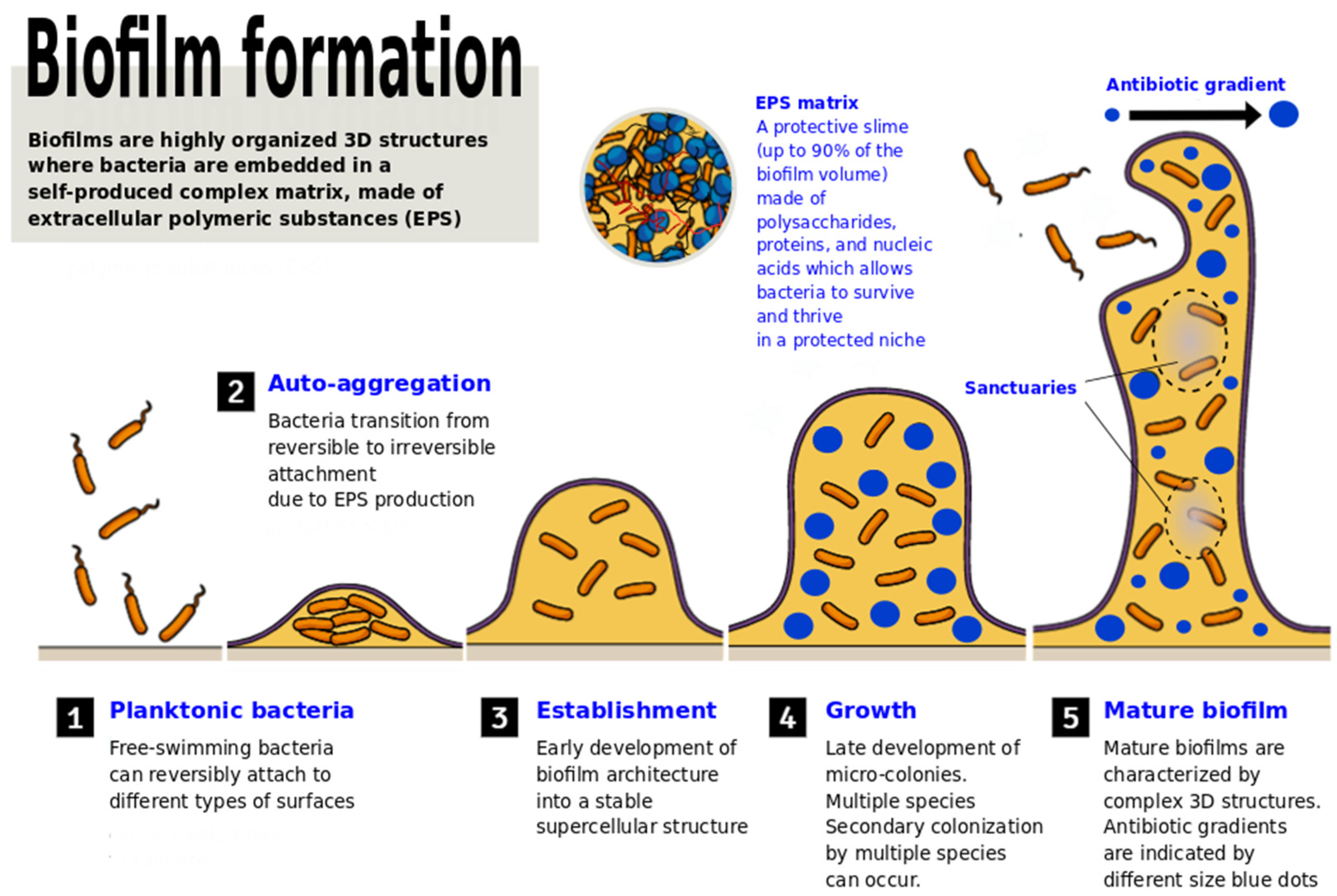
Bacterial populations inhabiting a variety of natural and human-associated niches have the ability to grow in the form of biofilms. A large part of pathological chronic conditions, and essentially all the bacterial infections associated with implanted medical devices or prosthetics, are caused by microorganisms embedded in a matrix made of polysaccharides, proteins, and nucleic acids. Biofilm infections are generally characterized by a slow onset, mild symptoms, tendency to chronicity, and refractory response to antibiotic therapy. Even though the molecular mechanisms responsible for resistance to antimicrobial agents and host defenses have been deeply clarified, effective means to fight biofilms are still required. Lactic acid bacteria (LAB), used as probiotics, are emerging as powerful weapons to prevent adhesion, biofilm formation, and control overgrowth of pathogens. Hence, using probiotics or their metabolites to quench and interrupt bacterial communication and aggregation, and to interfere with biofilm formation and stability, might represent a new frontier in clinical microbiology and a valid alternative to antibiotic therapies.

lactic acid bacteria biofilms probiotics quorum sensing antibiotic resistance

1. Introduction

Pathogenic bacterial biofilms are becoming one of the main concerns of the antibiotic era [1][2]. Biofilms are assemblages of microorganisms and the extracellular products they produce, that adhere on biotic or abiotic surfaces and are characterized by highly specialized interactions between them [3]. Biofilm-forming bacteria are embedded in a matrix of self-produced slime, constituted by extracellular polymeric substances (EPS) [4]. This growing mode can alter bacterial biological and physiological characteristics, such as reproduction, growth, gene transcription rate, and resistance towards antibiotics [5][6][7]. Schematically, the formation of a differentiated biofilm requires five maturation stages: (i) initial attachment of planktonic bacteria (reversible) to a surface; (ii) production and secretion of EPS and/or other means of docking, and specific adhesins (e.g., flagella, autotransporter proteins, fimbriae, curli fibers, and F-type conjugative pilus) that drive the transitional attachment from reversible to irreversible [8][9][10]; (iii) early-maturing of biofilm architecture as a super cellular structure; (iv) late-maturing of micro-colonies and evolution into a mature biofilm; and (v) detachment of cells from the biofilm and dispersion into the surrounding environment (Figure 1). All these processes are strictly regulated by different cell-to-cell signaling molecules responsible for population density-dependent gene expression that can deeply affect the process of biofilm formation [11][12].
Microbiolres 12 00068 g001
Figure 1. Schematic representation of the different steps required for the formation of a mature biofilm. The small and large blue dots represent areas with different antibiotic concentrations (denoting the presence of a gradient), and the grey zones are “sanctuaries” where bacteria can survive with a low concentration of antibiotics, which can favor the development of resistance.
The production of the EPS matrix, composed of polysaccharides, proteins, and nucleic acids (extracellular DNA—eDNA) allows for bacterial survival and proliferation in a protected niche with a constant nutrient supply and protection from the host immune system, disinfectants, and antibiotics [13][14]. Biofilms act as physical barriers, allowing bacteria to elude both immune detection and phagocytosis, while expressing genetic switches (or response regulators) that disturb immune cell activity [15]. Up to 80% of chronic infections worldwide are linked to biofilms and/or are caused by antibiotic resistant bacteria. Indeed, bacteria growing in a biofilm can be 100–1000 times more drug resistant compared to their planktonic counterpart [16].
The Antibiotic Resistance Threats Report (2019 AR Threats Report) by the American Centers for Disease Control and Prevention (CDC) reports that “more than 2.8 million antibiotic-resistant infections occur in the U.S. each year, and more than 35,000 people die as a result” [17]. The spread of antibiotic-resistant bacterial clones is a global threat to public health. The reasons behind this phenomenon span from unregulated antibiotic usage in livestock farming to malpractices or improper use of antibiotics in the treatment of human infections [18][19]. Different studies have shown that physicians tend to overprescribe antibiotics mainly due to pressure from patients or from the healthcare system, as well as financial incentives and attempts to maximize the number of patients treated. On the other hand, patients’ lack of knowledge and awareness, access to antibiotics without a prescription, or premature stopping of antibiotic therapies as a consequence of improved health conditions, are other resistance promoting factors [20][21][22][23][24].
Several in vivo and in vitro studies have shown that LAB possess the ability of contrasting biofilm formation and growth. LAB are probiotics and are not prone to trigger or promote the evolution of resistant pathogens. According to the European Food Safety Authority (EFSA), an important requirement of probiotics is, indeed, that they must not have antibiotic resistance genes which could spread through plasmids or transposons. Among LAB, members of the genera Lactobacillus and Bifidobacterium have emerged as the most commonly used probiotics [25].
In the majority of the scientific works on topical and oral probiotics, it is common to encounter a precise definition, originally given by World Health Organization (WHO): “Probiotics are live microorganisms which when administered in adequate amounts confer a health benefit on the host” [26][27]. To this clear statement corresponds a wide range of well-recognized and largely unquestioned “benefits” (e.g., recolonization of surfaces depleted of commensal bacteria after an antibiotic treatment, capacity to contrast and outcompete the growth of pathogenic microorganisms), plus a wider spectrum of unspecified, off-target, long-lasting, and sometimes, highly debated extra advantages (e.g., anti-carcinogenic effects, immune system modulation, mitigation of side effects of medicaments or invasive therapies). As a matter of fact, probiotics are often administered orally, but the benefits are not restricted to the gastrointestinal tract; changes and interactions affecting the microbiota of the skin, urinary tract, and mouth are well documented and indicative of broad range effects [28][29][30].
A relevant issue linked to the specific definition of probiotics reported above regards the quantification of the “sufficient amounts”. Despite the difficulty in defining this parameter, probiotics are commonly regarded as safe and are administered as billions of microbial cells. Although monitoring and continuous surveillance, as well as precaution, are mandatory, probiotics have the advantage of presenting no (or limited) side effects linked to overdosage [31][32]. Recently, Barzegari et al. (2020) have evidenced the possibility of using probiotics and their derivatives against biofilms and encouraged in vivo studies to define the best strain-related antibiofilm activity [33].
Although research on the topic is very active, further studies are needed to gain insights into the mechanisms by which probiotics and their metabolites can be used and properly applied to manage biofilm infections in humans.

2. The Battle of LAB against Pathogenic Biofilms

2.1. How Lactobacillus May Contrast Biofilm Formation and Stability

Methicillin-resistant Staphylococcus aureus (MRSA) is a multi-drug resistant (MDR) microorganism and one of the principal nosocomial pathogens worldwide [34]. Different strains belonging to the genus Lactobacillus (as well as Bifidobacterium) isolated from various sources have been shown to contrast the growth of S. aureus and even of clinical isolates of MRSA in vitro [35]. Their effects were mediated both by direct cell competitive exclusion and the production of short chain fatty acids or bacteriocin-like inhibitors. In addition, L. acidophilus was also reported to inhibit S. aureus biofilm formation and lipase production. In another study, L. fermentum TCUESC01, isolated from cocoa seeds, was shown to effectively inhibit S. aureus biofilm formation. The inhibition mechanism was based on the release of soluble molecules which suppressed the expression of two genes (icaA and icaR) with an important role in biofilm synthesis [36].
MDR Proteus mirabilis isolates show low antibiotic susceptibility and biofilm-forming activity that can cause serious urinary tract infections [37]. A recent study demonstrated that cultures and cell-free supernatants of L. casei DSM 20011 and L. reuteri DSM 20016 exhibited strong antimicrobial, anti-adherence, and antibiofilm formation activities against MDR P. mirabilis. In addition, supernatants of L. casei and L. reuteri significantly reduced mature biofilm formation and adherence (>60% compared to controls), indicating that these species of lactobacilli could be utilized to combat Proteus-associated urinary tract infections [38].
Dental caries has multifactorial causes and arises from an imbalance between the host and the microbiota of the mouth. For a long time, Streptococcus mutans in its biofilm form has been known to contribute to dental caries formation significantly; recently, the one pathogen –one disease approach has been deeply challenged, and the concurrent role of the entire microbiota in the health of the oral cavity tends to be more prominent [39]. The capacity of different Lactobacillus species to inhibit growth, biofilm formation, and gene expression of S. mutans has been evaluated. Susceptibility testing indicated antibacterial (pH-dependent) and antibiofilm activities of L. casei (ATCC 393), L. reuteri (ATCC 23272), L. plantarum (ATCC 14917), and L. salivarius (ATCC 11741) against S. mutans. All Lactobacillus species previously mentioned contrasted and limited the growth and virulence of S. mutans. Reduction in microcolony formation and exopolysaccharide structural changes were also highlighted by scanning electron microscopy. The highest antimicrobial activities were reported for L. casei and L. reuteri, whereas the lowest antimicrobial activities were observed with L. plantarum and L. salivarius. The highest antibiofilm and peroxide-dependent antimicrobial activities were reported for L. salivarius. Reduced expression of genes involved in exopolysaccharide production, acid tolerance, and quorum sensing were reported for all biofilm-forming cells treated with Lactobacillus spp. supernatants [40]. In a study on mixed biofilm formation by fungi and bacteria on silicone in vitro, Lactobacillus supernatant showed high efficiency against both microorganisms [41]. In the field of oral infections, the probiotic strain L. brevis CD2 was shown to inhibit the opportunistic anaerobe Prevotella melaninogenica (PM1), a well-known causative agent of periodontitis. The inhibitory effect of L. brevis CD2 on P. melaninogenica PM1 biofilms was evaluated in vitro using two different methods: the anaerobe was exposed to the supernatant of the strain in one case, or the two microorganisms were grown together to obtain single or mixed biofilms, in the second case. The inhibitory effect of CD2 on PM1 was also checked by the agar overlay method. The development of PM1 biofilm was strongly affected (56% decrease in OD570 value) by the CD2 supernatant after 96 h—with a dose-dependent biofilm reduction using several supernatant dilutions. Confocal microscopy on the mixed biofilms revealed the ability of CD2 to prevail over PM1, greatly reducing the biofilm of the latter. The authors hypothesized that the strong adherence ability of the CD2 strain and the release of metabolites may be responsible for reducing the PM1 biofilm [42].
The use of antibiotics for the treatment of cholera is associated with side effects, such as gut dysbiosis, due to the depletion of beneficial microbiota and the risk of spreading antibiotic resistance; hence, the search for alternative therapeutic agents is extremely active. Different strains of Lactobacillus spp., screened and isolated from fecal samples of healthy children in cholera endemic area, were tested for their abilities to prevent biofilm formation and to disperse the preformed biofilms of Vibrio cholerae and V. parahaemolyticus. The results showed that the culture supernatant (CS) of seven isolates of Lactobacillus spp. used in the study inhibited the biofilm formation of V. cholerae by more than 90% [43].
A recent study showed the role of L. gasseri in contrasting the adhesion of the protozoan parasite Trichomonas vaginalis to host cells, a critical virulence aspect of this pathogen [44]. The aggregation-promoting factor-2 (APF-2) produced by L. gasseri ATCC 9857 was found to be highly inhibitory in the adhesion of T. vaginalis to human vaginal ectocervical cells. This important finding highlights that lactobacilli remain of key importance for the development of specific therapeutic strategies, even towards non-bacterial pathogens.
As a matter of fact, probiotics are active against non-bacterial biofilms as well. For example, C. albicans biofilm is associated with denture-related stomatitis and oral candidiasis, especially in elderly people. A study investigating a C. albicans biofilm on a denture base resin treated with L. rhamnosus and L. casei showed that the probiotics’ surfactant exhibited strong antifungal activity against blastoconidia and biofilm of C. albicans. Even when the C. albicans biofilm was already formed and sequentially treated with L. rhamnosus and L. casei, inhibition of the biofilm on the denture surface was reported [45]. Therefore, L. rhamnosus and L. casei probiotics could have practical applications for preventing and treating denture-related stomatitis and other Candida infections, even in neonates [46][47].
It is not uncommon to register discrepancies between the effectiveness of probiotics in vitro and in vivo. Therefore, in vitro antimicrobial activity does not necessarily assure efficacy in animal infectious models. However, cases in which the in vitro and in vivo results were congruent are also reported. As an example, L. plantarum, which showed the highest inhibition activity against S. aureus in vitro, was also very effective topically in preventing skin wound infection in S. aureus-infected mice. Bacteriocin-producing Lactobacillus sakei 2a has been shown to protect gnotobiotic mice against experimental challenge with L. monocytogenes [48]. A recent study aimed at evaluating the effects of Lactobacillus administered intranasally on a murine model of P. aeruginosa pneumonia (strain PAO1). Two probiotic combinations were selected for in vivo testing (1-L.rff for L. rhamnosus and two L. fermentum strains, and 2-L.psb for L. paracasei, L. salivarius, and L. brevis) out of 50 clinical isolates screened for the ability to decrease the synthesis of two PAO1 produced QS-dependent virulence factors (elastase and pyocyanin). Intranasal priming with both probiotic blends acted as a prophylaxis and avoided fatal complications caused by PAO1 pneumonia in mice, showing encouraging results to move towards clinical trials [49].

2.2. How Bifodobacteria May Contrast Pathogenic Biofilms

Among the Bifidobacteria, Bifidobacterium bifidum BGN4 is a widely used probiotic strain that has been included as a major ingredient to produce nutraceutical products for the last 20 years [50]. The various bio-functional effects and potential for industrial application of B. bifidum BGN4 have been characterized and proven in vitro (i.e., phytochemical bio-catalysis, cell adhesion, anti-carcinogenic effects on cell lines, and immunomodulatory effects on immune cells) and in vivo experiments (see below).
A study investigated the effect of Bifidobacterium spp. on the interference with the production of quorum-sensing (QS) signals and biofilm formation by enterohemorrhagic E. coli (EHEC) O157:H7. In an AI-2 bioassay, cell extracts of different Bifidobacterium reference strains (B. longum ATCC 15707, B. adolescentis ATCC 15706, and B. breve ATCC 15700) were rather effective; they resulted in a 36% reduction in biofilm formation. Cell extracts of B. longum ATCC 15707 were also able to reduce the virulence of EHEC O157:H7 in the Caenorhabditis elegans nematode in vivo model [51]. Another study highlighted how B. lactis and B. infantis, alone or in combination, have an antagonist effect on biofilms of periodontopathogens, such as Porphyromonas gingivalis and Fusobacterium nucleatum, but minimal influence on Streptococcus oralis growth in vitro [52].
Bifidobacteria strains are often used in probiotic combination with other LAB. One of these combinations, constituted of L. rhamnosus GG, L. rhamnosus LC705, B. breve 99, and P. freudenreichii JS was shown to inhibit pathogen adhesion (including Salmonella enterica, Clostridium difficile, L. monocytogenes, and S. aureus) to human intestinal mucus (in vitro). The same combination with another bifidobacterial strain (B. lactis Bb12) was less effective [53].
The studies regarding the ability of Bifidobacteria to contrast pathogenic biofilms are not so numerous as the ones on lactobacilli. Some experimental works have also highlighted a lower effectiveness compared to other LAB. As an example, Miyazaki et al. (2010) highlighted that CS of a Lactobacillus strain has a strong bactericidal effect on auto aggregative E. coli, while no effect was reported for Bifidobacteria [54]. Discrepancies among laboratory results and experiments in animal models are known for Bifidobacteria as well. For example, the S. aureus 8325-4 strain was shown to be sensitive in vitro to L. acidophilus, while B. bifidum best inhibited experimental intravaginal staphylococcosis in mice caused by the same bacteria [35]. For B. bifidum BGN4, a wide spectrum of beneficial effects in vivo (i.e., suppressed allergic responses in mouse model and anti-inflammatory bowel disease) and in clinical studies (eczema in infants and adults with irritable bowel syndrome) have been demonstrated.

3. Conclusions

Specific probiotic combinations are demonstrating day-by-day to have a marked utility in the human field, and data on antibiofilm activity on various respiratory, genito-urinary, wound, and tissue pathogens, are starting to become convincing. However, there is still a long way to go, especially in their in vivo routine usage. Herein should encourage better investigation on probiotic–biofilm interactions and how to fight biofilm infections through the so-called “good bacteria”, such as bifidobacteria and lactobacilli, highlighting that there are “useful” or “good” biofilms as well. Mechanisms of action and antibiofilm activities must be considered as strain-related; therefore, we will need to focus our research on the development of such promising strains. It is often debated whether probiotics will become broadly used drugs or medicaments in the future; it is still too early to say, but given the uncertain longevity of antibiotics, it would be recommended to explore alternative means, and so far, probiotics represent one of the most promising.

References

  1. Khan, F.; Pham, D.T.N.; Oloketuyi, S.F.; Kim, Y.-M. Antibiotics Application Strategies to Control Biofilm Formation in Pathogenic Bacteria. Curr. Pharm. Biotechnol. 2020, 21, 270–286.
  2. Mann, R.; Holmes, A.; McNeilly, O.; Cavaliere, R.; Sotiriou, G.A.; Rice, S.A.; Gunawan, C. Evolution of biofilm-forming pathogenic bacteria in the presence of nanoparticles and antibiotic: Adaptation phenomena and cross-resistance. J. Nanobiotechnol. 2021, 19, 1–17.
  3. Moons, P.; Michiels, C.; Aertsen, A. Bacterial interactions in biofilms. Crit. Rev. Microbiol. 2009, 35, 157–168.
  4. Karygianni, L.; Ren, Z.; Koo, H.; Thurnheer, T. Biofilm Matrixome: Extracellular Components in Structured Microbial Communities. Trends Microbiol. 2020, 28, 668–681.
  5. Bakhtiari, N.M.; Gooraninezhad, S.; Karami, M. Biofilm-Producing Ability of Bovine Extraintestinal Pathogenic Escherichia coli and Its Correlation with Attachment Factors. Jundishapur J. Heal. Sci. 2018, 10.
  6. Cergole-Novella, M.C.; Pignatari, A.C.; Guth, B.E. Adhesion, biofilm and genotypic characteristics of antimicrobial resistant Escherichia coli isolates. Braz. J. Microbiol. 2015, 46, 167–171.
  7. Olsen, I. Biofilm-specific antibiotic tolerance and resistance. Eur. J. Clin. Microbiol. Infect. Dis. 2015, 34, 877–886.
  8. Fakruddin, M.D.; Bin Mannan, K.S.; Mazumdar, R.M. Correlation Between in vitro Biofilm Formation and Virulence Properties of Extra-Intestinal Pathogenic Escherichia Coli (Expec). Online J. Biol. Sci. 2014, 14, 261–270.
  9. Wood, T.K.; Barrios, A.F.G.; Herzberg, M.; Lee, J. Motility influences biofilm architecture in Escherichia coli. Appl. Microbiol. Biotechnol. 2006, 72, 361–367.
  10. Prüß, B.M.; Besemann, C.; Denton, A.; Wolfe, A.J. A Complex Transcription Network Controls the Early Stages of Biofilm Development by Escherichia coli. J. Bacteriol. 2006, 188, 3731–3739.
  11. Cai, P.; Sun, X.; Wu, Y.; Gao, C.; Mortimer, M.; Holden, P.A.; Redmile-Gordon, M.; Huang, Q. Soil biofilms: Microbial interactions, challenges, and advanced techniques for ex-situ characterization. Soil Ecol. Lett. 2019, 1, 85–93.
  12. Yi, L.; Li, J.; Liu, B.; Wang, Y. Advances in research on signal molecules regulating biofilms. World J. Microbiol. Biotechnol. 2019, 35, 130.
  13. Campoccia, D.; Mirzaei, R.; Montanaro, L.; Arciola, C.R. Hijacking of immune defences by biofilms: A multifront strategy. Biofouling 2019, 35, 1055–1074.
  14. Esanchez-Vizuete, P.; Eorgaz, B.; Eaymerich, S.; Le Coq, D.; Ebriandet, R. Pathogens protection against the action of disinfectants in multispecies biofilms. Front. Microbiol. 2015, 6, 705.
  15. González, J.F.; Hahn, M.; Gunn, J.S. Chronic biofilm-based infections: Skewing of the immune response. Pathog. Dis. 2018, 76.
  16. Sharma, D.; Misba, L.; Khan, A.U. Antibiotics versus biofilm: An emerging battleground in microbial communities. Antimicrob. Resist. Infect. Control 2019, 8, 76.
  17. CDC Antibiotic Resistance Threats in the United State; Department of Health and Human Services, CDC: Atlanta, GA, USA, 2019; pp. 1–113.
  18. Manyi-Loh, C.; Mamphweli, S.; Meyer, E.; Okoh, A. Antibiotic Use in Agriculture and Its Consequential Resistance in Environmental Sources: Potential Public Health Implications. Molecules 2018, 23, 795.
  19. Marquardt, R.R.; Li, S. Antimicrobial resistance in livestock: Advances and alternatives to antibiotics. Anim. Front. 2018, 8, 30–37.
  20. Bjorkman, I.; Berg, J.; Roing, M.; Erntell, M.; Lundborg, C.S. Perceptions among Swedish hospital physicians on prescribing of antibiotics and antibiotic resistance. BMJ Qual. Saf. 2010, 19, e8.
  21. Kumar, S.; Little, P.; Britten, N. Why do general practitioners prescribe antibiotics for sore throat? Grounded theory interview study. BMJ 2003, 326, 138.
  22. Machowska, A.; Lundborg, C.S. Drivers of Irrational Use of Antibiotics in Europe. Int. J. Environ. Res. Public Health 2018, 16, 27.
  23. Reynolds, L.; McKee, M. Factors influencing antibiotic prescribing in China: An exploratory analysis. Health Policy 2009, 90, 32–36.
  24. Sirota, M.; Round, T.; Samaranayaka, S.; Kostopoulou, O. Expectations for antibiotics increase their prescribing: Causal evidence about localized impact. Health Psychol. 2017, 36, 402–409.
  25. Ouwehand, A.C.; Tiihonen, K.; Saarinen, M.; Putaala, H.; Rautonen, N. Influence of a combination ofLactobacillus acidophilusNCFM and lactitol on healthy elderly: Intestinal and immune parameters. Br. J. Nutr. 2008, 101, 367–375.
  26. Binda, S.; Hill, C.; Johansen, E.; Obis, D.; Pot, B.; Sanders, M.E.; Tremblay, A.; Ouwehand, A.C. Criteria to Qualify Microorganisms as “Probiotic” in Foods and Dietary Supplements. Front. Microbiol. 2020, 11, 1662.
  27. Hill, C.; Guarner, F.; Reid, G.; Gibson, G.R.; Merenstein, D.J.; Pot, B.; Morelli, L.; Canani, R.B.; Flint, H.J.; Salminen, S.; et al. Expert Consensus Document: The International Scientific Association for Probiotics and Prebiotics consensus statement on the scope and appropriate use of the term probiotic. Nat. Rev. Gastroenterol. Hepatol. 2014, 11, 506–514.
  28. Collins, S.; Reid, G. Distant Site Effects of Ingested Prebiotics. Nutrients 2016, 8, 523.
  29. Fijan, S.; Frauwallner, A.; Langerholc, T.; Krebs, B.; Younes, J.A.T.H.; Heschl, A.; Turk, D.M.; Rogelj, I. Efficacy of Using Probiotics with Antagonistic Activity against Pathogens of Wound Infections: An Integrative Review of Literature. BioMed Res. Int. 2019, 2019, 7585486.
  30. Szántó, M.; Dózsa, A.; Antal, D.; Szabó, K.; Kemény, L.; Bai, P. Targeting the gut-skin axis—Probiotics as new tools for skin disorder management? Exp. Dermatol. 2019, 28, 1210–1218.
  31. Kothari, D.; Patel, S.; Kim, S.-K. Probiotic supplements might not be universally-effective and safe: A review. Biomed. Pharmacother. 2018, 111, 537–547.
  32. Lerner, A.; Shoenfeld, Y.; Matthias, T. Probiotics: If It Does Not Help It Does Not Do Any Harm. Really? Microorganisms 2019, 7, 104.
  33. Barzegari, A.; Kheyrolahzadeh, K.; Khatibi, S.M.H.; Sharifi, S.; Memar, M.Y.; Vahed, S.Z. The Battle of Probiotics and Their Derivatives against Biofilms. Infect. Drug Resist. 2020, 13, 659–672.
  34. Turner, N.A.; Sharma-Kuinkel, B.K.; Maskarinec, S.; Eichenberger, E.; Shah, P.P.; Carugati, M.; Holland, T.L.; Fowler, V.G. Methicillin-resistant Staphylococcus aureus: An overview of basic and clinical research. Nat. Rev. Genet. 2019, 17, 203–218.
  35. Sikorska, H.; Smoragiewicz, W. Role of probiotics in the prevention and treatment of meticillin-resistant Staphylococcus aureus infections. Int. J. Antimicrob. Agents 2013, 42, 475–481.
  36. Melo, T.A.; Dos Santos, T.F.; De Almeida, M.E.; Junior, L.A.G.F.; Andrade, E.F.; Rezende, R.P.; Marques, L.M.; Romano, C.C. Inhibition of Staphylococcus aureus biofilm by Lactobacillus isolated from fine cocoa. BMC Microbiol. 2016, 16, 250.
  37. Gomaa, S.; Serry, F.; Abdellatif, H.; Abbas, H. Elimination of multidrug-resistant Proteus mirabilis biofilms using bacteriophages. Arch. Virol. 2019, 164, 2265–2275.
  38. Shaaban, M.; Abd El-Rahman, O.A.; Al-Qaidi, B.; Ashour, H.M. Antimicrobial and Antibiofilm Activities of Probiotic Lactobacilli on Antibiotic-Resistant Proteus Mirabilis. Microorganisms 2020, 8, 960.
  39. Campbell, K. Oral microbiome findings challenge dentistry dogma. Nature 2021.
  40. Wasfi, R.; El-Rahman, O.A.A.; Zafer, M.M.; Ashour, H.M. Probiotic Lactobacillu ssp. inhibit growth, biofilm formation and gene expression of caries-inducingStreptococcus mutans. J. Cell. Mol. Med. 2018, 22, 1972–1983.
  41. Tan, Y.; Leonhard, M.; Moser, D.; Schneider-Stickler, B. Inhibition activity of Lactobacilli supernatant against fungal-bacterial multispecies biofilms on silicone. Microb. Pathog. 2017, 113, 197–201.
  42. Vuotto, C.; Barbanti, F.; Mastrantonio, P.; Donelli, G. Lactobacillus brevisCD2 inhibitsPrevotella melaninogenicabiofilm. Oral Dis. 2013, 20, 668–674.
  43. Kaur, S.; Sharma, P.; Kalia, N.; Singh, J.; Kaur, S. Anti-biofilm Properties of the Fecal Probiotic Lactobacilli Against Vibrio spp. Front. Cell. Infect. Microbiol. 2018, 8, 120.
  44. Phukan, N.; Brooks, A.; Simoes-Barbosa, A. A Cell Surface Aggregation-Promoting Factor from Lactobacillus gasseri Contributes to Inhibition of Trichomonas vaginalis Adhesion to Human Vaginal Ectocervical Cells. Infect. Immun. 2018, 86.
  45. Song, Y.-G.; Lee, S.-H. Inhibitory effects of Lactobacillus rhamnosus and Lactobacillus casei on Candida biofilm of denture surface. Arch. Oral Biol. 2017, 76, 1–6.
  46. Manzoni, P. Use of Lactobacillus casei Subspecies Rhamnosus GG and Gastrointestinal Colonization by Candida Species in Preterm Neonates. J. Pediatr. Gastroenterol. Nutr. 2007, 45, S190–S194.
  47. Matsubara, V.H.; Wang, Y.; Bandara, H.M.H.N.; Mayer, M.P.A.; Samaranayake, L.P. Probiotic lactobacilli inhibit early stages of Candida albicans biofilm development by reducing their growth, cell adhesion, and filamentation. Appl. Microbiol. Biotechnol. 2016, 100, 6415–6426.
  48. Bambirra, F.; Lima, K.; Franco, B.; Cara, D.; Nardi, R.; Barbosa, F.; Nicoli, J. Protective effect of Lactobacillus sakei 2a against experimental challenge with Listeria monocytogenes in gnotobiotic mice. Lett. Appl. Microbiol. 2007, 45, 663–667.
  49. Fangous, M.-S.; Gosset, P.; Galakhoff, N.; Gouriou, S.; Guilloux, C.-A.; Payan, C.; Vallet, S.; Héry-Arnaud, G.; Le Berre, R. Priming with intranasal lactobacilli prevents Pseudomonas aeruginosa acute pneumonia in mice. BMC Microbiol. 2021, 21, 195.
  50. Ku, S.; Park, M.S.; Ji, G.E.; You, H.J. Review on Bifidobacterium bifidum BGN4: Functionality and Nutraceutical Applications as a Probiotic Microorganism. Int. J. Mol. Sci. 2016, 17, 1544.
  51. Kim, Y.; Lee, J.W.; Kang, S.-G.; Oh, S.; Griffiths, M.W. Bifidobacterium spp. influences the production of autoinducer-2 and biofilm formation by Escherichia coli O157:H7. Anaerobe 2012, 18, 539–545.
  52. Valdez, R.M.A.; Ximenez-Fyvie, L.A.; Caiaffa, K.S.; dos Santos, V.R.; Cervantes, R.M.G.; Almaguer-Flores, A.; Duque, C. Antagonist effect of probiotic bifidobacteria on biofilms of pathogens associated with periodontal disease. Microb. Pathog. 2020, 150, 104657.
  53. Collado, M.C.; Jalonen, L.; Meriluoto, J.; Salminen, S. Protection mechanism of probiotic combination against human pathogens: In vitro adhesion to human intestinal mucus. Asia Pac. J. Clin. Nutr. 2006, 15, 570–575.
  54. Miyazaki, Y.; Yokota, H.; Takahashi, H.; Fukuda, M.; Kawakami, H.; Kamiya, S.; Hanawa, T. Effect of probiotic bacterial strains of Lactobacillus, Bifidobacterium, and Enterococcus on enteroaggregative Escherichia coli. J. Infect. Chemother. 2010, 16, 10–18.
More
Information
Subjects: Microbiology
Contributor MDPI registered users' name will be linked to their SciProfiles pages. To register with us, please refer to https://encyclopedia.pub/register :
View Times: 1.0K
Revisions: 2 times (View History)
Update Date: 05 Jan 2022
1000/1000
Video Production Service