Submitted Successfully!
To reward your contribution, here is a gift for you: A free trial for our video production service.
Thank you for your contribution! You can also upload a video entry or images related to this topic.
Version Summary Created by Modification Content Size Created at Operation
1 + 1737 word(s) 1737 2021-12-24 09:38:53 |
2 The format is correct. + 1 word(s) 1738 2021-12-30 03:11:06 | |
3 Format is correct. Meta information modification 1738 2021-12-30 09:26:41 |

Video Upload Options

Do you have a full video?

Confirm

Are you sure to Delete?
Cite
If you have any further questions, please contact Encyclopedia Editorial Office.
Pennati, R.; Manni, L.; Caicci, F.; Vanni, V. The Larva of the Ascidian Halocynthia roretzi. Encyclopedia. Available online: https://encyclopedia.pub/entry/17640 (accessed on 01 May 2024).
Pennati R, Manni L, Caicci F, Vanni V. The Larva of the Ascidian Halocynthia roretzi. Encyclopedia. Available at: https://encyclopedia.pub/entry/17640. Accessed May 01, 2024.
Pennati, Roberta, Lucia Manni, Federico Caicci, Virginia Vanni. "The Larva of the Ascidian Halocynthia roretzi" Encyclopedia, https://encyclopedia.pub/entry/17640 (accessed May 01, 2024).
Pennati, R., Manni, L., Caicci, F., & Vanni, V. (2021, December 30). The Larva of the Ascidian Halocynthia roretzi. In Encyclopedia. https://encyclopedia.pub/entry/17640
Pennati, Roberta, et al. "The Larva of the Ascidian Halocynthia roretzi." Encyclopedia. Web. 30 December, 2021.
The Larva of the Ascidian Halocynthia roretzi
Edit

The swimming larva represents the dispersal phase of ascidians, marine invertebrates belonging to tunicates. Due to its adhesive papillae, the larva searches the substrate, adheres to it, and undergoes metamorphosis, thereby becoming a sessile filter feeding animal. The papillae of H. roretzi, previously described as simple and conform, exhibit dynamic changes during settlement. This opens up new considerations on papillae morphology and evolution and deserves to be further investigated.

adhesive papillae adultation Botryllus schlosseri Ciona intestinalis tunicates

1. Introduction

Ascidians are sessile marine invertebrates belonging to the tunicates, a group that is evolutionarily close to vertebrates [1]. Some species are gregarious and form complex clusters of several organisms living in close proximity [2]. They can colonize different natural and artificial substrates and can modify the primary substrate providing habitats for other organisms. Thus, ascidians are considered essential members of the benthic communities contributing to increased ecosystem complexity and biodiversity [3] and have been proposed as valuable model organisms to test coastal water pollution [4][5][6]. Ascidians also have economic importance since some species, such as Halocynthia roretzi, are commercially reared for human consumption [7][8]. For this reason, H. roretzi is one of the most studied ascidians: its genome is compact with about 16,000 protein-coding genes [9] and its development is known in detail [10]. Other species (such as Ciona intestinalis) are potential biofuel sources [4]. Some ascidians (such as Botryllus schlosseri and Botrylloides violaceus) are considered invasive organisms in marinas and also fouling organisms in marine aquaculture, and their presence results in economic loss [11].
Ascidians show an extraordinary range of life history traits, with some species being solitary and others showing colonial organisation; the latter evolved several times during the diversification of the group [12][13]. Tunicate taxonomy has long been questioned; the group comprises approximately 3000 species that have traditionally been divided into three classes: Ascidiacea (sea squirts), Thaliacea (pelagic salps, doliolids and pyrosomes), and Appendicularia (larvaceans) [14]. Molecular based phylogenies revealed that Ascidiacea was paraphyletic [15][16] and the following three clades were proposed: (1) Stolidobranchia, (2) Appendicularia, and (3) Phlebobranchia plus Aplousobranchia plus Thaliacea [17].
Despite the gross anatomical similarities, ascidian larvae have been remodelled in different ways during evolution [18]. Colonial species, which produce yolked eggs, are ovoviviparous (or even viviparous) [19][20] and exhibit prolonged embryogenesis. Their larvae often undergo adultation, a mode of development in which adult structures differentiate precociously in the tadpole trunk. Adultation can manifest to different degrees, from the early appearance of siphons, a partial digestive tract, a few gill slits, a rudimentary heart, or one or more buds prior to asexual reproduction, up to involving more extensive development of adult structures [18][21]
The larva plays a crucial role in ascidian life since it selects the substrate to adhere to permanently. Indeed, after hatching, the tadpole larva swims for up to a few days, then attaches to a substrate and undergoes metamorphosis: the tail is retracted into the trunk, the larval tissues are resorbed, and adult structures differentiate from rudiments in the trunk [22]. The critical step of substrate selection and adhesion is mediated by three mucus-secreting organs, the adhesive papillae (or palps) that most ascidian larvae bear at their anterior tip. Generally, the papillae are composed of elongated mucus-secreting cells, called collocytes (CCs), primary sensory neurons (PSNs) and supporting axial columnar cells (ACCs) [22]. It was proposed that papillary sensory cells are mechanosensory neurons playing a central role in substrate selection [23]. Actually, the morphology of adhesive papillae is quite variable among species, and they can be classified into 10 types according to their histological characteristics [22][24]

2. The 3D Reconstruction of Halocynthia roretzi Larva

The H. roretzi larvae, 10 h post hatching at 12 °C, were found to be 1744 ± 18 µm long; the tail length is 1252 ± 15 µm and the trunk is 490 ± 6 µm (Figure 1). At the anterior tip of the trunk, three adhesive papillae are arranged according to the vertices of an equilateral triangle (two dorsal, one ventral) (Figure 1A,B′,G).
Figure 1. Anatomy of the larva of H. roretzi. (A) Whole mount larva (in block of resin, contrasted with OsO4). Right side view. t: tail; tr: trunk. The black square indicates the trunk region illustrated in (BE). (BF) Three-dimensional reconstructions. Colour code: blue, atrial chamber; dark green, ocellus; grey, nervous system; light green, notochord; red, muscles; yellow, endoderm (i.e., prospective pharynx). In B–D, the epidermis is transparent; in (E,F), the organs are transparent and their internal structures can be seen. (B) Right side view. (B′) Frontal view of the trunk showing the three papillae. (C) Left side view. (D) Dorsal view. (E) View of internal organs: trunk endoderm, nervous system and notochord. (F) Close view of the trunk region. (GN) Some cross histological sections, from anterior to posterior, utilized for the 3D reconstruction. Toluidine blue. Arrow in L indicates the area where the protostigmata will open during metamorphosis. a: atrium; drap, dlap, vap: dorsal right-, dorsal left-, and ventral-adhesive papilla, respectively; ep: epidermis; es: endodermal strand; itc: inner tunic compartment; m: muscle; me: mesenchyme; nc: notochord; ne: neck; otc: outer tunic compartment; pc: peribranchial chambers; oc: ocellus; ot: otolith; st: stomodeum (i.e., oral siphon primordium); sv: sensory vesicle; te: trunk endoderm; tf: tail fin; vg, visceral ganglion. The enlargement is the same in (BE) and in (GN).
In the posterior dorsal part of the trunk, the endoderm of the pharynx is in contact with the developing ectodermal peribranchial chambers; here, the protostigmata will open during metamorphosis (Figure 2). As is clearly visible in serial histological sections, the two peribranchial chambers are joined dorsally to the nervous system to form a small chamber representing the prospective atrial chamber of the adult (Figure 2D–I). The atrial chamber opens outside through the atrial siphon in the form of a shallow dent in the dorsal trunk Figure 1L and Figure 2A). However, at this stage, both the siphons are still covered with a thin layer of tunic, preventing circulation of seawater within the larva, which is lecithotrophic and does not filter (Figure 2D). During metamorphosis, the two peribranchial chambers and the atrial chamber will elongate as the pharynx grows, and the stigmata number will increase.
Figure 2. Atrial chamber in H. roretzi. See Figure 1 for colour code and abbreviations. (AC) Three-dimensional reconstruction. In B,C the organs are transparent. (A) Dorsal external view showing the dint in correspondence of the atrial siphon (arrowhead). (B) Dorsal view. (C) Frontal-posterior view enlightening the position of the atrial chamber relative to the notochord. (DI) Serial cross histological sections from anterior to posterior at the level of the atrial and peribranchial chambers. Toluidine blue. The enlargement is the same in (DI).

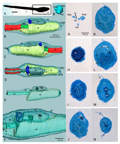
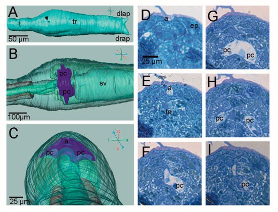
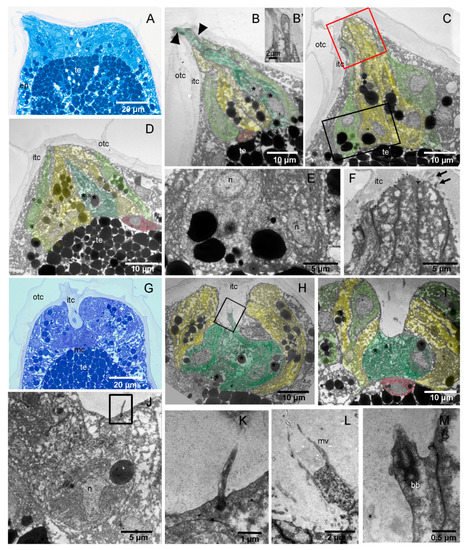
3. Adhesive Papillae

The swimming larva of H. roretzi bears three elongated conic papillae at the anterior tip of the trunk. The papillae lay over the basal lamina and are formed by a monolayer of differentiated ectodermal cells (Figure 3). Below the basal lamina, a conspicuous mesenchymal cell corresponding to each papilla can be consistently recognised (Figure 3B,D,I). In swimming larvae, the papillae are covered by the two layers of tunic (Figure 3A–C).
Figure 3. Histological and ultrastructural analysis of adhesive papillae in H. roretzi. (AF) Swimming larva: the papillae are conic. (GM) Adhering larva: the papillae have a central groove. (A,G) Longitudinal sections of the anterior trunk. Toluidine blue. (BF,HM) TEM images at different magnifications and different levels of papillae. Colour code: dark green, ACCs; light green, PSNs; red, mesenchyme cells; yellow, CCs. (BD): Three longitudinal sections of the same papilla. Note that, in (B), the finger-like protrusions (arrowheads) of ACCs are also visible in (B’). The black square area in (C) is enlarged in (E) to show a detail of a PNS basal cytoplasm. The red square area in (C) is enlarged in (F) to show a detail of CCS distal endings with microvilli. Asterisks: vesicles in ACC. (HI): Two longitudinal sections of the same papilla. The square area in (H) is enlarged in (L) to show ACC apical endings with microvilli, whereas the square area in (J) is enlarged in (K) to show a cilium of PSN. (M): Cilium basal body belonging to a PSN. bb: basal body of a cilium; mc: mesenchymal cell; mv: microvillus; n: nucleus. See also Figure 1 for abbreviations.

4. H. roretzi Exhibits Minimal Adultation

H. roretzi has been long considered very similar to the well-described larvae of the genera Ciona and Phallusia, which are phlebobranch model species [25]. These larvae are simple in their anatomy with no sign of adultation; the pharynx primordium occupies most of the trunk where the mesenchyme is organized in two lateral-ventral pockets [26]. The tail contains 40 notochord cells organized in a central row, flanked by 36 muscle cells on both sides. Dorsally to the notochord, the posterior neural tube is formed by a hollow of ependymal cells [22].

Indeed, it was already known that the larva of H. roretzi presents minimal caudalisation, since the tail has 42 rather than the conventional 36 or 38 tail muscle cells [27]. In this species, muscle cells are added to the posterior tip of the tail through the induction of more secondary muscle cells [28]

5. The Adhesive Papillae of H. roretzi Undergo Dynamic Changes during Adhesion

The papillae of H. roretzi were previously described as simple conic ones, similar to those of C. intestinalis and P. mammillata, even though a detailed description was never reported [22]. Indeed, in swimming larvae, the papillae of H. roretzi are formed by the same cell types of those of C. intestinalis. In particular, the ACCs bear finger-like protrusions and vesicle-rich CCs encircle the ACCs. The PNSs do not reach the papilla apex but are in a more proximal position. This is in accordance with what has been observed in other ascidians species such as Clavelina lepadiformis and Botrylloides leachii [29][30]. It was proposed that the neuron protrusions reach the substrate and are, therefore, stimulated only once the papillae are attached to the substrate and retracted [30]. Differently from C. intestinalis, the hyaline cap, which is formed by the inner tunic compartment on the tip of the papillae, is not recognisable in H. roretzi.
Interestingly, the morphological analysis reveals that during settlement, the papillae of H. roretzi undergo dynamic changes in shape, assuming a cup-like appearance characterized by a central groove. The ACCs become shorter than in swimming larvae and have nuclei with irregular shapes, suggesting that they undergo a deep rearrangement. Overall, the papillae are less extended and the PNSs now reach the anterior endings in a position where they can contact the substrate. It can be hypothesized that some cell components are also degenerating. These observations, which recall those reported for species with “scyphate papillae with axial protrusions” [22], suggest that the adhesive organs of H. roretzi are more complex than previously supposed and deserve to be further analysed also with a molecular approach.

References

  1. Delsuc, F.; Brinkmann, H.; Chourrout, D.; Philippe, H. Tunicates and not cephalochordates are the closest living relatives of vertebrates. Nature 2006, 439, 965–968.
  2. Sepúlveda, R.; Rozbaczylo, N.; Ibáñez, C.; Flores, M.; Cancino, J. Ascidian-associated polychaetes: Ecological implications of aggregation size and tube-building chaetopterids on assemblage structure in the Southeastern Pacific Ocean. Mar. Biodivers 2014, 45, 733–741.
  3. Voultsiadou, E.; Kyrodimou, M.; Antoniadou, C.; Vafidis, D. Sponge epibionts on ecosystem-engineering ascidians: The case of Microcosmus sabatieri. Estuar. Coast. Shelf Sci. 2010, 86, 598–606.
  4. Mercurio, S.; Messinetti, S.; Manenti, R.; Ficetola, G.F.; Pennati, R. Embryotoxicity characterization of the flame retardant tris(1-chloro-2-propyl)phosphate (TCPP) in the invertebrate chordate Ciona intestinalis. J. Exp. Zool. Part A Ecol. Integr. Physiol. 2021, 335, 339–347.
  5. Messinetti, S.; Mercurio, S.; Pennati, R. Effects of bisphenol A on the development of pigmented organs in the ascidian Phallusia mammillata. Invertebr. Biol. 2018, 137, 329–338.
  6. Messinetti, S.; Mercurio, S.; Pennati, R. Bisphenol A affects neural development of the ascidian Ciona robusta. J. Exp. Zool. Part A Ecol. Integr. Physiol. 2019, 331, 5–16.
  7. Lambert, G.; Karney, R.C.; Rhee, W.Y.; Carman, M. Wild and cultured edible tunicates: A review. Manag. Biol. Invasions 2016, 7, 59–66.
  8. Coleman, F.C.; Williams, S.L. Overexploiting marine ecosystem engineers: Potential consequences for biodiversity. Trends Ecol. Evol. 2002, 17, 40–44.
  9. Wang, K.; Dantec, C.; Lemaire, P.; Onuma, T.A.; Nishida, H. Genome-wide survey of miRNAs and their evolutionary history in the ascidian, Halocynthia roretzi. BMC Genom. 2017, 18, 314.
  10. Brozovic, M.; Martin, C.; Dantec, C.; Dauga, D.; Mendez, M.; Simion, P.; Percher, M.; Laporte, B.; Scornavacca, C.; Di Gregorio, A.; et al. ANISEED 2015: A digital framework for the comparative developmental biology of ascidians. Nucleic Acids Res. 2016, 44, D808–D818.
  11. Lambert, G. Invasive sea squirts: A growing global problem. J. Exp. Mar. Biol. Ecol. 2007, 342, 3–4.
  12. Brown, F.D.; Swalla, B.J. Evolution and development of budding by stem cells: Ascidian coloniality as a case study. Dev. Biol. 2012, 369, 151–162.
  13. Alié, A.; Hiebert, L.S.; Scelzo, M.; Tiozzo, S. The eventful history of nonembryonic development in tunicates. J. Exp. Zool. B Mol. Dev. Evol. 2021, 336, 250–266.
  14. Berrill, N.J.; Watson, D.M.S.; II-Studies in Tunicate development. Part V-The evolution and classification of Ascidians. Philos. Trans. R. Soc. Lond. B Biol. Sci. 1936, 226, 43–70.
  15. Rubinstein, N.D.; Feldstein, T.; Shenkar, N.; Botero-Castro, F.; Griggio, F.; Mastrototaro, F.; Delsuc, F.; Douzery, E.J.P.; Gissi, C.; Huchon, D. Deep Sequencing of Mixed Total DNA without Barcodes Allows Efficient Assembly of Highly Plastic Ascidian Mitochondrial Genomes. Genome Biol. Evol. 2013, 5, 1185–1199.
  16. Tsagkogeorga, G.; Turon, X.; Hopcroft, R.R.; Tilak, M.-K.; Feldstein, T.; Shenkar, N.; Loya, Y.; Huchon, D.; Douzery, E.J.P.; Delsuc, F. An updated 18S rRNA phylogeny of tunicates based on mixture and secondary structure models. BMC Evol. Biol. 2009, 9, 187.
  17. DeBiasse, M.B.; Colgan, W.N.; Harris, L.; Davidson, B.; Ryan, J.F. Inferring Tunicate Relationships and the Evolution of the Tunicate Hox Cluster with the Genome of Corella inflata. Genome Biol. Evol. 2020, 12, 948–964.
  18. Jeffery, W.R.; Swalla, B.J. Evolution of alternate modes of development in ascidians. BioEssays 1992, 14, 219–226.
  19. Zaniolo, G.; Manni, L.; Burighel, P. Ovulation and embryo-parent relationships in Botrylloides leachi (Ascidiacea, Tunicata). Invertebr. Reprod. Dev. 1994, 25, 215–225.
  20. Zaniolo, G.; Manni, L.; Brunetti, R.; Burighel, P. Brood pouch differentiation in Botrylloides violaceus, a viviparous ascidian (Tunicata). Invertebr. Reprod. Dev. 1998, 33, 11–23.
  21. Manni, L.; Lane, N.J.; Joly, J.-S.; Gasparini, F.; Tiozzo, S.; Caicci, F.; Zaniolo, G.; Burighel, P. Neurogenic and non-neurogenic placodes in ascidians. J. Exp. Zool. B. Mol. Dev. Evol. 2004, 302, 483–504.
  22. Burighel, P.; Cloney, R.A. Urochordata: Ascidiacea. In Microscopic Anatomy of Invertebrates, Vol 15. Hemichordata, Chaetognatha, and the Invertebrate Chordates; Harrison, F.W., Rupert, E.E., Eds.; Wiley-Liss, Inc.: New York, NY, USA, 1997; Volume 15.
  23. Dolcemascolo, G.; Pennati, R.; De Bernardi, F.; Damiani, F.; Gianguzza, M. Ultrastructural comparative analysis on the adhesive papillae of the swimming larvae of three ascidian species. Invertebr. Surviv. J. 2009, 6, S77–S86.
  24. Caicci, F.; Zaniolo, G.; Burighel, P.; Degasperi, V.; Gasparini, F.; Manni, L. Differentiation of papillae and rostral sensory neurons in the larva of the ascidian Botryllus schlosseri (Tunicata). J. Comp. Neurol. 2010, 518, 547–566.
  25. Hudson, C.; Yasuo, H. Similarity and diversity in mechanisms of muscle fate induction between ascidian species. Biol. Cell. 2008, 100, 265–277.
  26. Katz, M.J. Comparative anatomy of the tunicate tadpole, Ciona intestinalis. Biol. Bull. 1983, 164, 1–27.
  27. Passamaneck, Y.J.; Di Gregorio, A. Ciona intestinalis: Chordate development made simple. Dev. Dyn. 2005, 233, 1–19.
  28. Nishida, H. Determinative mechanisms in secondary muscle lineages of ascidian embryos: Development of muscle-specific features in isolated muscle progenitor cells. Development 1990, 108, 559–568.
  29. Pennati, R.; Zega, G.; Groppelli, S.; De Bernardi, F. Imunohistochemical analysis of the adhesive papillae of Botrylloides leachi (Chordata, Tunicata, Ascidiacea): Implications for their sensory function. Ital. J. Zool. 2007, 74, 325–329.
  30. Pennati, R.; Groppelli, S.; De Bernardi, F.; Mastrototaro, F.; Zega, G. Immunohistochemical analysis of adhesive papillae of Clavelina lepadiformis (Müller, 1776) and Clavelina phlegraea (Salfi, 1929) (Tunicata, Ascidiacea). Eur. J. Histochem. 2009, 53, e4.
More
Information
Contributors MDPI registered users' name will be linked to their SciProfiles pages. To register with us, please refer to https://encyclopedia.pub/register : , , ,
View Times: 850
Revisions: 3 times (View History)
Update Date: 31 Dec 2021
1000/1000