Submitted Successfully!
To reward your contribution, here is a gift for you: A free trial for our video production service.
Thank you for your contribution! You can also upload a video entry or images related to this topic.
Version Summary Created by Modification Content Size Created at Operation
1 + 2129 word(s) 2129 2021-12-06 03:05:52 |
2 format correct Meta information modification 2129 2021-12-17 04:37:15 | |
3 format correct Meta information modification 2129 2021-12-17 04:37:30 |

Video Upload Options

Do you have a full video?

Confirm

Are you sure to Delete?
Cite
If you have any further questions, please contact Encyclopedia Editorial Office.
Zhao, Y. Reactive Oxygen Species-Induced mtDNA Release. Encyclopedia. Available online: https://encyclopedia.pub/entry/17084 (accessed on 28 March 2024).
Zhao Y. Reactive Oxygen Species-Induced mtDNA Release. Encyclopedia. Available at: https://encyclopedia.pub/entry/17084. Accessed March 28, 2024.
Zhao, Yuanxin. "Reactive Oxygen Species-Induced mtDNA Release" Encyclopedia, https://encyclopedia.pub/entry/17084 (accessed March 28, 2024).
Zhao, Y. (2021, December 14). Reactive Oxygen Species-Induced mtDNA Release. In Encyclopedia. https://encyclopedia.pub/entry/17084
Zhao, Yuanxin. "Reactive Oxygen Species-Induced mtDNA Release." Encyclopedia. Web. 14 December, 2021.
Reactive Oxygen Species-Induced mtDNA Release
Edit

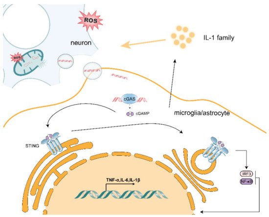
One of the most striking hallmarks shared by various neurodegenerative diseases, including Parkinson’s disease, Alzheimer’s disease and amyotrophic lateral sclerosis, is microglia-mediated and astrocyte-mediated neuroinflammation. Although inhibitions of both harmful proteins and aggregation are major treatments for neurodegenerative diseases, whether the phenomenon of non-normal protein or peptide aggregation is causally related to neuronal loss and synaptic damage is still controversial. Currently, excessive production of reactive oxygen species (ROS), which induces mitochondrial dysfunction in neurons that may play a key role in the regulation of immune cells, is proposed as a regulator in neurological disorders. In this review, we propose that mitochondrial DNA (mtDNA) release due to ROS may act on microglia and astrocytes adjacent to neurons to induce inflammation through activation of innate immune responses (such as cGAS/STING). Elucidating the relationship between mtDNA and the formation of a pro-inflammatory microenvironment could contribute to a better understanding of the mechanism of crosstalk between neuronal and peripheral immune cells and lead to the development of novel therapeutic approaches to neurodegenerative diseases.

mtDNA ROS cGAS/STING neuroinflammation

1. Possible Mechanisms of mtDNA Release

“Leaky” mtDNA from damaged mitochondria caused by mitochondrial dysfunction is a significant source of mitochondrial danger-associated molecular patterns (mtDAMPs). Mitochondria share several features with bacteria, including a double membrane structure and a circular genome with non-methylated CpG sites independent of nuclear DNA replication. Given this similarity, once released into the cytoplasmic or extracellular space, mtDNA fragments activate innate immunity and inflammation, such as DAMPs, which are similar to pathogen-associated molecular patterns (PAMPs). This occurs through a molecular cascade reaction that includes binding to Toll-like receptor 9 (TLR9) and subsequent activation of stimulators of the interferon gene (STING) pathway [1]. We speculate that accumulation of DAMPs in neurons may activate microglia and astrocytes and promote leukocyte infiltration.
ROS production is thought to be an upstream step in the oxidative damage of mitochondrial proteins, membranes and mtDNA. This is partly due to the fact that mitochondria are a major source of endogenous ROSs, which are produced in the mitochondrial matrix and escape from metabolic processes and electron transport chains during oxidative phosphorylation. ROSs are produced by electron transport chains and metabolic redox reactions, which can result in mtDNA mutations or deletions, oxidative damage to the respiratory chain, lipid peroxidation and overall mitochondrial dysfunction. When neurons are exposed to oxidative stress, their internal mitochondria produce ROS, leading to mitochondrial dysfunction and the possible release of oxidized mtDNA (Figure 1). Moreover, mtDNA is located in environments with high levels of ROS, and accumulated mtDNA can lead to organelle and cellular dysfunction. Thus, ROS-induced mitochondrial damage may cause the release of mtDNA, which ultimately leads to mitochondrial dysfunction and in turn, chronic inflammation and disease. As demonstrated by Zhao et al. [2], mtDNA damage can be aggravated by mitochondrial ROS. Mitochondrial Lon is a molecular chaperone and DNA-binding protein that plays a role in protein quality control and stress-response pathways. Lon levels regulate mtDNA metabolism and mitochondrial ROS production. Overexpression of Lon induces mitochondrial ROS to oxidize mtDNA, thereby allowing it to be released it into the cytoplasm [3].
Figure 1. Release of mtDNA from neurons causes activation of the cGAS/STING pathway in neighboring microglia and astrocytes, leading to increased transcription of pro-inflammatory factors.
As a DAMP, mtDNA can cause cellular stress. Many studies have been conducted on how mtDNA is released into the cytoplasm/extracellular space. Kim et al. found that cytoplasmic mtDNA was reduced both at basal levels and after H2O2 stimulation in the voltage-dependent anion channel 1/3(VDAC1/3) knockout mouse fibroblasts (MEF) when compared with wild-type MEF. Additionally, in the presence of mtDNA, VDAC1 trimer and higher-order oligomer formation was increased, suggesting that oxidatively stressed mitochondria release mtDNA fragments through pores formed in the outer mitochondrial membrane by VDAC oligomers [4]. Riley et al. treated osteosarcoma cell U2OS with ABT-737 (a BCL-xL, BCL-2 and BCL-w inhibitor), actinomycin D (ActD; an unstable transcription inhibitor) and qVD-OPh (a cysteine protease inhibitor) to promote mitochondrial apoptosis and mimic mitochondrial outer membrane permeabilization (MOMP). Moreover, mtDNA was observed by using a super-resolution Airyscan confocal microscopy in wild-type and BAX/BAK-deficient U2OS cells. They found that, under cysteine protease inhibitory conditions, which induced BAX/BAK-dependent MOMP, matrix-localized mtDNA was released from mitochondria [5]. Huang et al. transfected LPS into wild-type, Casp11−/− and GSDMD−/− mouse microvascular endothelial cells (MVEC) and found that intracellular LPS-induced mtDNA release was dependent on caspase-11 and Gasdermin D [6]. Similarly, Bao et al. detected mtDNA in the cytoplasm and mitochondrial fission, which is a sign of cellular stress, in hepatocellular carcinoma (HCC) cells treated with Drp1 [7]. Taken together, mtDNA can be released into the cytoplasm through pores formed by VDAC oligomerization, BAX/BAK-mediated MOMP and other means. In addition, Nakahira et al. extracted wild-type and NLRP3−/− mouse bone-marrow-derived macrophages, induced them with LPS, treated them with ATP and found that NALP3 also mediates mtDNA release [8]. It has been suggested that mtDNA can also be released by inflammatory vesicles. Oxidative stress in mitochondria leads to the accumulation of oxidized mtDNA fragments, which, along with ROS, are released in the cytosol as free molecules or engulfed into mitochondrial derived vesicles (MDVs). Guescini et al. demonstrated that, in human glioblastoma cells (U87MG) and astrocyte cells, exosomes can work as vesicular carriers of mtDNA [9]. Moreover, exosomes containing the complete mitochondrial genome were also detected by Sansone et al. [10]. Furthermore, Torralba et al. suggested that exosomes containing mtDNA can activate the cGAS/STING pathway in immune cells, which in turn elicits a downstream inflammatory response [11]. Accumulation of mtDNA in the cytoplasm will also spread to the extracellular space and act on nearby microglia and astrocytes (Figure 1) [12].

2. Effect of mtDNA on Microglia

The mtDNA released by neurons also has an effect on adjacent microglia. Liao et al. used mtDNA extracted from purified mitochondria, which were then transfected into cGAS knockout mice and tamoxifen-induced cGAS or HDAC3 knockout microglia, and demonstrated that the cGAS/STING pathway in microglia was activated by mtDNA and that HDAC3-regulated p65 acetylation and nuclear localization in microglia transcriptionally activated cGAS expression and enhanced cGAS/STING pathway activation, which exacerbated the inflammatory microenvironment [13]. Mathur et al. found that STINGgt/gt was not sensitive to ganciclovir (GCV)-induced interferon responses compared with microglia from wild-type mice [14]. Tsilioni extracted serum isolates from 20 clinical patients with brain-inflammation-induced extracellular vesicles, and the inflammatory group had more mtDNA in cultured microglia and secreted more of the pro-inflammatory cytokine interleukin IL-1β when compared with the normal group [15]. The accumulation of oxidative damage to linear mtDNA leads to the increased production of ROS, which activates redox-sensitive NF-κB to cause excessive neuroinflammation [16]. Thus, mtDNA can enhance microglia polarization toward M1 and exacerbate the inflammatory response. In contrast, Nasi et al. treated human microglia HMC3 cells with mtDNA extracted from Hela cells and found decreased mRNA expression of IL-1β [17]. However, the majority of studies have suggested that mtDNA promotes the secretion of pro-inflammatory factors by microglia, which thereby exacerbates neuroinflammation and thus furthers the development of neurodegenerative diseases.

3. Effect of mtDNA on Astrocytes

Mitochondrial oxidative stress is an important factor leading to the disruption of mtDNA integrity and mtDNA release, which in turn may act on surrounding astrocytes by entering the cytoplasm or being secreted extracellularly via vesicles/exosomes, permeability pores and other means. In a variety of neurodegenerative diseases, TAR DNA/RNA binding protein 43 (TDP-43) exhibits aberrant aggregation and localization within neurons and glial cells. It has been shown that TDP-43 can cause dysregulation of mitochondrial proteins, leading to mitochondrial oxidative stress [18]. Yu et al. used TDP-43 transgenic mice to demonstrate that TDP-43 can invade mitochondria and release mtDNA through permeability transition pores [19]; TDP-43 has also been shown to induce astrocyte inflammation [20]. Hu found that hypoxia-induced mtDNA damage could lead to apoptosis in mouse astrocytes [21]. Ignatenko et al. used replicative mtDNA decapping enzyme Twinkle (TwKO) inactivation to induce mtDNA depletion in mouse neurons and astrocytes and found that mtDNA deletion caused astrocyte overactivation to induce early onset neurological disease [22]. The above studies suggest that the mild stress caused by mtDNA causes astrocytes to secrete more inflammatory factors, thus promoting neuroinflammation, while exacerbating the development of neurodegenerative diseases. In contrast, when mtDNA is excessively damaged or even absent, it may cause astrocytes to undergo apoptosis, which would slow down inflammation and possibly delay the onset of neurodegenerative disease.

4. mtDNA Promotes Activation of the cGAS/STING Pathway

In neurodegenerative diseases, mtDNA serves as the initiation point of neuroinflammation and how it activates the inflammatory response. The mtDNA released by neuronal cells acts on nearby microglia and astrocytes via exocytosis. The cGAS/STING pathway plays a unique and critical role in neuroinflammation and neurodegenerative diseases as one of the important aberrant cytoplasmic DNA monitors. As the primary cytoplasmic DNA receptor for microglia and astrocytes, cGAS can be activated by mtDNA that is aberrantly localized in the cytoplasm. As shown in Figure 1, when mtDNA is released into the cytoplasm by stressed/damaged mitochondria, mtDNA binds to cGAS to form a complex that induces a conformational change in the active site of cGAS, which catalyzes the synthesis of cyclic GMP–AMP (cGAMP) from ATP and GTP. The cGAMP synthesized by cGAS contains two phosphodiester bonds, one between the 2′-OH of GMP and the 5′-phosphate of AMP and the other between the 3′-OH of AMP and the 5′-phosphate of GMP, and is referred to as 2′3′-cGAMP. Overall, 2′3′-cGAMP acts as a second messenger that binds and activates STING proteins on the endoplasmic reticulum membrane, inducing oligomerization to form tetramers and translocation from the endoplasmic reticulum to the endoplasmic reticulum–Golgi intermediate region. Upon cGAMP binding, STING undergoes a conformational change. The two wings of the protein are juxtaposed with the ligand located deep in the binding pocket, and the lid consisting of four antiparallel β-sheet chains is rearranged at the top of the binding pocket, producing a closed conformation. This conformational change leads to a 180° rotation of the ligand-binding region, resulting in the formation of STING oligomers by side-by-side stacking of dimeric STING molecules. After STING is activated, STING is transferred from the endoplasmic reticulum to the Golgi apparatus, where tetramerization of STING can serve as a signaling platform to recruit and activate TANK-binding kinase (TBK1), which in turn can phosphorylate the carboxy-terminal structural domain of STING, which subsequently recruits and phosphorylates interferon regulatory factor 3 (IRF3). Phosphorylated IRF3 forms a dimer that translocates to the nucleus and induces transcriptional expression of downstream type I interferon (IFN-I) and interferon-stimulated genes (ISGs), initiating the innate immune response [23]. Riley et al. treated wild-type and STING-deficient mouse endothelial SVEC cells with ABT-737 and the MCL-1 inhibitor S63845 to induce rapid mitochondrial apoptosis and found that MOMP-induced mtDNA release initiates a cGAS/STING-dependent type I interferon response [5]. Zhang et al. administered mtDNA extracted from liver tissue to wild-type and STING-deficient mice intraperitoneally and found that mtDNA activated IFN-β in a cGAS/STING pathway-dependent manner [24]. In addition, STING recruits IκB kinase (IKK), which phosphorylates the NF-κB inhibitor, IκBα, and phosphorylation of IκBα leads to NF-κB translocation to the nucleus, activating the classical NF-κB signaling pathway and inducing the expression of genes, such as tumor necrosis factor α (TNF-α) and interleukin 6 (IL-6) [23].

5. mtDNA/cGAS/STING Pathway Exacerbates Neurodegenerative Disease

The release of mtDNA due to mitochondrial dysfunction may be an important marker in neurodegenerative diseases. Leurs et al. compared the levels of free mtDNA in the cerebrospinal fluid of patients with relapsing-remitting multiple sclerosis (RRMS), patients with progressive multiple sclerosis (PMS), controls with various neurological diseases, and healthy controls and found that PMS patients had mtDNA concentrations that were nonspecifically elevated [25]. Podlesniy et al. [26] and Cervera-Carles et al. [27] also examined levels of free mtDNA in cerebrospinal fluid from clinical patients with AD versus asymptomatic patients at risk for AD and found that preclinical free mtDNA was significantly increased. Sliter et al. found that mtDNA-induced mitochondrial stress in the absence of Parkin or PINK1 led to a STING-mediated type I interferon response in mice by STINGgt/gtParkin−/− and PINK1−/− mice and their hybrids, demonstrating that Parkin and PINK1 prevent the development of a cytosolic response by clearing both damaged mitochondria to prevent increases in cytosol and circulating mtDNA to prevent inflammation and neurodegeneration, thereby alleviating symptoms of PD through mitochondrial autophagy [28]. Yu et al. used transgenic mice with the TDP-43 mutant allele A315T, STING-deficient mice, and the resulting progeny from crossing these two lines to demonstrate that TDP 43 releases mtDNA through the mPTP permeability pore and activates the cGAS/STING pathway thereby exacerbating ALS [29]. Jauhari et al. found in AANAT-KO mice that mtDNA release into the cytoplasm and its mediated inflammation are dependent on the cGAS/STING/IRF3 pathway [30]. mtDNA release due to mitochondrial Lon induced Cheng et al. also found that ROS induction by Lon-induced mtDNA release activates the STING/IFN pathway to induce an inflammatory response [3].

References

  1. Krysko, D.; Agostinis, P.; Krysko, O.; Garg, A.; Bachert, C.; Lambrecht, B.N.; Vandenabeele, P. Emerging role of damage-associated molecular patterns derived from mitochondria in inflammation. Trends Immunol. 2011, 32, 157–164.
  2. Zhao, M.; Wang, Y.; Li, L.; Liu, S.; Wang, C.; Yuan, Y.; Yang, G.; Chen, Y.; Cheng, J.; Lu, Y.; et al. Mitochondrial ROS promote mitochondrial dysfunction and inflammation in ischemic acute kidney injury by disrupting TFAM-mediated mtDNA maintenance. Theranostics 2021, 11, 1845–1863.
  3. Cheng, A.N.; Cheng, L.-C.; Kuo, C.-L.; Lo, Y.K.; Chou, H.-Y.; Chen, C.-H.; Wang, Y.-H.; Chuang, T.-H.; Cheng, S.-J.; Lee, A.Y.-L. Mitochondrial Lon-induced mtDNA leakage contributes to PD-L1–mediated immunoescape via STING-IFN signaling and extracellular vesicles. J. Immunother. Cancer 2020, 8, e001372.
  4. Kim, J.; Gupta, R.; Blanco, L.P.; Yang, S.; Shteinfer-Kuzmine, A.; Wang, K.; Zhu, J.; Yoon, H.E.; Wang, X.; Kerkhofs, M.; et al. VDAC oligomers form mitochondrial pores to release mtDNA fragments and promote lupus-like disease. Science 2019, 366, 1531–1536.
  5. Riley, J.S.; Quarato, G.; Cloix, C.; Lopez, J.; O’Prey, J.; Pearson, M.; Chapman, J.; Sesaki, H.; Carlin, L.M.; Passos, J.F.; et al. Mitochondrial inner membrane permeabilisation enables mt DNA release during apoptosis. EMBO J. 2018, 37, e99238.
  6. Huang, L.S.; Hong, Z.; Wu, W.; Xiong, S.; Gao, X.; Rehman, J.; Malik, A.B. mtDNA Activates cGAS Signaling and Suppresses the YAP-Mediated Endothelial Cell Proliferation Program to Promote Inflammatory Injury. Immunity 2020, 52, 475–486.e5.
  7. Bao, D.; Zhao, J.; Zhou, X.; Yang, Q.; Chen, Y.; Zhu, J.; Yuan, P.; Yang, J.; Qin, T.; Wan, S.; et al. Mitochondrial fission-induced mtDNA stress promotes tumor-associated macrophage infiltration and HCC progression. Oncogene 2019, 38, 5007–5020.
  8. Nakahira, K.; Haspel, J.A.; Rathinam, V.A.; Lee, S.-J.; Dolinay, T.; Lam, H.C.; Englert, J.A.; Rabinovitch, M.; Cernadas, M.; Kim, H.P.; et al. Autophagy proteins regulate innate immune responses by inhibiting the release of mitochondrial DNA mediated by the NALP3 inflammasome. Nat. Immunol. 2011, 12, 222–230.
  9. Li, T.; Tan, X.; Li, S.; Al-Nusaif, M.; Le, W. Role of Glia-Derived Extracellular Vesicles in Neurodegenerative Diseases. Front. Aging Neurosci. 2021, 13, 765395.
  10. Sansone, P.; Savini, C.; Kurelac, I.; Chang, Q.; Amato, L.B.; Strillacci, A.; Stepanova, A.; Iommarini, L.; Mastroleo, C.; Daly, L.; et al. Packaging and transfer of mitochondrial DNA via exosomes regulate escape from dormancy in hormonal therapy-resistant breast cancer. Proc. Natl. Acad. Sci. USA 2017, 114, E9066–E9075.
  11. Torralba, D.; Baixauli, F.; Villarroya-Beltri, C.; Fernández-Delgado, I.; Latorre-Pellicer, A.; Acín-Pérez, R.; Martín-Cófreces, N.B.; Jaso-Tamame, Á.L.; Iborra, S.; Jorge, I.; et al. Priming of dendritic cells by DNA-containing extracellular vesicles from activated T cells through anti-gen-driven contacts. Nat. Commun. 2018, 9, 2658.
  12. Gambardella, S.; Limanaqi, F.; Ferese, R.; Biagioni, F.; Campopiano, R.; Centonze, D.; Fornai, F. ccf-mtDNA as a Potential Link Between the Brain and Immune System in Neuro-Immunological Disorders. Front. Immunol. 2019, 10, 1064.
  13. Liao, Y.; Cheng, J.; Kong, X.; Li, S.; Li, X.; Zhang, M.; Zhang, H.; Yang, T.; Dong, Y.; Li, J.; et al. HDAC3 inhibition ameliorates ischemia/reperfusion-induced brain injury by regulating the microglial cGAS-STING pathway. Theranostics 2020, 10, 9644–9662.
  14. Mathur, V.; Burai, R.; Vest, R.T.; Bonanno, L.; Lehallier, B.; Zardeneta, M.E.; Mistry, K.N.; Do, D.; Marsh, S.; Abud, E.M.; et al. Activation of the STING-Dependent Type I Interferon Response Reduces Microglial Reactivity and Neuroinflammation. Neuron 2017, 96, 1290–1302.e6.
  15. Tsilioni, I.; Theoharides, T.C. Extracellular vesicles are increased in the serum of children with autism spectrum disorder, contain mitochondrial DNA, and stimulate human microglia to secrete IL-1β. J. Neuroinflamm. 2018, 15, 1–8.
  16. Nakanishi, H.; Hayashi, Y.; Wu, Z. The role of microglial mtDNA damage in age-dependent prolonged LPS-induced sickness behavior. Neuron Glia Biol. 2011, 7, 17–23.
  17. Nasi, M.; De Gaetano, A.; Bianchini, E.; De Biasi, S.; Gibellini, L.; Neroni, A.; Mattioli, M.; Pinti, M.; Tartaro, D.L.; Borella, R.; et al. Mitochondrial damage-associated molecular patterns stimulate reactive oxygen species production in human microglia. Mol. Cell. Neurosci. 2020, 108, 103538.
  18. Zuo, X.; Zhou, J.; Li, Y.; Wu, K.; Chen, Z.; Luo, Z.; Zhang, X.; Liang, Y.; Esteban, M.A.; Zhou, Y.; et al. TDP-43 aggregation induced by oxidative stress causes global mitochondrial imbalance in ALS. Nat. Struct. Mol. Biol. 2021, 28, 132–142.
  19. Yu, C.-H.; Davidson, S.; Harapas, C.R.; Hilton, J.B.; Mlodzianoski, M.J.; Laohamonthonkul, P.; Louis, C.; Low, R.R.J.; Moecking, J.; De Nardo, D.; et al. TDP-43 Triggers Mitochondrial DNA Release via mPTP to Activate cGAS/STING in ALS. Cell 2020, 183, 636–649.e18.
  20. Lee, S.; Kim, S.; Kang, H.Y.; Lim, H.R.; Kwon, Y.; Jo, M.; Jeon, Y.M.; Kim, S.R.; Kim, K.; Ha, C.M.; et al. The overexpression of TDP-43 in astrocytes causes neurodegeneration via a PTP1B-mediated inflammatory re-sponse. J. Neuroinflamm. 2020, 17, 299.
  21. Hu, J.; Bibli, S.I.; Wittig, J.; Zukunft, S.; Lin, J.; Hammes, H.-P.; Popp, R.; Fleming, I. Soluble epoxide hydrolase promotes astrocyte survival in retinopathy of prematurity. J. Clin. Investig. 2019, 129, 5204–5218.
  22. Ignatenko, O.; Chilov, D.; Paetau, I.; De Miguel, E.; Jackson, C.B.; Capin, G.; Paetau, A.; Terzioglu, M.; Euro, L.; Suomalainen, A. Loss of mtDNA activates astrocytes and leads to spongiotic encephalopathy. Nat. Commun. 2018, 9, 1–12.
  23. Motwani, M.; Pesiridis, S.; Fitzgerald, K.A. DNA sensing by the cGAS-STING pathway in health and disease. Nat. Rev. Genet. 2019, 20, 657–674.
  24. Zhang, X.; Wu, J.; Liu, Q.; Li, X.; Li, S.; Chen, J.; Hong, Z.; Wu, X.; Zhao, Y.; Ren, J. mtDNA-STING pathway promotes necroptosis-dependent enterocyte injury in intestinal ischemia reperfusion. Cell Death Dis. 2020, 11, 1050.
  25. E Leurs, C.; Podlesniy, P.; Trullas, R.; Balk, L.; Steenwijk, M.D.; Malekzadeh, A.; Piehl, F.; Uitdehaag, B.M.; Killestein, J.; Van Horssen, J.; et al. Cerebrospinal fluid mtDNA concentration is elevated in multiple sclerosis disease and responds to treatment. Mult. Scler. J. 2017, 24, 472–480.
  26. Podlesniy, P.; Figueiro-Silva, J.; Llado, A.; Antonell, A.; Sanchez-Valle, R.; Alcolea, D.; Lleo, A.; Molinuevo, J.L.; Serra, N.; Trullas, R. Low cerebrospinal fluid concentration of mitochondrial DNA in preclinical Alzheimer disease. Ann. Neurol. 2013, 74, 655–668.
  27. Cervera-Carles, L.; Alcolea, D.; Estanga, A.; Ecay-Torres, M.; Izagirre, A.; Clerigué, M.; Garcia-Sebastian, M.; Villanúa, J.; Escalas, C.; Blesa, R.; et al. Cerebrospinal fluid mitochondrial DNA in the Alzheimer’s disease continuum. Neurobiol. Aging 2017, 53, 192.e1–192.e4.
  28. Sliter, D.A.; Martinez, J.; Hao, L.; Chen, X.; Sun, N.; Fischer, T.D.; Burman, J.L.; Li, Y.; Zhang, Z.; Narendra, D.P.; et al. Parkin and PINK1 mitigate STING-induced inflammation. Nature 2018, 561, 258–262.
  29. Brambilla, G.; Gallotti, C. . Radiol. Med. 1987, 74, 49–58.
  30. Jauhari, A.; Baranov, S.V.; Suofu, Y.; Kim, J.; Singh, T.; Yablonska, S.; Li, F.; Wang, X.; Oberly, P.; Minnigh, M.B.; et al. Melatonin inhibits cytosolic mitochondrial DNA–induced neuroinflammatory signaling in accelerated aging and neurodegeneration. J. Clin. Investig. 2021, 131.
More
Information
Subjects: Neurosciences
Contributor MDPI registered users' name will be linked to their SciProfiles pages. To register with us, please refer to https://encyclopedia.pub/register :
View Times: 399
Entry Collection: Neurodegeneration
Revisions: 3 times (View History)
Update Date: 17 Dec 2021
1000/1000