Submitted Successfully!
To reward your contribution, here is a gift for you: A free trial for our video production service.
Thank you for your contribution! You can also upload a video entry or images related to this topic.
Version Summary Created by Modification Content Size Created at Operation
1 + 1924 word(s) 1924 2021-12-01 07:22:24 |
2 The format is correct. Meta information modification 1924 2021-12-06 03:04:28 | |
3 The format is correct. Meta information modification 1924 2021-12-06 03:05:18 | |
4 corrected the format -3 word(s) 1921 2021-12-06 03:17:04 | |
5 corrected the format Meta information modification 1921 2021-12-08 08:37:56 | |
6 content Meta information modification 1921 2021-12-08 08:38:38 |

Video Upload Options

Do you have a full video?

Confirm

Are you sure to Delete?
Cite
If you have any further questions, please contact Encyclopedia Editorial Office.
Montaseri, H.; Nkune, N.; Simelane, N. Photodynamic Therapy-Mediated Immune Responses in Three-Dimensional Tumor Models. Encyclopedia. Available online: https://encyclopedia.pub/entry/16750 (accessed on 27 July 2024).
Montaseri H, Nkune N, Simelane N. Photodynamic Therapy-Mediated Immune Responses in Three-Dimensional Tumor Models. Encyclopedia. Available at: https://encyclopedia.pub/entry/16750. Accessed July 27, 2024.
Montaseri, Hanieh, Nkune Nkune, Nokuphila Simelane. "Photodynamic Therapy-Mediated Immune Responses in Three-Dimensional Tumor Models" Encyclopedia, https://encyclopedia.pub/entry/16750 (accessed July 27, 2024).
Montaseri, H., Nkune, N., & Simelane, N. (2021, December 06). Photodynamic Therapy-Mediated Immune Responses in Three-Dimensional Tumor Models. In Encyclopedia. https://encyclopedia.pub/entry/16750
Montaseri, Hanieh, et al. "Photodynamic Therapy-Mediated Immune Responses in Three-Dimensional Tumor Models." Encyclopedia. Web. 06 December, 2021.
Photodynamic Therapy-Mediated Immune Responses in Three-Dimensional Tumor Models
Edit

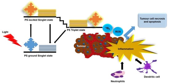
Photodynamic therapy (PDT) is a promising non-invasive phototherapeutic approach for cancer therapy that can eliminate local tumor cells and produce systemic antitumor immune responses. Significant efforts have been made in developing strategies to further investigate the immune mechanisms triggered by PDT. The majority of in vitro experimental models still rely on the two-dimensional (2D) cell cultures that do not mimic a three-dimensional (3D) cellular environment in the human body, such as cellular heterogeneity, nutrient gradient, growth mechanisms, and the interaction between cells as well as the extracellular matrix (ECM) and therapeutic resistance to anticancer treatments. In addition, in vivo animal studies are highly expensive and time consuming, which may also show physiological discrepancies between animals and humans. In this sense, there is growing interest in the utilization of 3D tumor models, since they precisely mimic different features of solid tumors. This entry summarizes the characteristics and techniques for 3D tumor model generation. Furthermore, researchers provide an entry of innate and adaptive immune responses induced by PDT in several in vitro and in vivo tumor models. Future perspectives are highlighted for further enhancing PDT immune responses as well as ideal experimental models for antitumor immune response sudies.

photodynamic therapy adaptive immunity 3D tumor models nanotechnology

1. Introduction

Photodynamic therapy (PDT) is a cancer modality that combines three essential components of a photosensitizer (PS), harmless light, and molecular oxygen [1]. It is based on the accumulation of a PS in pathological tissues, which can generate highly cytotoxic reactive oxygen species (ROS) upon its activation with a specific wavelength of light [2]. PDT presents unique advantages such as the selective uptake of PSs by tumor tissues, localized light exposure to the affected site, non-invasiveness feature, and simple procedure. Additionally, it endows low toxicity and high efficacy, with no drug resistance [3][4]. As shown in Figure 1, ROS generated by PDT can directly destruct the vasculature of the tumor by induction of apoptosis and/or necrosis, resulting in oxygen and nutrient depletion in the tumor [5]. As a result of this photodamage to the tumor and its microenvironment, a robust acute inflammatory response is produced at the tumor site [4][5]. The acute inflammatory response following PDT stimulates the immune system and causes the infiltration of host innate immune cells, which clear damaged cells in the treated area [5][6]. In a later stage, an adaptive immune memory may occur, allowing for a systemic response that can inhibit tumor recurrence and metastases in the long run [6].
Figure 1. Antitumor mechanisms induced by PDT.
When the immune monitoring function becomes dysfunctional, tumor cells can continue to grow and form malignant tumors [6][7]. Additionally, tumor cells can evade immune system barriers via multiple mechanisms, which is the main cause of the low clinical efficacy of the most antitumor treatments [8]. Generally, immunosuppressive cells and molecules counteract any antitumor effects in the body [9]. Depending on the influence of immunoregulatory factors in the tumor microenvironment, some tumors are less immunogenic and do not trigger any specific immune responses [7]. The low expression of transporter of antigenic peptide and major histocompatibility complex (MHC) molecules normally hinders antigen processing and presentation mechanisms, which in turn inhibits specific immune responses [7]. PDT can circumvent this dysfunction via the initiation of immunogenic tumor cell death modes, essentially immunogenic apoptosis and necrosis [6]. An innate immune response can be initiated by the exposure or release of danger stimuli from damaged cancer cells, which is known as damage-associated molecular patterns (DAMPs) [4][6]. These DAMPs alone or in combination with tumor antigens can be identified by antigen-presenting cells (APCs), which may trigger an adaptive immune response against the tumor [6].

2. Methods for 3D Tumor Generation

Over the recent years, several types of 3D tumor models have been established to generate spheroids in oncology. The main 3D culture models of cancer include multicellular tumor spheroids (MCTS), tumor tissue explant, and tumor on a chip [10]. MCTs are aggregates of tumor cells that resemble spheres and are generally cultivated in suspension or embedded gels using 3D culture methods [11]. This model can partly simulate the avascular layers (proliferation zone, quiescent zone, and necrotic zone) of solid tumors. It also has phenotypic characteristics that closely resemble the cellular microenvironment in the cancerous tissue to a great extent [12]. For instance, the unevenly diffused and distribution of oxygen and nutrient gradients within larger MCTS (critical size, 400 µm) often result in the formation of distinct layers, similar to those in poorly vascularized tumors [12][11]. Thus, MTCS have become prevalent because of the overall similarities between MCTS and tumors with respect to morphology, distinct metabolic and proliferation gradients [11]. MCTS can be exploited in various experimental research studies on tumor-specific processes such as angiogenesis, invasion, and metastasis, as well as assessment of responses to various therapies such as PDT and underlying mechanisms [11]. Several simple and reproducible methods have been successfully utilized to generate MCTs, which can be categorized into two distinguishable groups: scaffold-based and scaffold-free cell techniques [12]. The scaffold-free 3D cell culture method has been commonly used to produce MCTS and is comprised of techniques such as liquid overlay, the hanging drop, and bioreactor/agitation-based methods, such as shaker flasks and spinner vessels as well as hydrogels and magnetic levitation method [12][13].

3. PDT-Induced Antitumor Immune Responses

Ideal anticancer treatment should induce local tumor regression and systemic antitumor immune responses capable of obliterating distant metastases with minimal toxicity to healthy tissues [14]. From this perspective, PDT holds great promise, since it provides a strong and acute inflammatory response [9]. The local inflammatory responses lead to the infiltration of neutrophils into the tumor site and the generation of pro-inflammatory factors and cytokines [14]. Meanwhile, photodamaged cells show a systemic antitumor immune response, which then activates a secondary cause of tumor cell death [15]. PDT can trigger both innate and adaptive immune responses by subjecting PDT-treated tumor cells to complementary immune cells (Figure 2) [5][16][17].
Figure 2. An overview of PDT-induced innate and adaptive immune responses.

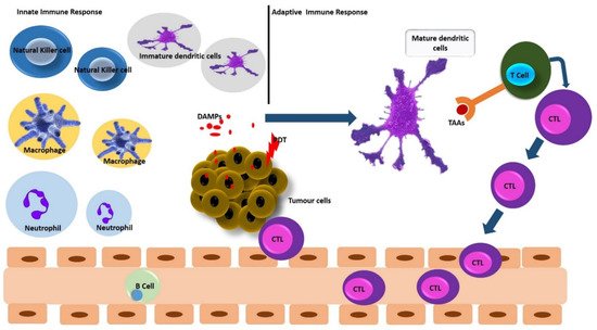
3.1. PDT and Innate Immune Response

The innate arm of the immune response system eliminates pathogenic agents by phagocytes (macrophages, neutrophils, and dendritic cells (DCs), the complement cascade, and natural killer (NK) cells [6]. Following an acute inflammatory response, PDT-induced activation of the innate immune system involves the release of cytokines, complement activation, infiltration, and activation of innate immune cells [2][9]. Then, post-treatment, the oxidative stress triggered by PDT leads to extended tumor tissue destruction [16]. Damaged tumor cells would exhibit damage-associated molecular patterns (DAMPs) or secrete DAMPs into the extracellular matrix [2][6][9]. Those DAMPs serve as harmful signals and can be recognized by antigen-presenting cells (mainly the DCs) [6]. DCs ingest and process tumor-associated antigens (TAAs) and present TAA-derived peptides to effector T cells, thereby coordinating an antitumor adaptive immunity, which could confer a prolonged systemic tumor immune control [9][18][19].

3.2. PDT and Adaptive Immune Response

The initiation of acute inflammation by PDT attracts neutrophils to the irradiated tumor area and secretes chemokines and granule proteins to promote DCs maturation and activation [14]. Activated DCs can stimulate naïve T cells to transform into cytotoxic tumor-specific T lymphocytes (CTLs) and antigens that can stimulate B cells to produce antibodies [4][9]. Upon exposure to DAMPs, which are released by PDT-damaged or dying cells, DCs can transit to a mature state and migrate to lymph nodes, which consequently present TAA-derived peptides to naïve T cells and generate CTLs to attack and obliterate residual cancer cells [6][16][17]. The mature DCSs are characterized by the overexpression of peptide-major histocompatibility (MHC) complexes on the cell surface, prime CD4+ T helper cells and CD8+ to CTLs, and trigger an adaptive immunity [5][15][17]

4. Enhancing PDT-Induced Antitumor Immune Responses

4.1. Intracellular Accumulation of PSs

PDT-induced cell death depends on the intracellular localization and binding sites of the PSs [20]. Photoexcitation of a mitochondrion-localized PS triggers the release of cytochrome C, which in turn activates apoptosis caspase [21]. Meanwhile, photodamage to the ER causes the release of Ca2+, which can potentially lead to apoptosis [22][23]. PDT can also cause subcellular organelle-specific stress, since subcellular organelle-dependent oxidative stress is linked to signaling pathways in immunogenic cell death [24][25]. Therefore, by formulating PSs with a high affinity to specific subcellular organelles, it may be possible to control both the PDT process and antitumor immune response in tumor cells to improve therapeutic efficacy.

4.2. ER-Targeted PDT

The ER is a vital organelle that performs several essential cellular processes and can influence cancer pathogenesis [22]. ER stress is the key initiator of intracellular signaling pathways that regulate immunogenic cell death (ICD) [26][27]). It was reported that ER is the accumulation site for PS drugs such as hypericin or meta-Tetra(hydroxyphenyl)chlorin (mTHPC) and can cause an extensive ROS-based ER stress upon photoactivation [28][29]. Such ER-accumulated PSs not only exert ROS-induced cellular destruction but also can cause ICD [28]. Thus, ER plays a key role in promoting PDT efficacy via direct effects on cancer cells and indirect effects on immunity [22].
However, one major problem that could potentially hamper the application of these PSs is their inherent low absorbance wavelength that cannot reach deep-seated cancer tissues [30]. As a result, in vivo PDT treatment using hypericin or mTHPC can only be applied for superficial tumor tissue. To fully explore their ability to eradicate deep tumor tissue, it would be ideal to conserve their ER localization and amplify their light absorption wavelength to the optimum near-infrared region through different derivatives [4]. In recent years, strides have been made to develop ICD inducers that can directly and effectively trigger ER stress [31]. Studies by Li et al. [31] developed a nanocomposite that consisted of ER-targeting pardaxin (FAL) peptides functionalized with indocyanine green (ICG) conjugated-hollow gold nanospheres (FAL-ICG-HAuNS), together with an oxygen-delivering hemoglobin (Hb) liposome (FAL-Hb lipo), which was designed to counteract hypoxia. Li et al. reported that light irradiation and the nanocomposite triggered robust ER stress and calreticulin (CRT) exposure on the surface, which stimulated dendritic cells [31].

4.3. Mitochondria Targeted PDT

Mitochondria are vital subcellular organelles that undertake critical roles in metabolism, including cellular proliferation. Their actions are complexly interlinked with signaling pathways and the apoptotic process [32]. Thus, great efforts have been made in developing mitochondria-targeted PSs to enhance PDT efficacy and improve cancer treatment outcomes. Yang et al. developed mitochondria-targeting gold nanoparticles combined with triphenylphosphonium (TPP) to increase 5-ALA PS cellular uptake to allow for enhanced ROS formation and improved the selective photodamage of breast cancer cells in PDT [33]. Xu et al. [34] investigated a dual-targeting nanophotosensitizer consisting of cationic porphyrin derivative (MitoTPP) with the polyethylene glycol (PEG)-functionalized and folic acid-modified nanographene oxide (NGO) that overexpressed folate receptor and subsequently localize in mitochondria [28]. Upon photoactivation, the released MitoTPP molecule produced cytotoxic singlet oxygen and induce enhanced oxidative stress in cells [34]. Soler et al. [35] demonstrated that photoactivated silicon phthalocyanine (Pc4) accumulated in mitochondria triggered apoptosis on activated CD3+ T cells that may be used in targeting T cell-related skin disorders [29].

4.4. Application of Nanotechnology

Nanomedicine is a rapidly evolving field that is transforming cancer diagnosis and treatment [36]. Nanoparticles (NPs) can be broadly defined as materials that have at least one dimension (1–100 nm) in the nanoscale regime, which thus provide unique chemical and physical properties [37][38]. NPs can overcome the current challenges of PDT and have emerged as a unique approach to improve the therapeutic efficacy of PDT [39]. Since NPs are hydrophilic, they can significantly enhance the solubility of conventional PSs and their cellular uptake [1]. NP-loaded PSs can passively accumulate in tumors due to enhanced permeability and retention (EPR), which is attributed to the leaky tumor vasculature and impaired lymphatic drainage of tumor tissues [40]. Moreover, the targeted delivery of PSs can be significantly enhanced by immobilizing targeting moieties on the surface of NPs, such as antibodies, aptamers, and peptides [41]. An active targeting NPs-PS system can increase the bioavailability of PSs in the affected area while minimizing unwanted side effects of PS drugs to adjacent healthy tissues [41].

References

  1. Kruger, C.A.; Abrahamse, H. Utilisation of Targeted Nanoparticle Photosensitiser Drug Delivery Systems for the Enhancement of Photodynamic Therapy. Molecules 2018, 23, 2628.
  2. dos Santos, A.F.; de Almeida, D.R.Q.; Terra, L.F.; Baptista, M.S.; Labriola, L. Photodynamic Therapy in Cancer Treatment—An Update Review. J. Cancer Metastatis Treat. 2019, 5, 25.
  3. Zhang, Y.; Wang, B.; Zhao, R.; Zhang, Q.; Kong, X. Multifunctional Nanoparticles as Photosensitizer Delivery Carriers for Enhanced Photodynamic Cancer Therapy. Mater. Sci. Eng. C Mater. Biol. Appl. 2020, 115, 111099.
  4. Yang, Y.; Hu, Y.; Wang, H. Targeting Antitumor Immune Response for Enhancing the Efficacy of Photodynamic Therapy of Cancer: Recent Advances and Future Perspectives. Oxid. Med. Cell. Longev. 2016, 2016, e5274084.
  5. Reginato, E.; Wolf, P.; Hamblin, M.R. Immune Response after Photodynamic Therapy Increases Anti-Cancer and Anti-Bacterial Effects. World J. Immunol. 2014, 4, 1–11.
  6. Beltrán Hernández, I.; Yu, Y.; Ossendorp, F.; Korbelik, M.; Oliveira, S. Preclinical and Clinical Evidence of Immune Responses Triggered in Oncologic Photodynamic Therapy: Clinical Recommendations. J. Clin. Med. 2020, 9, 333.
  7. Chen, H.; Dai, Z. Antitumor Immune Responses Induced by Photodynamic and Sonodynamic Therapy: A Narrative Review. J. Bio-X Res. 2021, 4, 77–86.
  8. Pitt, J.M.; Marabelle, A.; Eggermont, A.; Soria, J.-C.; Kroemer, G.; Zitvogel, L. Targeting the Tumor Microenvironment: Removing Obstruction to Anticancer Immune Responses and Immunotherapy. Ann. Oncol. 2016, 27, 1482–1492.
  9. Hwang, H.S.; Shin, H.; Han, J.; Na, K. Combination of Photodynamic Therapy (PDT) and Anti-Tumor Immunity in Cancer Therapy. J. Pharm. Investig. 2018, 48, 143–151.
  10. Nath, S.; Devi, G.R. Three-Dimensional Culture Systems in Cancer Research: Focus on Tumor Spheroid Model. Pharmacol. Ther. 2016, 163, 94–108.
  11. Lv, D.; Hu, Z.; Lu, L.; Lu, H.; Xu, X. Three-Dimensional Cell Culture: A Powerful Tool in Tumor Research and Drug Discovery. Oncol. Lett. 2017, 14, 6999–7010.
  12. Pinto, B.; Henriques, A.C.; Silva, P.M.A.; Bousbaa, H. Three-Dimensional Spheroids as In Vitro Preclinical Models for Cancer Research. Pharmaceutics 2020, 12, 1186.
  13. Chaicharoenaudomrung, N.; Kunhorm, P.; Noisa, P. Three-Dimensional Cell Culture Systems as an In Vitro Platform for Cancer and Stem Cell Modeling. World J. Stem Cells 2019, 11, 1065–1083.
  14. Mroz, P.; Hashmi, J.T.; Huang, Y.-Y.; Lange, N.; Hamblin, M.R. Stimulation of Anti-Tumor Immunity by Photodynamic Therapy. Expert Rev. Clin. Immunol. 2011, 7, 75–91.
  15. Vatansever, F.; Hamblin, M.R. Photodynamic Therapy and Antitumor Immune Response. Cancer Immunol. 2015, 383–399.
  16. Wachowska, M.; Muchowicz, A.; Demkow, U. Immunological Aspects of Antitumor Photodynamic Therapy Outcome. Cent. Eur. J. Immunol. 2015, 40, 481–485.
  17. Donohoe, C.; Senge, M.O.; Arnaut, L.G.; Gomes-da-Silva, L.C. Cell Death in Photodynamic Therapy: From Oxidative Stress to Anti-Tumor Immunity. Biochim. Biophys. Acta (BBA) Rev. Cancer 2019, 1872, 188308.
  18. Friedrich, J.; Seidel, C.; Ebner, R.; Kunz-Schughart, L.A. Spheroid-Based Drug Screen: Considerations and Practical Approach. Nat. Protoc. 2009, 4, 309–324.
  19. Ryu, N.-E.; Lee, S.-H.; Park, H. Spheroid Culture System Methods and Applications for Mesenchymal Stem Cells. Cells 2019, 8, 1620.
  20. Gift, M.; Ann, K.; Mfouo Tynga, I.; Abrahamse, H. A Review of Nanoparticle Photosensitizer Drug Delivery Uptake Systems for Photodynamic Treatment of Lung Cancer. Photodiagn. Photodyn. Ther. 2018, 22, 147–154.
  21. Kwiatkowski, S.; Knap, B.; Przystupski, D.; Saczko, J.; Kędzierska, E.; Knap-Czop, K.; Kotlińska, J.; Michel, O.; Kotowski, K.; Kulbacka, J. Photodynamic Therapy—Mechanisms, Photosensitizers and Combinations. Biomed. Pharmacother. 2018, 106, 1098–1107.
  22. Yuan, B.; Liu, J.; Guan, R.; Jin, C.; Ji, L.; Chao, H. Endoplasmic Reticulum Targeted Cyclometalated Iridium(III) Complexes as Efficient Photodynamic Therapy Photosensitizers. Dalton Trans. 2019, 48, 6408–6415.
  23. Zhuang, Z.; Dai, J.; Yu, M.; Li, J.; Shen, P.; Hu, R.; Lou, X.; Zhao, Z.; Tang, B.Z. Type I Photosensitizers Based on Phosphindole Oxide for Photodynamic Therapy: Apoptosis and Autophagy Induced by Endoplasmic Reticulum Stress. Chem. Sci. 2020, 11, 3405–3417.
  24. West, A.P.; Shadel, G.S.; Ghosh, S. Mitochondria in Innate Immune Responses. Nat. Rev. Immunol. 2011, 11, 389–402.
  25. Zhang, K.; Kaufman, R.J. From Endoplasmic-Reticulum Stress to the Inflammatory Response. Nature 2008, 454, 455–462.
  26. Garg, A.D.; Dudek, A.M.; Ferreira, G.B.; Verfaillie, T.; Vandenabeele, P.; Krysko, D.V.; Mathieu, C.; Agostinis, P. ROS-Induced Autophagy in Cancer Cells Assists in Evasion from Determinants of Immunogenic Cell Death. Autophagy 2013, 9, 1292–1307.
  27. Michaud, M.; Sukkurwala, A.Q.; Di Sano, F.; Zitvogel, L.; Kepp, O.; Kroemer, G. Synthetic Induction of Immunogenic Cell Death by Genetic Stimulation of Endoplasmic Reticulum Stress. OncoImmunology 2014, 3, e28276.
  28. Garg, A.D.; Agostinis, P. ER Stress, Autophagy and Immunogenic Cell Death in Photodynamic Therapy-Induced Anti-Cancer Immune Responses. Photochem. Photobiol. Sci. 2014, 13, 474–487.
  29. Agostinis, P.; Berg, K.; Cengel, K.A.; Foster, T.H.; Girotti, A.W.; Gollnick, S.O.; Hahn, S.M.; Hamblin, M.R.; Juzeniene, A.; Kessel, D.; et al. Photodynamic Therapy of Cancer: An Update. CA Cancer J. Clin. 2011, 61, 250–281.
  30. Md, S.; Haque, S.; Madheswaran, T.; Zeeshan, F.; Meka, V.S.; Radhakrishnan, A.K.; Kesharwani, P. Lipid Based Nanocarriers System for Topical Delivery of Photosensitizers. Drug Discov. Today 2017, 22, 1274–1283.
  31. Li, W.; Yang, J.; Luo, L.; Jiang, M.; Qin, B.; Yin, H.; Zhu, C.; Yuan, X.; Zhang, J.; Luo, Z.; et al. Targeting Photodynamic and Photothermal Therapy to the Endoplasmic Reticulum Enhances Immunogenic Cancer Cell Death. Nat. Commun. 2019, 10, 3349.
  32. Noh, I.; Lee, D.; Kim, H.; Jeong, C.-U.; Lee, Y.; Ahn, J.-O.; Hyun, H.; Park, J.-H.; Kim, Y.-C. Enhanced Photodynamic Cancer Treatment by Mitochondria-Targeting and Brominated Near-Infrared Fluorophores. Adv. Sci. 2018, 5, 1700481.
  33. Yang, Y.; Gao, N.; Hu, Y.; Jia, C.; Chou, T.; Du, H.; Wang, H. Gold Nanoparticle-Enhanced Photodynamic Therapy: Effects of Surface Charge and Mitochondrial Targeting. Ther. Deliv. 2015, 6, 307–321.
  34. Xu, J.; Zeng, F.; Wu, H.; Yu, C.; Wu, S. Dual-Targeting Nanosystem for Enhancing Photodynamic Therapy Efficiency. ACS Appl. Mater. Interfaces 2015, 7, 9287–9296.
  35. Soler, D.C.; Ohtola, J.; Sugiyama, H.; Rodriguez, M.E.; Han, L.; Oleinick, N.L.; Lam, M.; Baron, E.D.; Cooper, K.D.; McCormick, T.S. Activated T Cells Exhibit Increased Uptake of Silicon Phthalocyanine Pc 4 and Increased Susceptibility to Pc 4-Photodynamic Therapy-Mediated Cell Death. Photochem. Photobiol. Sci. 2016, 15, 822–831.
  36. Senapati, S.; Mahanta, A.K.; Kumar, S.; Maiti, P. Controlled Drug Delivery Vehicles for Cancer Treatment and Their Performance. Signal Transduct. Target. Ther. 2018, 3, 7.
  37. Tang, C.; Li, H. Application of Nanoparticles in the Early Diagnosis and Treatment of Tumors: Current Status and Progress. Tradit. Med. Res. 2020, 5, 34–43.
  38. Monge-Fuentes, V.; Muehlmann, L.A.; de Azevedo, R.B. Perspectives on the Application of Nanotechnology in Photodynamic Therapy for the Treatment of Melanoma. Nano Rev. 2014, 5, 24381.
  39. Nkune, N.; Kruger, C.; Abrahamse, H. Possible Enhancement of Photodynamic Therapy (PDT) Colorectal Cancer Treatment When Combined with Cannabidiol. Anti-Cancer Agents Med. Chem. 2020, 20, 137–148.
  40. Naidoo, C.; Kruger, C.A.; Abrahamse, H. Photodynamic Therapy for Metastatic Melanoma Treatment: A Review. Technol. Cancer Res. Treat. 2018, 17, 1533033818791795.
  41. Hong, E.J.; Choi, D.G.; Shim, M.S. Targeted and Effective Photodynamic Therapy for Cancer Using Functionalized Nanomaterials. Acta Pharm. Sin. B 2016, 6, 297–307.
More
Information
Subjects: Oncology
Contributors MDPI registered users' name will be linked to their SciProfiles pages. To register with us, please refer to https://encyclopedia.pub/register : , ,
View Times: 443
Revisions: 6 times (View History)
Update Date: 08 Dec 2021
1000/1000
Video Production Service