Submitted Successfully!
To reward your contribution, here is a gift for you: A free trial for our video production service.
Thank you for your contribution! You can also upload a video entry or images related to this topic.
Version Summary Created by Modification Content Size Created at Operation
1 + 3132 word(s) 3132 2021-11-29 04:32:23 |
2 format correction -10 word(s) 3122 2021-12-02 02:53:12 |

Video Upload Options

Do you have a full video?

Confirm

Are you sure to Delete?
Cite
If you have any further questions, please contact Encyclopedia Editorial Office.
Mehta, H. Non-Invasive Imaging and Proliferative Diabetic Retinopathy. Encyclopedia. Available online: https://encyclopedia.pub/entry/16649 (accessed on 16 April 2024).
Mehta H. Non-Invasive Imaging and Proliferative Diabetic Retinopathy. Encyclopedia. Available at: https://encyclopedia.pub/entry/16649. Accessed April 16, 2024.
Mehta, Hemal. "Non-Invasive Imaging and Proliferative Diabetic Retinopathy" Encyclopedia, https://encyclopedia.pub/entry/16649 (accessed April 16, 2024).
Mehta, H. (2021, December 01). Non-Invasive Imaging and Proliferative Diabetic Retinopathy. In Encyclopedia. https://encyclopedia.pub/entry/16649
Mehta, Hemal. "Non-Invasive Imaging and Proliferative Diabetic Retinopathy." Encyclopedia. Web. 01 December, 2021.
Non-Invasive Imaging and Proliferative Diabetic Retinopathy
Edit

Diabetic retinopathy (DR) is a leading cause of vision impairment in people aged 20–74 years. There is a trend of moving away from invasive (e.g., fundus fluorescein angiography) to non-invasive (e.g., wide-field optical coherence tomography (OCT), OCT angiography and colour fundus photography) imaging modalities to allow for more objective assessments that can be readily repeated in a time-efficient manner without compromising patient safety. Such quantitative assessments generating large amounts of data could benefit from artificial intelligence approaches to aid clinical decision making.

proliferative diabetic retinopathy optical coherence tomography angiography fundus imaging neovascularization non-invasive imaging artificial intelligence

1. Introduction

Diabetic retinopathy (DR) is a leading cause of vision impairment in people aged 20–74 years [1][2]. DR is a disease of global significance affecting populations in low, middle, and high-income countries. With the projected proportion of people living with diabetes expected to rise to 700 million by 2045, the global burden of diabetic-related eye disease will increase correspondingly, placing significant demand on healthcare systems [3].
The term DR describes microvascular and neural abnormalities seen in diabetes-affected eyes which may progress to proliferative diabetic retinopathy (PDR), characterised by the presence of neovascularization on the optic disc (NVD) or elsewhere (NVE) [4][5][6]. The new blood vessels that develop in PDR can be described by the histopathologic definition where there is a breach of the internal limiting membrane (ILM) and growth of the new vessels into the posterior hyaloid [7]. This is distinct from one of the features of severe non-proliferative DR (NPDR), where intraretinal microvascular abnormalities (IRMA) do not extend into the vitreous [8]. Important causes of vision loss in eyes with PDR include vitreous haemorrhage and tractional retinal detachment [9][10].
The disruption of the blood–retinal barrier (BRB) in DR states leads to an accumulation of blood components in the extravascular retinal parenchyma [11]. The grading of DR severity is based on the identification of features seen in the disease state which are hypothesised to result from breakdown in the BRB and underlying capillary nonperfusion or tissue hypoxia that drives an ischemic pathologic state which affects the structure and function of the retina [8][11][12]. Pathological features seen in DR include microaneurysms, retinal dot and blot haemorrhages, cotton wool spots, hard exudates, venous beading, IRMAs, attenuation or drop-out of retinal capillaries, and neovascularization at the vitreoretinal interface associated with tractional retinal detachment, pre-retinal haemorrhage, and vitreous haemorrhage (VH) (Table 1) [13][14][15].
Table 1. Summary of findings in diabetic retinopathy by non-invasive imaging.

DR Location or Feature

Imaging Findings

Retinal periphery

Capillary dropout (reduced vessel density)

Dilated and tortuous capillaries

Choriocapillaris

Flow voids increase as DR level worsens

Flow voids do not correlate with outer retinal changes

Reduced mean subfoveal choroidal and choriocapillaris thickness on OCT

FAZ

Enlargement

Loss of circularity

Slower blood flow velocity in perifoveal capillaries

Capillary Integrity

Loss of vessel density in peripapillary plexuses and in parafovea

Non-perfused parafoveal areas

Reduced FD of vascular tree in far peripheral retina on UWF angiography

Central non-perfused areas associated with macular thickening on OCT

Deep capillary plexus vessel density decreases with increasing severity of PDR

Vessel changes in the superficial retinal layers are present in later stages of PDR

Microaneurysms

Less easily detected on OCTA compared to FA

Turnover may be used as an objective measure for therapeutic response

Counts decrease following anti-VEGF treatment

Usually located in the deep plexus

IRMA

Do not breach the ILM

Greater calibre than adjacent capillaries

Some progress to NVE

DME

Chorioscleral interface may be not clearly identified in some areas

OCTA flow areas in deep plexus correspond to cystic changes

OCTA demontrates reperfusion following treatment with anti-VEGF

Increased vessel density of microaneurysms in perifoveal retina

NVD

Vessels originate outside of physiologic cup

Size and vascular pattern changes are visible in OCTA

Arise from retinal arteries or veins, posterior ciliary arteries, or the choroid

NVE

Located adjacent to areas of non-perfusion on OCTA

New classification systems based on NVE origin and branching pattern

PDR, proliferative diabetic retinopathy; OCTA, optical coherence tomography; FAZ, foveal avascular zone; IRMA, intraretinal microvascular abnormalities; DME, diabetic macular oedema; NVD, optic disc neovascularisation; NVE, neovascularisation; mERG, multifocal electroretinography; PRP, panretinal photocoagulation; FD, fractal dimension; UWF, ultrawide-field.
Classification systems have been developed which define features seen in DR to provide a standard for clinically staging the disease [13][16]. The Airlie House classification of DR stages based on colour fundus photography was published in 1968 and has since evolved into the modified Airlie House classification [13]. This system has been widely adopted into both clinical practice and are used in large multi-centre clinical research trials [15][17][18][19][20].

2. Invasive Fundus Imaging Modalities to Guide Diagnosis of PDR

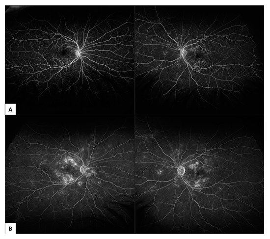
2.1. Fundus Fluorescein Angiography (FFA)

FFA is an invasive procedure whereby fluorescein dye is injected in a vein such as the antecubital fossa and then retinal perfusion and leakage associated with disease states affecting the microvasculature can be assessed with a series of fundus photographs [21]. FFA is the gold standard imaging technique for the assessment of PDR, characterised by fluorescein leakage from neovascular tissue, increasing in intensity and area over time. FFA can also identify areas of peripheral retinal and macular non-perfusion (Figure 1).
Figure 1. Ultrawide field (UWF) fluorescein fundus angiography (FFA) in non-proliferative diabetic retinopathy (NPDR) captured on 200° Optos (Optos California, Optos PLC, Dunfermline, United Kingdom): (A) showing early phase leakage at 1-minute following fluorescein dye injection; (B) showing late phase leakage at 6 minutes following fluorescein dye injection.
Limitations of FFA imaging are that patients require intravenous injection of an exogenous dye. This may be contraindicated in some diseases with end-stage microvascular disease including diabetic nephropathy. Some patients may develop life threatening severe allergy. The FFA procedure takes longer to perform and may be less well-tolerated by patients when compared to other non-invasive imaging procedures, limiting the number of times the test can be repeated.
Furthermore, FFA has a relatively low spatial resolution compared with newer OCT-A machines [22]. Importantly, FFA is unable to separately distinguish superficial from deep capillary networks due to its limits of two-dimensions, and retinal details become progressively obscured over the time-course of FFA imaging due to extravasation of the dye [23]. Whilst FFA may better identify microaneurysms, some of which may be missed by OCT-A in some studies, it is unable to identify the histological layer of positive findings, nor reliably follow-up identified areas of retina in serially aligned scans [22].

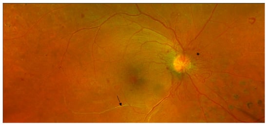
2.2. Indocyanine Green Angiography (ICG-A)

ICG-A is an invasive test similar to FFA with a different dye. Indocyanine green (ICG) dye has been histologically localised to both the choroidal intravascular space as well as extravasating into the choroidal stroma and accumulating within the RPE [24]. ICG therefore passes through local blood and pigmentary obscurations, adding to its utility in imaging the deep retinal and choroidal layers.
ICG-A can identify hypo-fluorescence in zones which represent a filling delay or defect at the level of the choriocapillaris which have been observed in DR [25]. Retinal avascular zones are more likely to be identified by FFA compared to ICG-A. Studies in DR have shown no leakage of ICG on angiographic studies in eyes with known diffuse diabetic macular oedema [26].
Limitations of ICG-A are similar to FFA; the principal of these being that repeated imaging is limited by the potential morbidity from an invasive dye test. Additionally, images are only able to be captured in two-dimensions. ICG-A is not routinely recommended for the diagnosis or management of PDR.

2.3. Ophthalmic B-Scan Ultrasonography

Ophthalmic B-scan ultrasonography has a role in imaging in the context of DR where VH obscures the direct view of the retina [27]. B-scan ultrasound can demonstrate whether a tractional retinal detachment accompanies other retinal pathology such as VH, a thickened posterior hyaloid, posterior vitreous detachment, or sub-retinal haemorrhage. Although examination with a B-scan may be considered less invasive by comparison to FFA and ICG angiography, it does require contact and manipulation of the ultrasound probe position with the patients closed eye making it more invasive than other non-touch, non-invasive imaging modalities.

3. Non-Invasive Fundus Imaging Modalities to Guide Diagnosis and Treatment of PDR

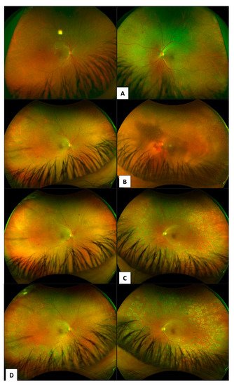
3.1. Colour Fundus Imaging

Seminal studies from the 1980s that established a gold-standard evidence base to inform management of DR include the Diabetic Retinopathy Study (DRS) [20], the Diabetic Retinopathy Vitrectomy Study (DRVS) [14][28], and the Early Treatment Diabetic Retinopathy Study (ETDRS) [29]. These early studies used 30-degree fundus cameras to image the posterior pole of the retina. The ETDRS defined seven-standard, central 30-degree photographic fields for viewing which was used for grading the severity of DR.
Pathologic features of DR which are captured by colour fundus imaging include haemorrhages, microaneurysms, IRMA, venous beading, cotton wool spots, hard exudates, drusen, retinal thickening, papillary swelling, new vessels on the disc (Figure 2) and NVE, pre-retinal and subretinal haemorrhage, vitreous haemorrhage, and tractional retinal detachment (Table 1). The extended modified Airlie House DR classification grading scale characterises the location and extent of these specific retinal lesions, which have been shown to be highly predictive of disease severity and the risk of DR progression over time. It is the accepted gold standard for grading which is used widely in research settings including large multi-centre clinical trials. Aspects of the classification system also form a basis for estimated risk of progression of DR to guide follow-up and treatment decisions in clinical practice.
Figure 2. Non-invasive Optos (Optos California, Optos PLC, Dunfermline, UK) colour fundus photograph of proliferative diabetic retinopathy. Sheathed retinal arteriole inferior to macula (arrow). Neovascularisation of the disc (*). Scattered intraretinal haemorrhages. Previous grid retinal laser nasal to disc (right eye).
The modified Airlie House classification uses non-simultaneous stereoscopic pairs of seven-standard photographic fields plus additional photographs to better define the grading scale for hard exudates, soft exudates, arteriovenous nicking, retinal elevation, and vitreous haemorrhage. The modified Airlie House classification also separates arterial abnormalities previously pooled as venous characteristics. The seven-standard ETDRS photographic fields are; field one centred on the optic disc, field two centred on the macula, field three temporal to the macula, fields four to seven are tangential to horizontal lines passing through the upper and lower poles of the disc and to a vertical line passing through the disc centre. Besides the seven-standard fields, an optional eighth field was added in the ETDRS for follow-up to document new vessels, fibrous proliferations, pre-retinal haemorrhage, or vitreous haemorrhage outside of the standard defined seven fields [13].
Since the advent of commercially available UWF fundus imaging systems, numerous studies have compared images captured from UWF to the stereoscopic seven-standard ETDRS fields. The level of agreeability between the imaging fields for grading DR severity has been demonstrated to be high, but UWF images have also been demonstrated to provide additional prognostic value with regard to the risks of DR onset, progression and outcomes compared to areas only visualised by seven-standard field ETDRS photography.
Newer imaging modalities, such as models of the Optos fundus camera (Optos plc, Dunfermline, Scotland, UK), enable mydriatic non-simultaneous stereoscopic 200-degree UWF images of the retina [21]. High-resolution UWF scanning laser ophthalmoscopes enable approximately 80–85% of the retinal surface to be captured in a single image. The benefits of this newer technology pertain to added efficiencies with reducing total imaging time, the number of images required, greater ease of image evaluation, reduced rates of poor-resolution images and a reduction in the areas of retina missed by imaging as result of operator error. Such benefits and commercial availability of these UWF imaging systems have meant that they are now widespread in clinical practice (Figure 3) and are used by newer clinical trials.
Figure 3. Case series of patient showing progression of proliferative diabetic retinopathy. Diagnosis (A) to 5 years follow-up patient re-presents with vitreous haemorrhage (B). (C) 1-month post vitrectomy and pan-retinal endolaser photocoagulation for management of VH due to PDR. (D) right eye showing grid laser 360 PRP and left eye showing PRP delivered with endolaser during vitrectomy.
Limitations of technology at the time when the original ETDRS criteria was developed meant that the far retinal periphery was not able to be systematically captured by imaging systems. Over the past thirty years since the ETDRS, numerous retinal imaging studies and clinical observations have demonstrated the significance of PPL as pathologic features of DR which are not evaluated by seven-standard field ETDRS photography. A lesion is considered to be predominantly peripheral if more than half of the area of the lesion being graded is in the peripheral retinal field compared to the modified seven-standard ETDRS photographic fields. A 4-year follow-up study by Silva et al. [21] found that eyes which had no PDR at baseline but did have PPLs on UWF imaging had a 3.2 increased risk of DR progression compared to eyes without PPLs (p = 0.005). Additionally, eyes with PPL were found to have a 4.7-fold increased risk for progression to PDR over 4 years even after correcting for diabetes duration and HbA1c levels [21]. This finding is supported by other studies similarly using UWF imaging [7][15][30].
Intraoperative visualization of areas of traction and source of PDR can assist vitreo-retinal surgery (Figure 4 and Figure 5).
Figure 4. Proliferative diabetic retinopathy presents in (A) with vitreous haemorrhage in the right eye. Post vitrectomy Optos (Optos California, Optos PLC, Dunfermline, UK) colour fundus photograph 200° (B) showing targeted endolaser to areas of neovascularisation during vitrectomy (C).
Figure 5. Traction retinal detachment and vitreous haemorrhage in proliferative diabetic retinopathy. Colour fundus photograph (A); Fluorescein angiography (B); Intraoperative video stills from recording during vitrectomy surgery showing pre-retinal, posterior hyaloid haemorrhage (C).

3.2. Optical Coherence Tomography (OCT)

OCT is a non-invasive imaging technique with three-dimensional volume information which provides micron level high-resolution cross-sectional retinal images. Segmentation of OCT allows for clearer differentiation of retinal layers, however earlier OCT used a shorter wavelength of 800 nm, which had limited penetration beyond the RPE to the choriocapillaris. Newer models have longer wavelength capabilities to 1050 nm which allow better visualization of the choroidal structures [3]. OCT is now the current clinical standard for observing the structural changes seen in diabetic macular oedema (DMO) [3][31].
Commercially available spectral-domain (SD-OCT) machines; Heidelberg Spectralis (Heidelberg Engineering, Heidelberg, Germany), Zeiss Cirrus (Carl Zeiss Meditec, Dublin, CA, USA), swept-source OCT (SS-OCT, Topcon, Tokyo, Japan), and Optovue RTVue (Optovue Inc., Fremont, CA, USA) machines, and one time-domain OCT Zeiss Stratus (Carl Zeiss Meditec) are used by clinical studies described in the body of this review [32].
With OCT technology it is possible to non-invasively evaluate cross-sectional images of the posterior vitreous and retinal layers to assess the ILM and posterior hyaloid for breaches in keeping with diagnostic criterion for NVE in the PDR stage of disease [23]. Assessment of the vitreoretinal interface plays an important role in management decisions for DR, both for prognostication and timeliness for interventional therapies. In assessment of eyes with contraction of fibrovascular tissue and tractional retinal detachment for consideration of vitrectomy, imaging of the extent of the traction in particular macular involvement is essential [33]. Macular threatening traction or direct involvement is an indication for vitrectomy and membrane segmentation and delamination to relieve the traction to restore the macular anatomy [34]. Inner retinal traction and disruption of neural layers is well imaged with OCT [35][36].
Techniques used for imaging an extended field with earlier OCT technology have been described by Mishra et al. [37] in 2017, where the authors used a wide-field 12 mm × 12 mm radial swept source OCT together with a convex +90 diopter double aspheric noncontact slit-lamp lens placed between the eye and the OCT machine to theoretically expand the imaging field by increasing the imaging light incidence angle. This technique however has been shown to have a higher incidence of rim artefacts and reflection on OCT images, and, as each pixel is magnified without the enhanced hardware capabilities of the OCT system, there is some loss of retinal architectural detail and resolution [37]. Whilst this technique provides a panoramic OCT view of a large segment of fundus to define the vitreoretinal relationship at one time, there are compromises in resolution and image quality which may be further degraded by poor media or lens clarity, poor patient fixation and smaller pupil size. OCT and OCTA technologies have been further enhanced since the years of recruitment in these studies (2016–2017) [8][37] with faster scan rates, wider fields of view, denser scan volumes, pseudocolour imaging acquisition modules and enhanced depth range of OCT imaging now available.

3.3. OCTA

The OCT-A uses multiple, sequential OCT B-scans in cross-section to produce reconstructed images of the network of blood vessels enabling three different vascular layers to be visualised: the superficial capillary plexus, the deep capillary plexus, and the choriocapillaris. OCT-A allows quantitative, serial assessments of areas of capillary non-perfusion, including high-resolution images which are aligned to allow ease of monitoring disease progression at specific identified retinal areas. This feature makes OCT-A better suited to follow-up for DR with less risk to the patient when compared to invasive imaging modalities [22]. There is currently a lack of clinical studies which examine the role of OCT-A in DR peripheral lesions. Most recent (2020) studies in PDR patients using OCT-A principally examine the posterior pole [12][38].
A classification system distinguishing NVE by topographic distribution, origin, and morphologic features has been described by Pan et al. [8][39]. This system relies on a combination of FA and OCTA to describe NVE as either originating from the venous side (Type 1) with tethering to the posterior hyaloid surface, from capillary networks (Type 2), or from intraretinal microvascular abnormalities (type 3) where the posterior hyaloid was still attached to the retina, as opposed to vitreoschisis or partial PVD in Type 1 NVE. The clinical implications of this classification system are that the three types of NVE may have varying topographical distribution features, and the authors have described distinguishable risk for complications including vitreous haemorrhage and traction retinal detachment, and differing responsiveness to treatment observed in a recently published study [39]. Limitations of the study [8] describing this classification of NVE according to OCTA features include the cross-sectional study design with a small cohort (35 eyes), and the reliance on only a small field of OCTA view (6 mm × 6 mm); therefore, NVE in the periphery were not included.
Recently, OCT-A has been suggested to more accurately measure areas of capillary dropout across the total, superficial, and deep capillary plexus in regions of ischaemia compared to images obtained by FFA [23].
Wide-field swept-source OCT-A has been used to provide information on microvascular perfusion in DR and may have a role in detecting peripheral capillary dropout, however it is not selective to excluding larger vessels so may not distinguish between different stages of DR progression [12].
OCT-A may have a role in better understanding the integrity of microvascular perfusion and how this relates to the maintenance of oxygen delivery to the retina, which has not yet been adequately explored in patients with PDR. Vitrectomy has been shown to improve rates of hypoxia and reduce VEGF production from ischaemic areas of retina [40]. Reduced VEGF levels reduces the stimulus for neovascularization processes as in diabetic neovascularization. Vitrectomy results in changes in oxygen flux across the retina and removes VEGF away from the retina at a faster rate than eyes without vitrectomy [40][41]. Therefore, vitrectomy could have additional benefits in diabetic eyes with retinal ischaemia [42].

References

  1. Keel, S.; Xie, J.; Foreman, J.; van Wijngaarden, P.; Taylor, H.R.; Dirani, M. The Prevalence of Diabetic Retinopathy in Australian Adults with Self-Reported Diabetes: The National Eye Health Survey. Ophthalmology 2017, 124, 977–984.
  2. Mathur, R.; Bhaskaran, K.; Edwards, E.; Lee, H.; Chaturvedi, N.; Smeeth, L.; Douglas, I. Population trends in the 10-year incidence and prevalence of diabetic retinopathy in the UK: A cohort study in the Clinical Practice Research Datalink 2004–2014. BMJ Open 2017, 7, e014444.
  3. Amoaku, W.M.; Ghanchi, F.; Bailey, C.; Banerjee, S.; Banerjee, S.; Downey, L.; Gale, R.; Hamilton, R.; Khunti, K.; Posner, E.; et al. Diabetic retinopathy and diabetic macular oedema pathways and management: UK Consensus Working Group. Eye 2020, 34, 1–51.
  4. Ramchandran, R.; Bawany, M.H.; Ding, L.; Sharma, G.; Wykoff, C.C.; Kuriyan, A.E. Automated vessel density detection in fluorescein angiography images correlates with vision in proliferative diabetic retinopathy. Investig. Ophthalmol. Vis. Sci. 2020, 61, 5312.
  5. Chhablani, J.; Sharma, A.; Goud, A.; Peguda, H.K.; Rao, H.L.; Begum, V.U.; Barteselli, G. Neurodegeneration in type 2 diabetes: Evidence from spectral-domain optical coherence tomography. Investig. Ophthalmol. Vis. Sci. 2015, 56, 6333–6338.
  6. Duh, E.J.; Sun, J.; Stitt, A.W. Diabetic retinopathy: Current understanding, mechanisms, and treatment strategies. JCI Insight 2017, 2, e93751.
  7. Fan, W.; Nittala, M.G.; Velaga, S.B.; Hirano, T.; Wykoff, C.C.; Ip, M.; Lampen, S.I.; van Hemert, J.; Fleming, A.; Verhoek, M.; et al. Distribution of Nonperfusion and Neovascularization on Ultrawide-Field Fluorescein Angiography in Proliferative Diabetic Retinopathy (RECOVERY Study): Report 1. Am. J. Ophthalmol. 2019, 206, 154–160.
  8. Pan, J.; Chen, D.; Yang, X.; Zou, R.; Zhao, K.; Cheng, D.; Huang, S.; Zhou, T.; Yang, Y.; Chen, F. Characteristics of Neovascularization in Early Stages of Proliferative Diabetic Retinopathy by Optical Coherence Tomography Angiography. Am. J. Ophthalmol. 2018, 192, 146–156.
  9. Rush, R.B.; Del Valle Penella, A.; Reinauer, R.M.; Rush, S.W.; Bastar, P.G. Internal Limiting Membrane Peeling during Vitrectomy for Diabetic Vitreous Hemorrhage: A Randomized Clinical Trial. RETINA 2020, 41, 1118–1126.
  10. Marcus, D.M.; Singh, H.; Farooq, A.; Starnes, D.; Walia, H. Endolaserless vitrectomy with intravitreal aflibercept injection (IAI) for proliferative diabetic retinopathy (PDR)-related vitreous hemorrhage (LASER LESS TRIAL). Investig. Ophthalmol. Vis. Sci. 2017, 58, 5036.
  11. Curtis, T.M.; Gardiner, T.A.; Stitt, A.W. Microvascular lesions of diabetic retinopathy: Clues towards understanding pathogenesis? Eye 2009, 23, 1496–1508.
  12. Tan, B.; Chua, J.; Lin, E.; Cheng, J.; Gan, A.; Yao, X.; Wong, D.W.; Sabanayagam, C.; Wong, D.; Chan, C.M.; et al. Quantitative Microvascular Analysis with Wide-Field Optical Coherence Tomography Angiography in Eyes with Diabetic Retinopathy. JAMA Netw. Open 2020, 3, e1919469.
  13. Early Treatment Diabetic Retinopathy Study Research Group. Grading diabetic retinopathy from stereoscopic color fundus photographs—An extension of the modified Airlie House classification: ETDRS report number 10. Ophthalmology 1991, 98, 786–806.
  14. The Diabetic Retinopathy Vitrectomy Study Research Group. Early vitrectomy for severe vitreous hemorrhage in diabetic retinopathy: Two-year results of a randomized trial—Diabetic Retinopathy Vitrectomy Study report 2. Arch. Ophthalmol. 1985, 103, 1644–1652.
  15. Silva, P.S.; Cruz, A.J.D.; Ledesma, M.G.; van Hemert, J.; Radwan, A.; Cavallerano, J.; Aiello, L.M.; Sun, J.K. Diabetic Retinopathy Severity and Peripheral Lesions Are Associated with Nonperfusion on Ultrawide Field Angiography. Ophthalmology 2015, 122, 2465–2472.
  16. Wilkinson, C.; Ferris, F.; Klein, R.; Lee, P.; Agardh, C.D.; Davis, M.; Dills, D.; Kampik, A.; Pararajasegaram, R.; Verdaguer, J.T. Proposed international clinical diabetic retinopathy and diabetic macular edema disease severity scales. Ophthalmology 2003, 110, 1677–1682.
  17. Gross, J.G.; Glassman, A.R.; Liu, D.; Sun, J.K.; Antoszyk, A.N.; Baker, C.W.; Bressler, N.M.; Elman, M.J.; Ferris, F.L.; Gardner, T.W.; et al. Five-Year Outcomes of Panretinal Photocoagulation vs Intravitreous Ranibizumab for Proliferative Diabetic Retinopathy. JAMA Ophthalmol. 2018, 136, 1138–1148.
  18. Preti, R.C.; Vasquez Ramirez, L.M.; Ribeiro Monteiro, M.L.; Pelayes, D.E.; Takahashi, W.Y. Structural and functional assessment of macula in patients with high-risk proliferative diabetic retinopathy submitted to panretinal photocoagulation and associated intravitreal bevacizumab injections: A comparative, randomised, controlled trial. Ophthalmologica 2013, 230, 1–8.
  19. Nikkhah, H.; Ghazi, H.; Razzaghi, M.R.; Karimi, S.; Ramezani, A.; Soheilian, M. Extended targeted retinal photocoagulation versus conventional pan-retinal photocoagulation for proliferative diabetic retinopathy in a randomized clinical trial. Int. Ophthalmol. 2017, 38, 313–321.
  20. The Diabetic Retinopathy Study Research Group. Photocoagulation Treatment of Proliferative Diabetic Retinopathy: Clinical application of Diabetic Retinopathy Study (DRS) findings, DRS Report Number 8. Ophthalmology 1981, 88, 583–600.
  21. Silva, P.S.; Cavallerano, J.; Haddad, N.M.N.; Kwak, H.; Dyer, K.H.; Omar, A.F.; Shikari, H.; Aiello, L.M.; Sun, J.K. Peripheral Lesions Identified on Ultrawide Field Imaging Predict Increased Risk of Diabetic Retinopathy Progression over 4 Years. Ophthalmology 2015, 122, 949–956.
  22. Elnahry, A.G.; Ramsey, D.J. Automated Image Alignment for Comparing Microvascular Changes Detected by Fluorescein Angiography and Optical Coherence Tomography Angiography in Diabetic Retinopathy. Semin. Ophthalmol. 2021, 36, 757–764.
  23. Ishibazawa, A.; Nagaoka, T.; Yokota, H.; Takahashi, A.; Omae, T.; Song, Y.S.; Takahashi, T.; Yoshida, A. Characteristics of retinal neovascularization in proliferative diabetic retinopathy imaged by optical coherence tomography angiography. Investig. Ophthalmol. Vis. Sci. 2016, 57, 6247–6255.
  24. Chang, A.A.; Morse, L.; Handa, J.T.; Morales, R.B.; Tucker, R.; Hjelmeland, L.; A Yannuzzi, L. Histologic localization of indocyanine green dye in aging primate and human ocular tissues with clinical angiographic correlation. Ophthalmology 1998, 105, 1060–1068.
  25. Shiragami, C.; Shiraga, F.; Matsuo, T.; Tsuchida, Y.; Ohtsuki, H. Risk factors for diabetic choroidopathy in patients with diabetic retinopathy. Graefe’s Arch. Clin. Exp. Ophthalmol. 2002, 240, 436–442.
  26. Costa, R.A.; Calucci, D.; Orefice, J.L. Indocyanine Green Angiography for The Detection Of Macular “Treatable Lesions” In Patients with Diabetic Retinopathy. Investig. Ophthalmol. Vis. Sci. 2011, 52, 583.
  27. Mohamed, I.E.; Mohamed, M.A.; Yousef, M.; Mahmoud, M.Z.; Alonazi, B. Use of ophthalmic B-scan ultrasonography in determining the causes of low vision in patients with diabetic retinopathy. Eur. J. Radiol. Open 2018, 5, 79–86.
  28. Diabetic Retinopathy Vitrectomy Study Research Group. Early Vitrectomy for Severe Vitreous Hemorrhage in Diabetic Retinopathy. Four-year results of a randomized trial. Diabetic Retinopathy Study report 5. Arch. Ophthalmol. 1990, 108, 958–964.
  29. Flynn, H.W.; Chew, E.Y.; Simons, B.D.; Barton, F.B.; Remaley, N.A.; Ferris, F.L. Pars Plana Vitrectomy in the Early Treatment Diabetic Retinopathy Study. Ophthalmology 1992, 99, 1351–1357.
  30. Babiuch, A.; Wykoff, C.C.; Hach, J.; Srivastava, S.; E Talcott, K.; Yu, H.J.; Nittala, M.; Sadda, S.; Ip, M.S.; Le, T.; et al. Longitudinal panretinal microaneurysm dynamics on ultra-widefield fluorescein angiography in eyes treated with intravitreal aflibercept for proliferative diabetic retinopathy in the recovery study. Br. J. Ophthalmol. 2020, 105, 1111–1115.
  31. Shimura, M.; Kitano, S.; Muramatsu, D.; Fukushima, H.; Takamura, Y.; Matsumoto, M.; Kokado, M.; Kogo, J.; Sasaki, M.; Morizane, Y.; et al. Real-world management of treatment-naïve diabetic macular oedema: 2-year visual outcome focusing on the starting year of intervention from STREAT-DMO study. Br. J. Ophthalmol. 2020, 104, 1755–1761.
  32. Gross, J.G.; Glassman, A.R.; Jampol, L.M.; Inusah, S.; Aiello, L.P.; Antoszyk, A.N.; Baker, C.W.; Berger, B.B.; Bressler, N.M.; Browning, D.; et al. Panretinal Photocoagulation vs Intravitreous Ranibizumab for Proliferative Diabetic Retinopathy: A randomized clinical trial. JAMA 2015, 314, 2137–2146.
  33. Su, C.C.; Yang, C.H.; Yeh, P.T.; Yang, C.M. Macular tractional retinoschisis in proliferative diabetic retinopathy: Clinical characteristics and surgical outcome. Ophthalmologica 2013, 231, 23–30.
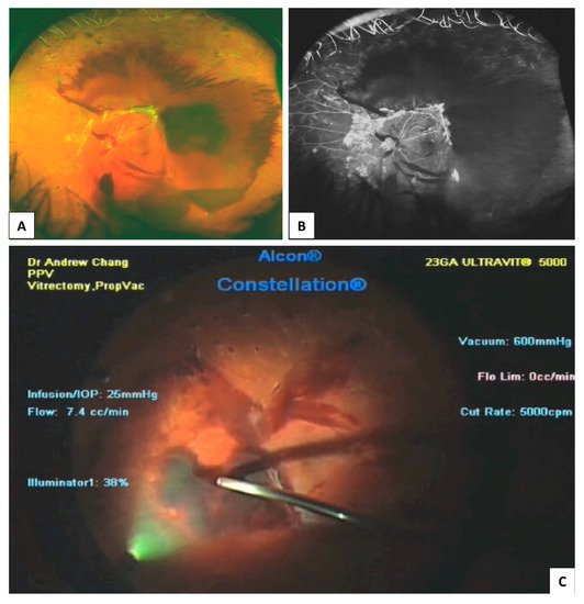
  34. Cruz-Inigo, Y.J.; Acaba, L.A.; Berrocal, M.H. Surgical management of retinal diseases: Proliferative diabetic retinopathy and traction retinal detachment. Dev. Ophthalmol. 2014, 54, 196–203.
  35. Ophir, A.; Martinez, M.R.; Mosqueda, P.; Trevino, A. Vitreous traction and epiretinal membranes in diabetic macular oedema using spectral-domain optical coherence tomography. Eye 2010, 24, 1545–1553.
  36. Chhablani, J.K.; Kim, J.S.; Cheng, L.; Kozak, I.; Freeman, W. External limiting membrane as a predictor of visual improvement in diabetic macular edema after pars plana vitrectomy. Graefe’s Arch. Clin. Exp. Ophthalmol. 2012, 250, 1415–1420.
  37. Mishra, D.K.; Shanmugam, M.P.; Ramanjulu, R.; Sagar, P. Comparison of standard and “innovative wide-field” optical coherence tomography images in assessment of vitreoretinal interface in proliferative diabetic retinopathy: A pilot study. Indian J. Ophthalmol. 2020, 69, 99–102.
  38. Um, T.; Seo, E.J.; Kim, Y.J.; Yoon, Y.H. Optical coherence tomography angiography findings of type 1 diabetic patients with diabetic retinopathy, in comparison with type 2 patients. Graefe’s Arch. Clin. Exp. Ophthalmol. 2020, 258, 281–288.
  39. Pan, J.; Chen, F.; Chen, D.; Yang, X.; Wang, J.; Chen, Z.; He, X.; Zhou, T.; Zheng, J.; Chen, H. Novel Three Types of Neovascularization Elsewhere Determine the Differential Clinical Features of Proliferative Diabetic Retinopathy. Retina 2020, 41, 1265–1274.
  40. Stefansson, E. Physiology of retinal oxygenation. Acta Ophthalmol. 2015, 93.
  41. Stefánsson, E. Ocular Oxygenation and the Treatment of Diabetic Retinopathy. Surv. Ophthalmol. 2006, 51, 364–380.
  42. Sharma, T.; Fong, A.; Lai, T.Y.; Lee, V.; Das, S.; Lam, D. Surgical treatment for diabetic vitreoretinal diseases: A review. Clin. Exp. Ophthalmol. 2016, 44, 340–354.
More
Information
Subjects: Ophthalmology
Contributor MDPI registered users' name will be linked to their SciProfiles pages. To register with us, please refer to https://encyclopedia.pub/register :
View Times: 542
Revisions: 2 times (View History)
Update Date: 02 Dec 2021
1000/1000