Submitted Successfully!
To reward your contribution, here is a gift for you: A free trial for our video production service.
Thank you for your contribution! You can also upload a video entry or images related to this topic.
Version Summary Created by Modification Content Size Created at Operation
1 + 2081 word(s) 2081 2021-11-23 04:30:35 |
2 The format is correct Meta information modification 2081 2021-11-24 02:49:46 |

Video Upload Options

Do you have a full video?

Confirm

Are you sure to Delete?
Cite
If you have any further questions, please contact Encyclopedia Editorial Office.
Teng, Y. Exosomes to Head and Neck Cancer. Encyclopedia. Available online: https://encyclopedia.pub/entry/16306 (accessed on 19 April 2024).
Teng Y. Exosomes to Head and Neck Cancer. Encyclopedia. Available at: https://encyclopedia.pub/entry/16306. Accessed April 19, 2024.
Teng, Yong. "Exosomes to Head and Neck Cancer" Encyclopedia, https://encyclopedia.pub/entry/16306 (accessed April 19, 2024).
Teng, Y. (2021, November 23). Exosomes to Head and Neck Cancer. In Encyclopedia. https://encyclopedia.pub/entry/16306
Teng, Yong. "Exosomes to Head and Neck Cancer." Encyclopedia. Web. 23 November, 2021.
Exosomes to Head and Neck Cancer
Edit

Head and neck squamous cell carcinoma (HNSCC) represents an aggressive and heterogenous group of cancers whose pathologies remain largely unresolved. Exosomes are a subtype of extracellular vesicles secreted by a variety of cells that have begun to spark significant interest in their roles in cancer. As membranous vesicles, spanning from 30–150 nm in diameter, exosomes mediate the transport of various molecules, such as proteins, nucleic acids, and lipids, intercellularly throughout the body. In doing so, exosomes not only act to deliver materials to cancer cells but also as signals that can confer their progression. Accumulating evidence shows the direct correlation between exosomes and the aggressiveness of HNSCC. 

exosomes head and neck squamous cell carcinoma

1. Introduction

Head and neck cancer is the sixth most common cancer worldwide [1][2][3]. More than 90% of all head and neck cancers are classified as head and neck squamous cell carcinoma (HNSCC), with a high frequency of tumor recurrence/metastasis and low patient survival. HNSCC comprises a heterogeneous group of cancers derived from the mucosal epithelium of the oral cavity, pharynx, and larynx and is typically linked to tobacco consumption, alcohol abuse, and human papillomavirus (HPV) infection. Despite advances in HNSCC surgical treatment, chemoradiotherapy, and immunotherapy, more than two-thirds of HNSCC patients still have no way to effectively control their clinical progression, facing a 5-year survival rate of about 60% [4]. Therefore, exploring new therapeutic targets for the treatment of HNSCC is of great significance.
Exosomes are small membranous vesicles that act as intercellular messengers through the cargo they carry. These discoid vesicles are 30–150 nm in diameter and secreted by living cells from early endosomes containing a typical lipid bilayer structure [5]. Their distribution is widespread, and they are found in various bodily fluids, such as serum, plasma, saliva, urine, and amniotic fluid [6]. Through their transportation of lipids, proteins, and nucleic acids, exosomes play an indispensable role in transferring material and information between cells and are consequently expected to serve as early diagnostic markers for various diseases [7][8].

2. Exosomes

2.1. Biogenesis of Exosomes

The biological origins of exosomes begin with early endosomes, which form through plasma membrane invagination. During this process, extracellular components and cell membrane proteins are encapsulated, and inward budding of the early endosome’s membrane leads to the formation of an exosomal vesicle. Early endosomes not only exchange materials with other organelles but fuse with different endosomes to form late endosomes. Late endosomes with intraluminal vesicles are known as multivesicular bodies (MVBs), and these are responsible for fusing with cell membranes to release the exosomes into the extracellular environment (Figure 1) [8][9].
Figure 1. Schematic presentation of the process by which exosomes are produced and released from the cell to the TME.

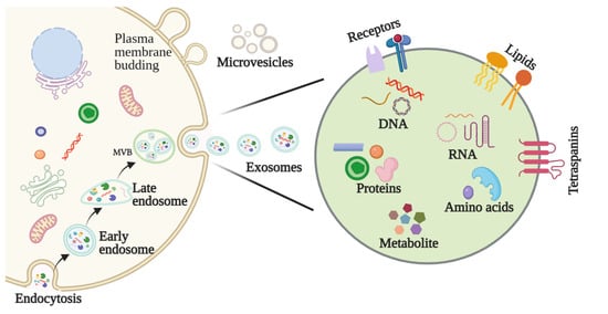
2.2. Features and Components of Exosomes

Exosomes, microvesicles, microparticles, and apoptotic bodies are subtypes of extracellular vesicles (EVs) [10]. While these vesicles share similar characteristics, they differ in their morphologies, biological properties, biogenesis, and functional roles. Exosomes are structurally similar to their cells of origin, possess a lipid bilayer structure, and typically consist of lipids, proteins, nucleic acids, and metabolites [11]. Exosomes also notably possess specific surface proteins that can be used to discern whether they are tumor derived (Figure 1) [12].

2.3. Methods for Exosome Isolation and Characterization

The isolation of pure exosomes is the first critical step in studying their mechanisms of action and their application in biomedical sciences. Various techniques have been adopted to facilitate the isolation of exosomes, including ultracentrifugation, size-based filtration, size-exclusion chromatography, polymer precipitation, and microfluidics-based isolation. Because exosome membranes are known to contain large quantities of proteins, immunoaffinity methods are also suitable for isolating exosomes [13].
Characterization of the physicochemical properties of exosomes, including their size, shape, surface charge, density, and porosity, is also important to their functional determinations. Many techniques have been routinely used to characterize exosomes, including dynamic light scattering (DLS), tunable resistive pulse sensing (TRPS), flow cytometry, electron microscopy, nanoparticle tracking analysis (NTA), and atomic force microscopy (AFM) [13]. Due to the inherent heterogeneity of exosomes and the advantages and limitations of each technique, more sophisticated techniques are necessary for the isolation and characterization of exosomes.

3. The Function of Exosomes in HNSCC

3.1. Exosomes Affect HNSCC Growth

Angiogenesis is critical to tumor cell proliferation, and, in one study, TGF-β-containing HNSCC-derived exosomes were found to promote angiogenesis in vitro and in vivo [14]. Additionally, Wang et al. reported that CAF-derived exosomes containing miR-3188 can influence the proliferation of HNSCC cells in vitro and in vivo, which suggests a potential therapeutic value for exosome-delivered miR-3188 in inhibiting HNSCC growth [15]. While investigating the diagnostic potential of salivary exosomal miRNAs as screening biomarkers for oral squamous cell carcinoma (OSCC), researchers also found that exogenous exosome miR-24-3p increases recipient malignant cell proliferation through targeting of PER1 protein [16].

3.2. Exosomes Are Involved in HNSCC Invasion and Metastasis

Epithelial to mesenchymal transformation (EMT) is an important feature of tumor progression and promotes the invasion and metastasis of tumor cells into the stroma. Exosome-mediated intercellular communication contributes significantly to this process and investigating it has led to breakthroughs in the field of cancer metastasis [17]. One study has shown that PO-243 exosomes from the plasma of HNSCC patients undergoing photodynamic therapy can alter the mesenchymal characteristics of tumor cells, directing them towards epithelial phenotypes [18]. Another study evaluated immunocaptured CD44v3+ tumor cell-derived exosomes from the plasma of 44 HNSCC patients and 7 healthy donors and found higher immunosuppressive protein levels in CD44v3+ exosomes compared with CD44v3(-) exosomes. The relative fluorescence intensity of these markers was associated with higher disease stages and lymph node metastasis [19].

3.3. Exosomes Regulate the HNSCC Microenvironment

3.3.1. Exosomal Modulation of the Pre-Metastatic Niche (PME)

How a tumor induces PMN formation in a specific organ remains to be determined. The suppressive nature of immune cells in the TME is critical to the regulation of anti-tumor immune responses. One of the possible mechanisms is that TDEs mediate tumor PMN remodeling to establish a supportive and receptive niche to promote tumor cell colonization and metastasis. Maybruck et al. have found, however, that head and neck cancer cells can induce a suppressive phenotype in human CD8+ T cells through the release of TDEs [20]. Specifically, the group revealed through mass spectrometry that the immunoregulatory protein galectin-1 was present in these exosomes and played a key role in inducing this suppressive phenotype. The purification of exosomal RNA and subsequent CD8+ T cell suppression analysis also implicated RNAs in T cell dysfunction. This study suggests that tumor immunosuppressive exosomes could be a potential therapeutic target to preserve T cell function in anti-tumor immune responses [20].

3.3.2. Exosomal Modulation of Tumor Hypoxia

Related studies have shown that the levels of PD-L1-containing exosomes in the plasma of HNSCC patients, though not serum, correlated with the patients’ disease activity, stage, and lymph node status [21]. Additionally, exosomes secreted by OSCC cells have been reported to activate macrophages through p38, Akt, and SAPK/JNK signaling shortly after their uptake [22]. Specifically, the exosomal-derived adhesive glycoprotein THBS1 was found to participate in the polarization of macrophages to an M1-like phenotype. Treating OSCC cells with culture media from exosome-activated macrophages also significantly promoted OSCC cell motility [22]. Still, further study is warranted to delineate the ways exosomes contribute to macrophages’ functional phenotypes and understand their associated microenvironmental consequences.

3.3.3. Exosomal Modulation of Immune Escape and Suppression

Paula Silva et al. evaluated the potential effects of EVs originating from HNSCC cells on immune system response [23]. In this study, monocyte-derived dendritic cells (mono-DCs) were treated with EVs derived from oropharyngeal squamous cell carcinoma (OPHSCC) and OSCC cell lines [23]. Impairment of mono-DC maturation and migration was observed following EV internalization. Gene expression profiling in mDCs treated with EVs further reveals disrupted immune responses possibly targeted by miRNAs (e.g., miR-17-5p and miR-21) within EVs [23].

3.4. Exosomes Promote Drug Resistance in HNSCC

One way by which exosomes contribute to tumor drug resistance is through their sequestering and efflux of cytotoxic drugs, effectively shielding cells from the drug’s effect and preventing its intracellular accumulation [24]. Exosomes can also contribute to external therapeutic resistance through their mediation of intercellular communication and delivery of mRNAs, miRNAs, DNAs, and/or proteins, for example [25]. Qin et al. have found that CAFs are inherently resistant to cisplatin and that exosomes act to transfer functional miR-196a from CAFs to HNSCC cells. In doing so, HNSCC cell proliferation and resistance to apoptosis are conferred through the targeting of CDKN1B and ING5, two suggested tumor suppressors [26]. Hence, the group put forward that miR-196a may represent a promising biomarker and/or potential therapeutic target for cisplatin-resistant cancers [26].

4. Role of Exosomes in the Diagnosis and Treatment of HNSCC

4.1. Exosomes as a Potential Biomarker in HNSCC

Early diagnosis and treatment are critical determinants in a cancer patient’s prognosis. The application of biomarkers in HNSCC detection, as well as factors such as staging, treatment efficacy, and prognosis, have consequently garnered attention in recent years [27]. Biomarkers represent a diverse range of molecules, and abnormalities in their levels or makeup can be detected in bodily fluids, like urine, saliva, and blood, as well as tumors themselves. In particular, exosomal biomarkers may be roughly divided into nucleic acids, proteins, lipids, and metabolites (Figure 2).

4.2. Exosomes as Therapeutic Targets in HNSCC

Despite recent improvements in HNSCC treatment, the long-term survival rate of advanced HNSCC patients remains less than 50% [28]. Given their diverse roles in cancer support, from proliferation to metastasis, exosomes and the cargo they carry constitute an innumerable set of potential targetable points for influencing HNSCC progression [29][30]. Many studies have indicated that exosomes are involved in anticancer therapy resistance due to their critical role in cellular communication and the TME.
Many pharmacological agents are being explored to investigate how best to block exosome excretion and trafficking. For example, GW4869 is a cell-permeable, symmetrical dihydroimidazolo-amide compound that acts as a non-competitive inhibitor of membrane neutral sphingomyelinase (nSMase). GW4869 has been relatively extensively studied and reported to inhibit exosomes, as it can interfere with lipid composition that is necessary for exosome shedding [31]. Indomethacin, a non-steroidal anti-inflammatory drug, has been shown to downregulate transcription of the ABCA3 transporter [32]. ABCA3 is an intracellular protein involved in lipid transport, and its inhibition impairs exosomes release. Thus, indomethacin may also be considered a potential agent for exosome inhibition. Due to the involvement of Ras during exosome release, Ras inhibitors are particularly associated with exosome trafficking. Among them, manumycin A, a farnesyltransferase inhibitor blocking the Ras pathway, has been studied as an inhibitor of exosome secretion [33]. However, secretion control, in vivo biological distribution, targeting specificity, and cell penetration in exosome-targeting cancer therapies remain challenging to HNSCC and other solid tumors. Interestingly, engineered exosomes have been applied to develop therapeutic anticancer vaccines for HPV-associated tumors [34]. Because a mutant of a human immunodeficiency virus 1 (HIV-1) Nef protein (Nefmut) has the potential to act as an exosome-anchoring element upon fusion with heterologous proteins, exosomes engineered in vitro were used to upload high amounts of HPV-E7 fused to Nefmut.

4.3. Exosomes as Drug Carriers for HNSCC Treatment

Exosomes are recognized as one of the most promising anti-tumor therapeutic vectors due to their bilayer structure, biocompatibility, low immunogenicity, and ability to interact with target cells [35]. Given their limited bioavailability, however, synthetic exosome-mimics have been used in their place to mediate endogenous and exogenous delivery for cancer treatment [12]. In particular, Cohen et al. evaluated the parameters of exosomes derived from mesenchymal stem cells (MSC-exo) and A431 squamous cell carcinoma cells (A431-exo) as drug vectors and found MSC-exo to exhibit significantly greater tumor penetration and distribution in A431 tumor-bearing mice [36].

5. Prospects for Exosomes in Anticancer Therapy

Even with recent advances, HNSCC remains a major clinical challenge. Through their intercellular transmission of materials and signals in the local and distant TME, exosomes play a crucial role in promoting HNSCC proliferation, invasion, metastasis, drug resistance, and survival. Many studies have already pointed to exosomes as promising tumor biomarkers, and, through their employment in patients, improvements to HNSCC detection and treatment regimens may soon be a reality. Plant- and human tissue-derived exosomes have undergone clinical trials in lung cancer, melanoma, and colorectal [37][38][39][40]. Most recently, MSC exosomes containing siRNA targeting oncogenic KrasG12D mutations are being employed against pancreatic cancer in a clinical trial (NCT03608631). Still, exosomes have yet to be clinically applied in HNSCC therapies. This may be due in part to the high cost of purification and isolation of exosomes and a lack of understanding of the complex effects resulting from HNSCC-derived exosomes [41]. Therefore, it is imperative that scientists continue to improve purification and isolation technologies and delineate exosome’s specific mechanisms in HNSCC progression in order to harness them as tools to improve patient outcomes.

References

  1. Siegel, R.L.; Miller, K.D.; Jemal, A. Cancer Statistics, 2017. CA Cancer J. Clin. 2017, 67, 7–30.
  2. Ferlay, J.; Colombet, M.; Soerjomataram, I.; Mathers, C.; Parkin, D.M.; Pineros, M.; Znaor, A.; Bray, F. Estimating the global cancer incidence and mortality in 2018: GLOBOCAN sources and methods. Int. J. Cancer 2019, 144, 1941–1953.
  3. Bray, F.; Ferlay, J.; Soerjomataram, I.; Siegel, R.L.; Torre, L.A.; Jemal, A. Global cancer statistics 2018: GLOBOCAN estimates of incidence and mortality worldwide for 36 cancers in 185 countries. CA Cancer J. Clin. 2018, 68, 394–424.
  4. Johnson, D.E.; Burtness, B.; Leemans, C.R.; Lui, V.W.Y.; Bauman, J.E.; Grandis, J.R. Head and neck squamous cell carcinoma. Nat. Rev. Dis. Primers 2020, 6, 92.
  5. Steinbichler, T.B.; Dudas, J.; Riechelmann, H.; Skvortsova, I.I. The role of exosomes in cancer metastasis. Semin. Cancer Biol. 2017, 44, 170–181.
  6. Pegtel, D.M.; Gould, S.J. Exosomes. Annu. Rev. Biochem. 2019, 88, 487–514.
  7. Wang, X.; Guo, J.; Yu, P.; Guo, L.; Mao, X.; Wang, J.; Miao, S.; Sun, J. The roles of extracellular vesicles in the development, microenvironment, anticancer drug resistance, and therapy of head and neck squamous cell carcinoma. J. Exp. Clin. Cancer Res. 2021, 40, 35.
  8. McAndrews, K.M.; Kalluri, R. Mechanisms associated with biogenesis of exosomes in cancer. Mol. Cancer. 2019, 18, 52.
  9. Kalluri, R.; Lebleu, V. The biology, function, and biomedical applications of exosomes. Science 2020, 367, eaau6977.
  10. McBride, J.D.; Rodriguez-Menocal, L.; Badiavas, E.V. Extracellular Vesicles as Biomarkers and Therapeutics in Dermatology: A Focus on Exosomes. J. Investig. Dermatol. 2017, 137, 1622–1629.
  11. Abels, E.R.; Breakefield, X.O. Introduction to Extracellular Vesicles: Biogenesis, RNA Cargo Selection, Content, Release, and Uptake. Cell. Mol. Neurobiol. 2016, 36, 301–312.
  12. Li, S.P.; Lin, Z.X.; Jiang, X.Y.; Yu, X.Y. Exosomal cargo-loading and synthetic exosome-mimics as potential therapeutic tools. Acta Pharmacol. Sin. 2018, 39, 542–551.
  13. Gurunathan, S.; Kang, M.H.; Jeyaraj, M.; Qasim, M.; Kim, J.H. Review of the Isolation, Characterization, Biological Function, and Multifarious Therapeutic Approaches of Exosomes. Cells 2019, 8, 307.
  14. Ludwig, N.; Yerneni, S.S.; Azambuja, J.H.; Razzo, B.M.; Hinck, C.S.; Pietrowska, M.; Hinck, A.; Whiteside, T.L. Abstract B34: TGF-β-rich tumor-derived exosomes promote a proangiogenic phenotype in HNSCC. Clin. Cancer Res. 2020, 26, B34.
  15. Wang, X.; Qin, X.; Yan, M.; Shi, J.; Xu, Q.; Li, Z.; Yang, W.; Zhang, J.; Chen, W. Loss of exosomal miR-3188 in cancer-associated fibroblasts contributes to HNC progression. J. Exp. Clin. Cancer Res. 2019, 38, 151.
  16. He, L.; Ping, F.; Fan, Z.; Zhang, C.; Deng, M.; Cheng, B.; Xia, J. Salivary exosomal miR-24-3p serves as a potential detective biomarker for oral squamous cell carcinoma screening. Biomed. Pharmacother. 2020, 121, 109553.
  17. Wee, I.; Syn, N.; Sethi, G.; Goh, B.C.; Wang, L. Role of tumor-derived exosomes in cancer metastasis. Biochim. Biophys. Acta Rev. Cancer 2019, 1871, 12–19.
  18. Theodoraki, M.N.; Yerneni, S.S.; Brunner, C.; Theodorakis, J.; Hoffmann, T.K.; Whiteside, T.L. Plasma-derived Exosomes Reverse Epithelial-to-Mesenchymal Transition after Photodynamic Therapy of Patients with Head and Neck Cancer. Oncoscience 2018, 5, 75–87.
  19. Theodoraki, M.N.; Matsumoto, A.; Beccard, I.; Hoffmann, T.K.; Whiteside, T.L. CD44v3 protein-carrying tumor-derived exosomes in HNSCC patients’ plasma as potential noninvasive biomarkers of disease activity. Oncoimmunology 2020, 9, 1747732.
  20. Maybruck, B.T.; Pfannenstiel, L.W.; Diaz-Montero, M.; Gastman, B.R. Tumor-derived exosomes induce CD8(+) T cell suppressors. J. Immunother. Cancer 2017, 5, 65.
  21. Theodoraki, M.N.; Yerneni, S.S.; Hoffmann, T.K.; Gooding, W.E.; Whiteside, T.L. Clinical Significance of PD-L1(+) Exosomes in Plasma of Head and Neck Cancer Patients. Clin. Cancer Res. 2018, 24, 896–905.
  22. Xiao, M.; Zhang, J.; Chen, W.; Chen, W. M1-like tumor-associated macrophages activated by exosome-transferred THBS1 promote malignant migration in oral squamous cell carcinoma. J. Exp. Clin. Cancer Res. 2018, 37, 143.
  23. Silva, E.D.P.; Marti, L.C.; Andreghetto, F.M.; de Sales, R.O.; Hoberman, M.; Dias, B.D.S.; Diniz, L.F.A.; dos Santos, A.M.; Moyses, R.A.; Curioni, O.A.; et al. Extracellular vesicles cargo from head and neck cancer cell lines disrupt dendritic cells function and match plasma microRNAs. Sci. Rep. 2021, 11, 18534.
  24. Dong, X.; Bai, X.; Ni, J.; Zhang, H.; Duan, W.; Graham, P.; Li, Y. Exosomes and breast cancer drug resistance. Cell Death Dis. 2020, 11, 987.
  25. Steinbichler, T.B.; Dudas, J.; Skvortsov, S.; Ganswindt, U.; Riechelmann, H.; Skvortsova, I.I. Therapy resistance mediated by exosomes. Mol. Cancer 2019, 18, 58.
  26. Qin, X.; Guo, H.Y.; Wang, X.N.; Zhu, X.Q.; Yan, M.; Wang, X.; Xu, Q.; Shi, J.B.; Lu, E.Y.; Chen, W.T.; et al. Exosomal miR-196a derived from cancer-associated fibroblasts confers cisplatin resistance in head and neck cancer through targeting CDKN1B and ING5. Genome. Biol. 2019, 20, 12.
  27. Qu, X.Y.; Li, J.W.; Chan, J.; Meehan, K. Extracellular Vesicles in Head and Neck Cancer: A Potential New Trend in Diagnosis, Prognosis, and Treatment. Int. J. Mol. Sci. 2020, 21, 8260.
  28. Gollin, S.M. Cytogenetic alterations and their molecular genetic correlates in head and neck squamous cell carcinoma: A next generation window to the biology of disease. Genes Chromosomes Cancer 2014, 53, 972–990.
  29. Ebnoether, E.; Muller, L. Diagnostic and Therapeutic Applications of Exosomes in Cancer with a Special Focus on Head and Neck Squamous Cell Carcinoma (HNSCC). Int. J. Mol. Sci. 2020, 21, 4344.
  30. Brand, M.; Laban, S.; Theodoraki, M.N.; Doescher, J.; Hoffmann, T.K.; Schuler, P.J.; Brunner, C. Characterization and Differentiation of the Tumor Microenvironment (TME) of Orthotopic and Subcutaneously Grown Head and Neck Squamous Cell Carcinoma (HNSCC) in Immunocompetent Mice. Int. J. Mol. Sci. 2020, 22, 247.
  31. Catalano, M.; O’Driscoll, L. Inhibiting extracellular vesicles formation and release: A review of EV inhibitors. J. Extracell. Vesicles 2019, 9, 1703244.
  32. Aung, T.; Chapuy, B.; Vogel, D.; Wenzel, D.; Oppermann, M.; Lahmann, M.; Weinhage, T.; Menck, K.; Hupfeld, T.; Koch, R.; et al. Exosomal evasion of humoral immunotherapy in aggressive B-cell lymphoma modulated by ATP-binding cassette transporter A3. Proc. Natl. Acad. Sci. USA 2011, 108, 15336–15341.
  33. Datta, A.; Kim, H.; Lal, M.; McGee, L.; Johnson, A.; Moustafa, A.A.; Jones, J.C.; Mondal, D.; Ferrer, M.; Abdel-Mageed, A.B. Manumycin A suppresses exosome biogenesis and secretion via targeted inhibition of Ras/Raf/ERK1/2 signaling and hnRNP H1 in castra-tion-resistant prostate cancer cells. Cancer Lett. 2017, 408, 73–81.
  34. Di Bonito, P.; Accardi, L.; Galati, L.; Ferrantelli, F.; Federico, M. Anti-Cancer Vaccine for HPV-Associated Neoplasms: Focus on a Therapeutic HPV Vaccine Based on a Novel Tumor Antigen Delivery Method Using Endogenously Engineered Exosomes. Cancers 2019, 11, 138.
  35. Li, Y.; Zhang, Y.; Li, Z.; Zhou, K.; Feng, N. Exosomes as Carriers for Antitumor Therapy. ACS Biomater. Sci. Eng. 2019, 5, 4870–4881.
  36. Cohen, O.; Betzer, O.; Elmaliach-Pnini, N.; Motiei, M.; Sadan, T.; Cohen-Berkman, M.; Dagan, O.; Popovtzer, A.; Yosepovich, A.; Barhom, H.; et al. ‘Golden’ exosomes as delivery vehicles to target tumors and overcome intratumoral barriers: In vivo tracking in a model for head and neck cancer. Biomater. Sci. 2021, 9, 2103–2114.
  37. Dai, S.; Wei, D.; Wu, Z.; Zhou, X.; Wei, X.; Huang, H.; Li, G. Phase I clinical trial of autologous ascites-derived exosomes combined with GM-CSF for colorectal cancer. Mol. Ther. 2008, 16, 782–790.
  38. Besse, B.; Charrier, M.; Lapierre, V.; Dansin, E.; Lantz, O.; Planchard, D.; Le Chevalier, T.; Livartoski, A.; Barlesi, F.; Laplanche, A.; et al. Dendritic cell-derived exosomes as maintenance immunotherapy after first line chemotherapy in NSCLC. Oncoimmunology 2015, 5, e1071008.
  39. Cully, M. Exosome-based candidates move into the clinic. Nat. Rev. Drug Discov. 2021, 20, 6–7.
  40. Santos, P.; Almeida, F. Exosome-Based Vaccines: History, Current State, and Clinical Trials. Front. Immunol. 2021, 12, 711565.
  41. Cao, J.; Zhang, M.; Xie, F.; Lou, J.; Zhou, X.; Zhang, L.; Fang, M.; Zhou, F. Exosomes in head and neck cancer: Roles, mechanisms and applications. Cancer Lett. 2020, 494, 7–16.
More
Information
Subjects: Cell Biology
Contributor MDPI registered users' name will be linked to their SciProfiles pages. To register with us, please refer to https://encyclopedia.pub/register :
View Times: 371
Revisions: 2 times (View History)
Update Date: 24 Nov 2021
1000/1000