Submitted Successfully!
To reward your contribution, here is a gift for you: A free trial for our video production service.
Thank you for your contribution! You can also upload a video entry or images related to this topic.
Version Summary Created by Modification Content Size Created at Operation
1 + 3466 word(s) 3466 2021-11-02 06:51:28 |
2 h -74 word(s) 3392 2021-11-11 07:26:44 | |
3 h Meta information modification 3392 2021-11-12 07:18:25 |

Video Upload Options

Do you have a full video?

Confirm

Are you sure to Delete?
Cite
If you have any further questions, please contact Encyclopedia Editorial Office.
Pan, M. Detection of Food Allergens. Encyclopedia. Available online: https://encyclopedia.pub/entry/15896 (accessed on 27 April 2024).
Pan M. Detection of Food Allergens. Encyclopedia. Available at: https://encyclopedia.pub/entry/15896. Accessed April 27, 2024.
Pan, Mingfei. "Detection of Food Allergens" Encyclopedia, https://encyclopedia.pub/entry/15896 (accessed April 27, 2024).
Pan, M. (2021, November 11). Detection of Food Allergens. In Encyclopedia. https://encyclopedia.pub/entry/15896
Pan, Mingfei. "Detection of Food Allergens." Encyclopedia. Web. 11 November, 2021.
Detection of Food Allergens
Edit

Food allergies have seriously affected the life quality of some people and even endangered their lives. At present, there is still no effective cure for food allergies. Avoiding the intake of allergenic food is still the most effective way to prevent allergic diseases. Therefore, it is necessary to develop rapid, accurate, sensitive, and reliable analysis methods to detect food allergens from different sources. Aptamers are oligonucleotide sequences that can bind to a variety of targets with high specifificity and selectivity, and they are often combined with different transduction technologies, thereby constructing various types of aptamer sensors. In recent years, with the development of technology and the application of new materials, the sensitivity, portability, and cost of flfluorescence sensing technology have been greatly improved. Therefore, aptamer-based flfluorescence sensing technology has been widely developed and applied in the specifific recognition of food allergens.

allergen detection aptamer

1. Introduction

Food allergies, an adverse reaction to antigenic substances in food mediated by the immune system, have been recognized as a global health issue with increasing prevalence in the field of food safety [1][2]. Most food allergies are immunoglobulin (Ig) E-mediated type I (immediate type) hypersensitivity reactions [3]. An epidemiological survey by the institute of infectious diseases shows that about 6–9.3% of children and 3.4–5.0% of adults have food allergies, which means the incidence of food allergies in infants and children is generally higher than that of adults [4][5][6]. However, there is still no standard cure for food allergies except avoiding eating foods that contain allergens. Therefore, the development of rapid and effective detection methods for allergens in food matrices is a topic of concern in the whole society.
In the past few decades, many mature techniques have been widely used in the detection of food allergens, such as the enzyme-linked immunosorbent assay (ELISA), liquid chromatography-mass spectrometry (LC-MS), and polymerase chain reaction (PCR) [7][8][9]. The ELISA method has been widely used in the detection of food allergens due to its high specificity and sensitivity. Nevertheless, due to the influence of various external conditions such as food processing methods, there would be false positive and false negative results [10][11]. Moreover, PCR method is usually used for monitoring allergic components in food processing due to its high specificity and high automation. However, PCR technology is not suitable for identifying allergen proteins with unascertained genes, which limits its scope of application [12][13]. Furthermore, HPLC and LC-MS are standard strategies for the quantitative analysis of allergens in various food matrices. Because of the precision requirements of the instruments, these methods usually require strict sample pre-treatment processes, a larger sample volume, and a longer analysis time, resulting in a higher detection cost [14]. Currently, biosensors with high sensitivity and specificity, such as surface-enhanced Raman spectroscopy (SERS), electrochemical biosensors, and quartz crystal microbalance (QCM) biosensors, can rapidly analyze and screen food allergens and allow on-site analysis, which are considered effective detection technology [15][16][17]. However, these biosensors usually require expensive instruments, proficient operators, and higher requirements for the surrounding environment. Therefore, there is an urgent need to develop rapid, accurate, sensitive, and easy-to-operate detection methods to quantify allergens in food matrices.
Nucleic acid aptamer is a nucleic acid sequence that can specifically recognize the target, screened by systematic evolution of ligands by exponential enrichment (SELEX) in vitro [18]. The combination of aptamer and target is achieved through single-stranded oligonucleotide deoxyribonucleic acid (DNA) or ribonucleic acid (RNA) folded into a specific three-dimensional structure (stem-loop, hairpin and G-quadruplex and other spatial conformations) [19][20][21]. Regardless of the technical requirements for the preparation of aptamers, the convenience and timeliness far exceed those of antibodies. Moreover, the screened aptamers can be artificially synthesized, which is easy to achieve standardization. In recent years, aptamers have received extensive attention due to their veracity, high specificity, and affinity, and they have been used in disease diagnosis and treatment, drug delivery, food safety testing, and environmental monitoring [22][23][24]. In terms of food safety, the application of aptamers to the detection of allergens in food matrices is expected to achieve the goal of accurate, rapid, and low-cost detection of allergens.
Fluorescence detection technology, due to its low cost, high sensitivity, simple performance, has attracted wide attention [25][26][27]. Combining fluorescence detection technology with aptamers, the development of biosensors with high sensitivity and simple detection procedures provides a feasible strategy for the detection of food allergens [28]. Aptamer-based fluorescence sensing detection is a relatively common analysis method. The fluorophore is combined with the aptamer in a labeled or non-labeled manner, and the analyte concentration and other information are reflected by the interaction of the excitation light and the identification element [29][30]. Furthermore, fluorescence intensity, decay rate, spectral properties, and fluorescence anisotropy can be used alone or in combination as signal detection means.

2. Classification of Food Allergens

A great variety of food allergens exist widely in nature. According to the source of food allergens, they can be classified into animal allergens, plant allergens, and fungal allergens. Table 1 lists the classification of major food allergens, allergy symptoms, and other information. Specific information about food allergies is also discussed in the following sections.
Table 1. The major allergens in food matrices and their allergenic properties.
Food Major Allergens Molecular Mass (kDa) Types of Proteins The Structure of Proteins Allergy Symptoms Reference
Fish Pan h 1 10–13 Calbindin Contains 3 EF-hand regions (a motif composed of a 12-residue loop with a 12-residue-α-helix domain on each side), 2 of which can bind calcium. Blushing, hives, nausea, stomach pain, and intestinal bleeding. [31]
Shellfish Cra c 1 33–39 Protein bound to actin Adopting an α-helix structure, two molecules are entangled with each other to form a parallel dimeric α-helix structure. Nausea, diarrhea, abdominal pain, and muscle paralysis. [32]
Cra c 2 38–45 Phosphoglycoprotein Arginine kinase consists of an N-terminal domain (1–111) and a C-terminal domain (112–357). The N-terminal domain is all α-helices, and the C-terminal domain is an 8-strand anti-parallel β-sheet structure surrounded by 7 α-helices. [33]
Milk Bos d 8 57–37.5 Phosphate calcium binding protein Consists of 4 independent proteins: αs1-casein, αs2-casein, β-casein, and κ-casein. Skin rash, urticaria, eczema, vomiting, diarrhea, abdominal cramps, etc. [34]
Bos d 4 14.4 Combine with metal ions and participate in lactose synthesis With a two-piece structure containing α-single loop and 310 helix larger subdomain. [35]
Bos d 5 18 Lipid transporter Consists of two subunits connected by non-covalent bonds, mainly in the form of dimers. [36]
Egg Gal d1 28 Phosphoglycoprotein Contains 3 independent homologous structural energy domains, and 3 functional domains are arranged consecutively in space. Eczema, dermatitis, urticaria, vomiting, diarrhea, gastroesophageal reflux, etc. [37]
Gal d2 45 Phosphoglycoprotein Containing 4 free sulfhydryl groups, composed of 385 amino acid residues, these amino acid residues are twisted and folded to form a spherical structure with high secondary structure, most of which are α-helix and β-sheet. [38]
Gal d3 77 Iron-binding glycoprotein Consisting of 686 amino acids, including 12 disulfide bonds, the N-terminal and C-terminal 2 domains each contain a binding site for Fe3+. [39]
Gal d4 14.3 Basic globulin A single peptide chain composed of 18 kinds of 129 amino acid residues, with 4 pairs of disulfide bonds to maintain the enzyme configuration, with lysine at the N-terminus and leucine at the C-terminus. [40]
Peanut Ara h 1 63.5 7S Globulin The secondary structure contains β-turns, and the quaternary structure is a trimeric complex formed by 3 monomers. Angioedema, hypotension, asthma, anaphylactic shock, etc. [41]
Ara h 2 17–20 2S Albumin A monomeric protein. [42]
Ara h 3 57 11S Globulin The N-terminal and C-terminal domains of the monomer form contain 2 ciupin folds (composed of two sets of parallel β-turns, random coils and 3 α-helices). [43]
Wheat Tri a 36 40 Gluten - Wheat exercise stimulates allergies, urticaria, dermatitis, bread asthma, nausea, and diarrhea. [44]
Soybean Gly m 5 150–200 7S Globulin Trimer composed of α’-subunit, α-subunit and β subunit. Red and itchy skin, asthma and allergic rhinitis, abdominal pain, diarrhea, etc. [45]
Gly m 6 320–360 11S Globulin A hexamer composed of the interaction of G1, G2, G3, G4, and G5 subunits. [46]
Nuts Ana o 1 50 7S legumin Exist as a trimer in natural state. Metallic taste in the mouth, edema of the tongue or throat, difficulty breathing and swallowing, urticaria all over the body, flushing of the skin, cramping abdominal pain, nausea. [47]
Jug r 2 44 Consists of 593 amino acid residues. [48]
Cor a 11 48 Consists of 401 amino acid residues, with two potential N-glycosylation sites (Asn38 and Asn254) and a leader peptide of 46 amino acids. [49]
Ana o 3 14 2S albumin Composed of 5 helical structures, containing 2 subunits, connected by cysteine disulfide bonds. [50]
Jug r 1 15–16 Consists of 142 amino acid residues. [51]
Jug r 4 58.1 11S globulin Except for the first 23 amino acid residues which are predicted as signal peptides, the remaining part has a total of 507 amino acid residues. [52]
Cor a 9 40 Composed of 515 amino acid residues, the sequence homology with Ara h 3 is about 45%. [53]
Pru du 6 350 Exist in the form of hexamers, each monomer subunit is composed of one acid chain of 40 to 42 kDa and one alkaline chain of 20 kDa. [54]

3. Detection of Animal Food Allergens

Seafood allergy is not only an important public health issue, but a serious food safety issue that affects the quality of life and may even be life threatening [55]. For people with seafood allergies, avoiding foods containing seafood allergens is still the best option. Therefore, the monitoring of allergens is a process that requires strict supervision [56]. In order to evaluate seafood allergens, new detection methods with high sensitivity and high efficiency are required.
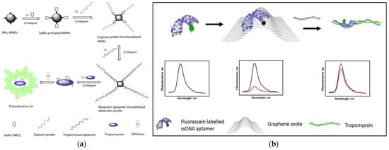
As we all know, magnetic separation is easy to operate and can effectively reduce or eliminate the interference from complex matrices in food. Therefore, based on functionalized magnetic nanoparticles (MNPs) as a separation carrier, Zhang et al. developed a simple and versatile label-free aptamer-based fluorescent sensor for the sensitive detection of TM (Figure 1a) [57]. In the study, OliGreen dye was selected as a fluorescent signal probe. The aptamer hybridizes with the capture probe bound to the surface of the MNPs to form an aptamer-MNPs complex as detection probe. When interacting with the target, the conformation of the complex changes, resulting in the release of the aptamer from the surface of the MNPs. So, the released aptamer in the supernatant produced a significant fluorescence enhancement signal, which is because the combination of OliGreen dye and ssDNA will produce ultrasensitive and specific fluorescence enhancement phenomenon. It is worth noting that when the commercially available OliGreen dye is in the free state, the fluorescence is weak or no fluorescence, but the fluorescence will increase by more than 1000 times once combined with the aptamer ssDNA. Under the optimal conditions, the linear range was 0.4–5 μg mL−1 (R2 = 0.996), with a limit of detection LOD of 77 ng mL−1. In addition, the highly selective aptamer-based fluorescent sensor was successfully applied to the detection of TM in food matrix. Wu et al. also developed a similar sensor with a LOD of 4.2 nM and the concentration linear from 0.5–50 μg mL−1 [58]. Recently, Chinappan et al. developed an aptamer-based fluorescent-labeled sensor for the detection of TM. (Figure 1b) [59]. Graphene oxide (GO) is used as a platform for screening the minimum length of aptamer sequences that can bind to the target with high affinity. A fluorescein dye labeled GO quenches the truncated aptamer by π-stacking and hydrophobic interactions. After the addition of TM, the fluorescence was restored due to the competitive binding of the aptamer to GO. More importantly, the aptamer selected in this study is a truncated ligand fragment, which has four times higher affinity than the full-sequence aptamer, with a LOD of 2.5 nM. The developed aptamer-based fluorescence sensor can complete the detection within 30 min. The performance of the sensor was confirmed in the addition experiment of chicken broth, and a high percentage recovery rate (~97 ± 10%) was achieved. Compared with the above studies, the sensitivity and specificity of this work have been greatly improved.
Figure 1. (a) Schematic of preparation of magnetic-assisted fluorescent aptamer for tropomyosin detection. Reproduced with permission from [57]. Copyright Sensors and Actuators B-Chemical, 2018. (b) Schematic of graphene oxide-based fluorescent aptamer biosensor for TM detection A: Changes in the fluorescence intensity of the aptamer released from the GO surface; B: The linear correlation of the fluorescence intensity of TMT2 (at 515 nm) with the concentration of TM. Reproduced with permission from [59]. Copyright Food Chemistry, 2020.
Fluorescence resonance energy transfer (FRET) is a mechanism widely used in the preparation of biosensors, which is an energy transfer phenomenon between two fluorescent molecules that are very close 
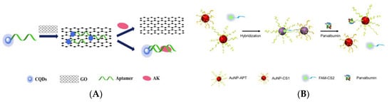
[60]. Zhou et al. designed an aptamer-based “on-off-on” fluorescent biosensor based on FRET and used developed carboxyl functionalized carbon quantum dots (cCQDs) and GO for the detection of shellfish allergen arginine kinase (AK) (Figure 2A) [61]. The cCQDs-aptamer probe and GO self-assemble for the first time through a specific π-π interaction, so that the fluorescence of cCQDs is effectively quenched. After the addition of AK, cCQDs-aptamer is released from the GO surface and then forms the cCQDs-aptamers-AK complex, which restores the fluorescence of cCQDs. The aptamer-based FRET sensor can perform sensitive detection in the AK concentration range of 0.001–10 μg mL−1, with a LOD of 0.14 ng mL−1 (S/N = 3) and a limit of quantification (LOQ) of 0.27 ng mL−1 (S/N = 10). Furthermore, in a control experiment with a blank sample, it was found that the sensor has high specificity. This reliable, precise, highly specific, and easy-to-operate aptamer sensor may provide a new perspective for the application of fluorescence sensing technology in the field of food safety.
Figure 2. (A) Schematic of a “on-off-on” fluorescence aptasensor for AK detection. Reproduced with permission from [61]. Copyright Microchemical Journal, 2020. (B) Schematic of a dual-mode fluorescence sensor for PV detection based on AuNP color changes and FAM-CS2 fluorescence changes. (C) a: Schematic of the aptamer selection procedure by capturing GO-SELEX; b: Affinity of Apt5 towards PV; c: Specificity of Apt5 towards PV. Reproduced with permission from [62]. Copyright Microchemical Journal, 2020.
In recent years, biosensors based on dual signals or functions have received widespread attention due to the diversity of detection. Dual-mode nanosensors usually use colorimetric and fluorescent reporters to achieve convenient visual inspection and highly sensitive fluorescent detection [63][64]. Wang et al. developed a dual-mode aptamer-based fluorescent sensor for the detection of PV, the major allergen of fish (Figure 2B) [62]. In Figure 2C(a), aptamer towards PV was obtained by in vitro screening of random ssDNA library containing a 40-mer randomized region using the triple-mode GO-SELEX. The aptamer-modified gold nanoparticle (AuNP-APT), complementary short-strand modified gold nanoparticles (AuNP-CS1), and fluorescent dye-labeled complementary short-strands (FAM-CS2) were assembled by DNA hybridization. After the addition of PV, the competitive interaction with aptamer leads to the decomposition of the aptamer sensor, resulting in the color shift of the AuNPs solution and the recovery of the FAM-CS2 fluorescence signal. The results showed that the aptamer sensor showed a good colorimetric response (2.5–20 μg mL−1) and linear fluorescence correlation (2.38–40 μg mL−1) in the PV concentration range. In addition, the affinity and specificity of the aptamer sensor were also investigated, as shown in Figure 2C(b,c). Therefore, aptamer 5 with good affinity (KD = 7.66 × 10⁻⁷ M) and specificity is the best aptamer for aptasensor construction. They also studied the feasibility of aptamer sensor in real fish samples, revealing the potential in field of monitoring and quantitative detection of food allergens.
Recently, as an alternative to antibodies, the use of peptide aptamers as biosensors has attracted more attention. Peptide aptamers usually contain 10–20 amino acids, which the high selective recognition ability is equivalent to that of antibodies. Phadke et al. used ribosome display technology to select two fluorescent peptide aptamers Cas1 and Cas2. for the detection of α-casein [65]. Among them, 7-nitrobenzofurazan (NBD)-modified aminophenylalanine is coupled to the translated peptides to prepare fluorescent peptide aptamers. This is because the peptide can quench the fluorescence of NBD. Once the peptide recognizes the target α-casein, the NBD-modified phenylalanine is released, and its fluorescence will instantly increase. It is worth noting that although the fluorescence of the two aptamers increased slightly in the presence of the control protein β-lactoglobulin, the modification of Cas1 with polyethylene glycol (PEG-Cas1) inhibited this phenomenon, which is because PEG-Cas1 may inhibit the interaction between aptamer and β-lactoglobulin. The aptamer sensor with a LOD of 0.04 mM, is equivalent to that of the kit. Moreover, the system can detect α-casein in short time (20–25 s) when compared with the 15 min required by immunochromatography kits. In addition, it is found that when using PEG-Cas1 to detect casein, the instant increase in fluorescence can be observed even with the naked eye. This study has contributed to improving the specificity of aptamers.
In order to reduce the incidence of milk allergy, hypoallergenic formula (HF) has been commercialized as a substitute for milk [66]. Nevertheless, in some cases, infants who consume these formula milk powder still have allergic reactions because of residual β-lactoglobulin in HF [67]. Therefore, it is necessary to establish a method that can detect the lower concentration of β-lactoglobulin. Shi et al. used carbon dots (CDs) as a fluorescent signal and Fe3O4 NPs as a magnetic separator to establish a fluorescent-labeled assay for the detection of β-lactoglobulin [68]. The assay is based on the hybridization between aptamers immobilized on Fe3O4 NPs and CDs-labeled complementary oligonucleotides (cDNA). In the presence of β-lactoglobulin, the aptamer preferentially binds to β-lactoglobulin, and part of the CDs-cDNA is released into the solution. After magnetic separation, the fluorescence signal of the supernatant increased with the increase of β-lactoglobulin concentration. Based on this, the aptamer assay with the range of 0.25–50 ng mL−1 and a LOD of 37 pg mL
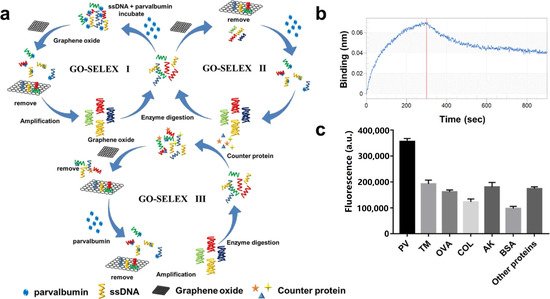
−1 has been successfully applied to the detection of trace β-lactoglobulin in HF. In the study of Qi et al., the binding mechanism of aptamer and β-lactoglobulin and the detection principle of fluorescent surface-enhanced Raman scattering (fluorescent-SERS) dual-mode aptamer sensor were thoroughly studied, which provides a theory basis and application potential for the development of aptamer sensors [69]. In Figure 3a, the circular dichroism of Lg-18, thermodynamic parameters analysis, secondary structure of Lg-18, and the result of molecular docking between aptamer Lg-18 and β-lactoglobulin were performed to illustrate the successful selection of aptamer. The specific response principle of the dual-mode aptamer sensor is shown in Figure 3b. The fluorescent-SERS aptamer sensor shows a wider linear range (10–5000 ng mL−1), and the LOD is 0.05 ng mL−1. Furthermore, under the interference of other proteins, the aptamer sensor showed excellent specificity.
Figure 3. (a) Schematic of Lg-18 and β-lactoglobulin binding. A: Circular dichroism analysis of Lg-18 before and after binding. B: Analysis of thermodynamic parameters in the combined process. C: Secondary structure of Lg-18 predicted by Mfold online software. D: Analysis of molecular docking results of aptamer Lg-18 and β-lactoglobulin (b) Schematic of aptamer-based fluorescent Raman dual-mode biosensor for detection of β-lactoglobulin. Reproduced with permission from [69]. Copyright Sensors and Actuators B-Chemical, 2021.
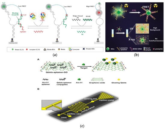
Lys, as an allergen in egg and a biomarker of many diseases, its detection and quantification are of great significance in clinical diagnosis 
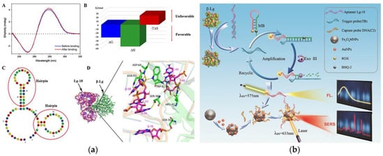
[70]. Sapkota et al. developed an aptamer sensor based on single-molecule FRET (smFRET) for the detection of Lys (Figure 4a) [71]. One of the arms has a blocking chain (B1), which is extended by 15 nucleotides to partially hybridize to the aptamer. The aptamer sensor remains open and almost no FRET efficiency occurs when Lys is not detected. After the addition of Lys, the aptamer binds to Lys and is displaced from the sensor, resulting in a foothold-mediated replacement of B1 by another chain, H1. At this time, the binding of Lys triggers the conformational transition state from low FRET to high FRET. Using this strategy, they demonstrated that the aptamer sensor can detect Lys at concentration as low as 30 nM, with a dynamic range extends to ~2 μM, and is almost free of interference from similar biomolecules. In addition, the smFRET method requires only a small number of aptamers, which offers the advantage of cost-effectiveness. In fluorescence detection, the presence of background fluorescence induced by ultraviolet-visible light in biological samples can lead to inaccurate detection results [72]. However, Ou et al. developed an X-ray nanocrystal scintillator aptamer sensor for sensitive detection of Lys based on the characteristics of weak scattering and almost no absorption of biological chromophores under X-ray irradiation (Figure 4b) [73]. In this study, aptamer-labeled lanthanide-doped nanocrystalline scintillators are designed to detect Lys quickly and sensitively through FRET. The use of low-dose X-rays as the excitation source and nanocrystals containing heavy atoms can achieve efficient luminescence, which endows the aptamer fluorescence sensor with high sensitivity (LOD: 0.94 nM), specificity, and sample recovery. In addition, this technology can provide a new generation of high-efficiency strategy without autofluorescence interference for the sensing and detection of biomarkers in biomedical applications.
Figure 4. (a) Schematic of aptamer-based fluorescent biosensor for Lys detection. Reproduced with permission from [71]. Copyright sensors, 2020. (b) Schematic of aptamer sensor based on nanocrystal scintillator for detecting Lys without autofluorescence. Reproduced with permission from [73]. Copyright Analytical Chemistry, 2019. (c) A: Schematic of the Qdots-aptamer-GO quenching sensing principle; B: Schematic of the designed microfluidic chip. Reproduced with permission from [74]. Copyright Biosensors & Bioelectronics, 2016.

References

  1. Sathe, S.K.; Liu, C.Q.; Zaffran, V.D. Food allergy. Annu. Rev. Food Sci. Technol. 2016, 7, 191–220.
  2. Carter, C.A.; Frischmeyer-Guerrerio, P.A. The genetics of food allergy. Curr. Allergy Asthm. Rep. 2018, 18, 2.
  3. Anvari, S.; Miller, J.; Yeh, C.Y.; Davis, C.M. IgE-Mediated food allergy. Clin. Rev. Allergy Immunol. 2019, 57, 244–260.
  4. Zhou, J.R.; Qi, Q.Q.; Wang, C.; Qian, Y.F.; Liu, G.M.; Wang, Y.B.; Fu, L.L. Surface plasmon resonance (SPR) biosensors for food allergen detection in food matrices. Biosens. Bioelectron. 2019, 142, 111449.
  5. Gupta, M.; Cox, A.; Nowak-Wegrzyn, A.; Wang, J.L. Diagnosis of food allergy. Immunol. Allergy Clin. 2018, 38, 39–52.
  6. Quake, C.; Nadeau, K.C. The role of epigenetic mediation and the future of food allergy research. Semin. Cell. Dev. Biol. 2015, 43, 125–130.
  7. He, S.F.; Li, X.; Wu, Y.; Wu, S.D.; Wu, Z.H.; Yang, A.S.; Tong, P.; Yuan, J.L.; Gao, J.Y.; Chen, H.B. A novel sandwich enzyme-linked immunosorbent assay with covalently bound monoclonal antibody and gold probe for sensitive and rapid detection of bovine beta-lactoglobulin. Anal. Bionanl. Chem. 2018, 410, 3693–3703.
  8. Kim, M.J.; Park, S.B.; Kang, H.B.; Lee, K.M.; Kim, H.Y. Development of ultrafast PCR for rapid detection of buckwheat allergen DNA (fag e 1) in processed foods. Food Control 2021, 130, 108334.
  9. Xiong, W.L.; Parker, C.H.; Boo, C.C.; Fiedler, K.L. Comparison of allergen quantification strategies for egg, milk, and peanut in food using targeted LC-MS/MS. Anal. Bioanal. Chem. 2021, 413, 5755–5766.
  10. Yu, Z.W.; Wang, Y.Q.; Li, Z.X.; Pramod, S.N.; Zhang, L.J.; Lin, H. Development of ELISA method for detecting crustacean major allergen tropomyosin in processed food samples. Food Anal. Method. 2019, 12, 2719–2729.
  11. Liu, C.Q.; Zaffran, V.D.; Gupta, S.; Roux, K.H.; Sathe, S.K. Pecan (Carya illinoinensis) detection using a monoclonal antibody-based direct sandwich enzyme-linked immunosorbent assay. LWT-Food Sci. Technol. 2019, 116, 108516.
  12. Kim, M.J.; Kim, H.I.; Kim, J.H.; Suh, S.M.; Kim, H.Y. Rapid on-site detection of shrimp allergen tropomyosin using a novel ultrafast PCR system. Food Sci. Biotechnol. 2019, 28, 591–597.
  13. Linacero, R.; Sanchiz, A.; Ballesteros, I.; Cuadrado, C. Application of real-time PCR for tree nut allergen detection in processed foods. Crit. Rev. Food Sci. 2020, 60, 1077–1093.
  14. De Angelis, E.; Pilolli, R.; Monaci, L. Coupling SPE on-line pre-enrichment with HPLC and MS/MS for the sensitive detection of multiple allergens in wine. Food Control 2017, 73, 814–820.
  15. Gezer, R.G.; Liu, G.L.; Kokini, J.L. Development of a biodegradable. sensor platform from gold coated zein nanophotonic films to detect peanut allergen, Ara h1, using surface enhanced raman spectroscopy. Talanta 2016, 150, 224–232.
  16. Mishra, G.K.; Barfidokht, A.; Tehrani, F.; Mishra, R.K. Food safety analysis using electrochemical biosensors. Foods 2018, 7, 141.
  17. Bragazzi, N.L.; Amicizia, D.; Panatto, D.; Tramalloni, D.; Valle, I.; Gasparini, R. Quartz-crystal microbalance (QCM) for public health: An overview of its applications. Adv. Protein Chem. Str. 2015, 101, 149–211.
  18. Xia, X.H.; He, Q.; Dong, Y.; Deng, R.J.; Li, J.H. Aptamer-based homogeneous analysis for food control. Curr. Anal. Chem. 2020, 16, 4–13.
  19. Abbaspour, A.; Norouz-Sarvestani, F.; Noon, A.; Soltani, N. Aptamer-conjugated silver nanoparticles for electrochemical dual-aptamer-based sandwich detection of staphylococcus aureus. Biosens. Bioelectron. 2015, 68, 149–155.
  20. Yan, C.; Zhang, J.; Yao, L.; Xue, F.; Lu, J.F.; Li, B.G.; Chen, W. Aptamer-mediated colorimetric method for rapid and sensitive detection of chloramphenicol in food. Food Chem. 2018, 260, 208–212.
  21. Song, S.H.; Gao, Z.F.; Guo, X.; Chen, G.H. Aptamer-based detection methodology studies in food safety. Food Anal. Methods 2019, 12, 966–990.
  22. Liu, L.S.; Lu, X.L.; Zhao, Y.X. Aptamer-based strategies for cancer diagnosis and therapy. J. Nanosci. Nanotechnol. 2016, 16, 6611–6621.
  23. Gray, B.P.; Kelly, L.; Ahrens, D.P.; Barry, A.P.; Kratschmer, C.; Levy, M.; Sullenger, B.A. Tunable cytotoxic aptamer-drug conjugates for the treatment of prostate cancer. Proc. Natl. Acad. Sci. USA 2018, 115, 4761–4766.
  24. Cheng, L.; Zhang, J.; Lin, Y.; Wang, Q.; Zhang, X.X.; Ding, Y.H.; Cui, H.F.; Fan, H. An electrochemical molecular recognition-based aptasensor for multiple protein detection. Anal. Biochem. 2015, 491, 31–36.
  25. Markova, L.V.; Myshkin, N.K.; Makarenko, V.M. Fluorescence method for quick transformer oil monitoring. Chem. Technol. Fuels Oils 2016, 52, 194–202.
  26. Zienkiewicz, K.; Alche, J.D.; Zienkiewicz, A.; Tormo, A.; Castro, A.J. Identification of olive pollen allergens using a fluorescence-based 2D multiplex method. Electrophoresis 2015, 36, 1043–1050.
  27. Wang, T.; Zeng, L.H.; Li, D.L. A review on the methods for correcting the fluorescence inner-filter effect of fluorescence spectrum. Appl. Spectrosc. Rev. 2017, 52, 883–908.
  28. Fu, L.L.; Qian, Y.F.; Zhou, J.R.; Zheng, L.; Wang, Y.B. Fluorescence-based quantitative platform for ultrasensitive food allergen detection: From immunoassays to DNA sensors. Compr. Rev. Food Sci. Saf. 2020, 19, 3343–3364.
  29. Zhang, G.X.; Liu, Y.L.; Yang, M.; Huang, W.S.; Xu, J.H. An aptamer-based, fluorescent and radionuclide dual-modality probe. Biochimie 2020, 171, 55–62.
  30. Ma, P.F.; Guo, H.L.; Duan, N.; Ma, X.Y.; Yue, L.; Gu, Q.H.; Wang, Z.P. Label free structure-switching fluorescence polarization detection of chloramphenicol with truncated aptamer. Talanta 2021, 230, 122349.
  31. Dijkema, D.; Emons, J.A.M.; Van de Ven, A.A.J.M.; Elberink, J.O. Fish allergy: Fishing for novel diagnostic and therapeutic options. Clin. Rev. Allergy Immunol. 2020.
  32. Wai, C.Y.Y.; Leung, N.Y.H.; Leung, P.S.C.; Chu, K.H. Modulating shrimp tropomyosin-mediated allergy: Hypoallergen DNA vaccines induce regulatory T cells to reduce hypersensitivity in mouse model. Int. J. Mol. Sci. 2019, 20, 4656.
  33. Xing, P.; Yu, H.Q.; Li, M.; Xiao, X.J.; Jiang, C.L.; Mo, L.H.; Zhang, M.; Yang, P.C.; Liu, Z.G. Characterization of arginine kinase, anovel allergen of dermatophagoides farinae (Der f 20). Am. J. Transl. Res. 2015, 7, 2815–2823.
  34. Wasik, M.; Nazimek, K.; Nowak, B.; Askenase, P.W.; Bryniarski, K. Delayed-type hypersensitivity underlying casein allergy is suppressed by extracellular vesicles carrying miRNA-150. Nutrients 2019, 11, 907.
  35. Cong, Y.J.; Zhou, S.Y.; Li, L.F. Identification of the critical amino acid residues of immunoglobulin E and immunoglobulin G epitopes in alpha-lactalbumin by alanine scanning analysis. J. Food Sci. 2016, 81, T2597–T2603.
  36. Surucu, O.; Abaci, S. Electrochemical determination of beta-lactoglobulin in whey proteins. J. Food Meas. Charact. 2020, 14, 11–19.
  37. Suprun, M.; Getts, R.; Grishina, G.; Suarez-Farinas, M.; Sampson, H.A. Epitope-specific antibody binding on major hen’s egg-white allergen, ovomucoid, using novel multiplex immunoassay. Allergy 2019, 74, 256–257.
  38. Bermingham, M.; Filep, S.; Reid-Black, K.; Thorpe, C.; Wuenschmann, S.; Chapman, M.; Hindley, J. Two-site immunoassays for the quantification of major egg allergens ovomucoid (Gal D 1) and ovalbumin (Gal D 2). Allergy 2019, 74, 438.
  39. Hwang, H.S.; Kim, B.S.; Park, H.; Park, H.Y.; Choi, H.D.; Kim, H.H. Type and branched pattern of N-glycans and their structural effect on the chicken egg allergen ovotransferrin: A comparison with ovomucoid. Glycoconj. J. 2014, 31, 41–50.
  40. Tan, L.; Chong, K.W.; Goh, S.H. Lysozyme and mucolytics—The hidden allergen. Singap. Med. J. 2020, 61, 497.
  41. Ramesh, M.; Yuenyongviwat, A.; Konstantinou, G.N.; Lieberman, J.; Pascal, M.; Masilamani, M.; Sampson, H.A. Peanut T-cell epitope discovery: Ara h 1. J. Allergy Clin. Immunol. 2016, 137, 1764–1771.
  42. Shroba, J.; Barnes, C.; Nanda, M.; Dinakar, C.; Ciaccio, C. Ara h2 levels in dust from homes of individuals with peanut allergy and individuals with peanut tolerance. Allergy Asthma Proc. 2017, 38, 192–196.
  43. Pandey, A.K.; Varshney, R.K.; Sudini, H.K.; Pandey, M.K. An improved enzyme-linked immunosorbent assay (ELISA) based protocol using seeds for detection of five major peanut allergens Ara h 1, Ara h 2, Ara h 3, Ara h 6, and Ara h 8. Front. Nutr. 2019, 6, 68.
  44. Gomes, R.; Loureiro, C.; Pita, C.; Todo-Bom, A. Tri a 19 and wheat exercised-dependent urticaria. Allergy 2015, 70, 605–606.
  45. Schmidt, M.A.; Hymowitz, T.; Herman, E.M. Breeding and characterization of soybean Triple Null; a stack of recessive alleles of Kunitz Trypsin Inhibitor, Soybean Agglutinin, and P34 allergen nulls. Plant Breed. 2015, 134, 310–315.
  46. Geng, T.; Stojsin, D.; Liu, K.; Schaalje, B.; Postin, C.; Ward, J.; Wang, Y.C.; Liu, Z.L.; Li, B.; Glenn, K. Natural variability of allergen levels in conventional soybeans: Assessing variation across north and south america from five production years. J. Agric. Food Chem. 2017, 65, 463–472.
  47. Archila, L.D.; Chow, I.T.; McGinty, J.W.; Renand, A.; Jeong, D.; Robinson, D.; Farrington, M.L.; Kwok, W.W. Ana o 1 and Ana o 2 cashew allergens share cross-reactive CD4(+) T cell epitopes with other tree nuts. Clin. Exp. Allergy 2016, 46, 871–883.
  48. Schein, C.; Teuber, S.S.; Cheng, H.; Grimm, C.C.; Maleki, S.J. Antibodies to the physiochemical-consensus sequence of Jug r 2 containing glutamine-rich repeats bind allergens in peanuts and other tree nuts. J. Allergy Clin. Immunol. 2013, 131, AB21.
  49. Iwan, M.; Vissers, Y.M.; Fiedorowicz, E.; Kostyra, H.; Kostyra, E.; Savelkoul, H.F.J.; Wichers, H.J. Impact of maillard reaction on immunoreactivity and allergenicity of the hazelnut allergen Cor a 11. J. Agric. Food Chem. 2011, 59, 7163–7171.
  50. Mattison, C.P.; Grimm, C.C.; Li, Y.C.; Chial, H.J.; McCaslin, D.R.; Chung, S.Y.; Bren-Mattison, Y.; Wasserman, R.L. Identification and characterization of Ana o 3 modifications on arginine-111 residue in heated cashew nuts. J. Agric. Food Chem. 2017, 65, 411–420.
  51. Sato, S.; Yamamoto, M.; Yanagida, N.; Ito, K.; Ohya, Y.; Imai, T.; Nagao, M.; Borres, M.P.; Moverare, R.; Ebisawa, M. Jug r 1 sensitization is important in walnut-allergic children and youth. J. Aller. Clin. Immunol. Pract. 2017, 5, 1784–1786.
  52. Blankestijn, M.A.; Jager, C.F.D.; Blom, W.M.; Otten, H.G.; de Jong, G.A.H.; Gaspari, M.; Houben, G.F.; Knulst, A.C.; Verhoeckx, K.C.M. A subset of walnut allergic adults is sensitized to walnut 11S globulin Jug r 4. Clin. Exp. Allergy 2018, 48, 1206–1213.
  53. Masthoff, L.J.N.; Mattsson, L.; Zuidmeer-Jongejan, L.; Lidholm, J.; Andersson, K.; Akkerdaas, J.H.; Versteeg, S.A.; Garino, C.; Meijer, Y.; Kentie, P.; et al. Sensitization to Cor a 9 and Cor a 14 is highly specific for a hazelnut allergy with objective symptoms in Dutch children and adults. J. Allergy Clin. Immunol. 2013, 132, 393–399.
  54. Kabasser, S.; Hafner, C.; Chinthrajah, S.; Sindher, S.B.; Kumar, D.; Kost, L.E.; Long, A.J.; Nadeau, K.C.; Breiteneder, H.; Bublin, M. Identification of Pru du 6 as a potential marker allergen for almond allergy. Allergy 2021, 76, 1463–1472.
  55. Davis, C.M.; Gupta, R.S.; Aktas, O.N.; Diaz, V.; Kamath, S.D.; Lopata, A.L. Clinical management of seafood allergy. J. Aller. Clin. Immunol. Pract. 2020, 8, 37–44.
  56. Tong, W.S.; Yuen, A.W.T.; Wai, C.Y.Y.; Leung, N.Y.H.; Chu, K.H.; Leung, P.S.C. Diagnosis of fish and shellfish allergies. J. Asthma Allergy. 2018, 11, 247–260.
  57. Zhang, Y.X.; Wu, Q.P.; Sun, M.; Zhang, J.M.; Mo, S.P.; Wang, J.; Wei, X.H.; Bai, J.L. Magnetic-assisted aptamer-based fluorescent assay for allergen detection in food matrix. Sensor. Actuators B-Chem. 2018, 263, 43–49.
  58. Zhang, Y.X.; Wu, Q.P.; Wei, X.H.; Zhang, J.M.; Mo, S.P. DNA aptamer for use in a fluorescent assay for the shrimp allergen tropomyosin. Microchim. Acta 2017, 184, 633–639.
  59. Chinnappan, R.; Rahamn, A.A.; AlZabn, R.; Kamath, S.; Lopata, A.L.; Abu-Salah, K.M.; Khalid, M.; Zourob, M. Aptameric biosensor for the sensitive detection of major shrimp allergen, Tropomyosin. Food Chem. 2020, 314, 126133.
  60. Shokoufi, N.; Heshi, S.V. Enhancmentation of photo-thermal lens of fluorescence molecules by fluorescence resonance energy transfer mechanism. J. Fluoresc. 2021, 31, 587–593.
  61. Zhou, J.R.; Ai, R.; Weng, J.J.; Li, L.F.; Zhou, C.S.; Ma, A.J.; Fu, L.L.; Wang, Y.B. A “on-off-on” fluorescence aptasensor using carbon quantum dots and graphene oxide for ultrasensitive detection of the major shellfish allergen arginine kinase. Microchem. J. 2020, 158, 105171.
  62. Wang, Y.B.; Li, H.; Zhou, J.R.; Qi, Q.Q.; Fu, L.L. A colorimetric and fluorescent gold nanoparticle-based dual-mode aptasensor for parvalbumin detection. Microchem. J. 2020, 159, 105413.
  63. Shi, Y.P.; Pan, Y.; Zhang, H.; Zhang, Z.M.; Li, M.J.; Yi, C.Q.; Yang, M.S. A dual-mode nanosensor based on carbon quantum dots and gold nanoparticles for discriminative detection of glutathione in human plasma. Biosens. Bioelectron. 2014, 56, 39–45.
  64. Liu, T.; Li, N.; Dong, J.X.; Zhang, Y.; Fan, Y.Z.; Lin, S.M.; Luo, H.Q.; Li, N.B. A colorimetric and fluorometric dual-signal sensor for arginine detection by inhibiting the growth of gold nanoparticles/carbon quantum dots composite. Biosens. Bioelectron. 2017, 87, 772–778.
  65. Phadke, C.; Tada, S.; Kono, I.; Hiyama, A.; Takase, Y.; Gayama, S.; Aigaki, T.; Ito, Y.; Uzawa, T. Instantaneous detection of alpha(s)-casein in cow’s milk using fluorogenic peptide aptamers. Anal. Methods-UK 2020, 12, 1368–1373.
  66. Lozano-Ojalvo, D.; Perez-Rodriguez, L.; Pablos-Tanarro, A.; Lopez-Fandino, R.; Molina, E. Pepsin treatment of whey proteins under high pressure produces hypoallergenic hydrolysates. Innov. Food Sci. Emerg. 2017, 43, 154–162.
  67. Shi, M.L.; Cen, Y.; Sohail, M.; Xu, G.H.; Wei, F.D.; Ma, Y.S.; Xu, X.M.; Ma, Y.J.; Song, Y.Y.; Hu, Q. Aptamer based fluorometric beta-lactoglobulin assay based on the use of magnetic nanoparticles and carbon dots. Microchim. Acta 2018, 185, 40.
  68. Qi, S.; Duan, N.; Sun, Y.H.; Zhou, Y.; Ma, P.F.; Wu, S.J.; Wang, Z.P. High-affinity aptamer of allergen beta-lactoglobulin: Selection, recognition mechanism and application. Sensor Actuators B-Chem. 2021, 340, 129956.
  69. Panahipour, L.; Tabatabaei, A.A.; Gruber, R. Hypoallergenic infant formula lacks transforming growth factor beta activity and has a lower anti-inflammatory activity than regular infant formula. J. Dairy Sci. 2020, 103, 6771–6781.
  70. Wu, J.; Hou, Y.; Wang, P.Y.; Wang, Z.N.; Li, Y.J.; Wang, S.; Yang, M. Detection of lysozyme with aptasensor based on fluorescence resonance energy transfer from carbon dots to graphene oxide. Luminescence 2016, 31, 1207–1212.
  71. Sapkota, K.; Dhakal, S. FRET-based aptasensor for the selective and sensitive detection of lysozyme. Sensors 2020, 20, 914.
  72. Li, X.L.; Yu, Z.S. Determination of selenium in biological samples with an energy -dispersive X-ray fluorescence spectrometer. Appl. Radiat. Isot. 2016, 111, 45–49.
  73. Ou, X.Y.; Chen, Y.Y.; Xie, L.L.; Chen, J.; Zan, J.; Chen, X.F.; Hong, Z.Z.; He, Y.; Li, J.; Yang, H.H. X-ray nanocrystal scintillator-based aptasensor for autofluorescence-free detection. Anal. Chem. 2019, 19, 10149–10155.
  74. Jimenez-Lopez, J.C.; Foley, R.C.; Brear, E.; Clarke, V.C.; Lima-Cabello, E.; Florido, J.F.; Singh, K.B.; Alche, J.D.; Smith, P.M.C. Characterization of narrow-leaf lupin (Lupinus angustifolius L.) recombinant major allergen IgE-binding proteins and the natural beta-conglutin counterparts in sweet lupin seed species. Food Chem. 2018, 244, 60–70.
More
Information
Subjects: Others
Contributor MDPI registered users' name will be linked to their SciProfiles pages. To register with us, please refer to https://encyclopedia.pub/register :
View Times: 564
Revisions: 3 times (View History)
Update Date: 12 Nov 2021
1000/1000