| Version | Summary | Created by | Modification | Content Size | Created at | Operation |
|---|---|---|---|---|---|---|
| 1 | Mohd Imran | + 1778 word(s) | 1778 | 2021-09-30 10:00:42 | | | |
| 2 | Catherine Yang | Meta information modification | 1778 | 2021-09-30 11:02:28 | | |
Video Upload Options
Mutagenic complications can cause disease in both present as well as future generations. The disorders are caused by exogenous and endogenous agents that damage DNA beyond the normal repair mechanism. Rapid industrialization and the population explosion have contributed immensely to changes in the environment, leading to unavoidable exposure to mutagens in our daily life.
1. Introduction
The alteration of DNA leading to a heritable change in the nucleotide sequence is called mutation. The agents that cause these alterations are referred to as mutagens and are derived from sources of endogenous and exogenous origin [1]. Some of the important endogenous reactions that cause the production of mutagens include oxidation, methylation, deamination, and depurination. Although the body has mechanisms to repair damaged DNA, oxidative damages are not perfectly repaired, leading to mutations [2].
The mutagens derived from exogenous sources include food products, environmental pollutants, drugs, pesticides, viruses, and irradiation [1]. The human diet is known to contain a great variety of natural mutagens and carcinogens. Some of the important food mutagens identified are pyrrolizidine alkaloids, polycyclic aromatics, aflatoxins, nitrosamines, and heterocyclic amines [3][4].
Mutations can be classified as genomic, point and chromosomal. Genomic mutations refer to changes in the number of chromosomes, such as loss or gain of a single chromosome. Point mutation means changes in the nucleotide sequence in one or a few codons and includes base substitution (one base is substituted by another), deletion or addition of one or more bases from one or more codons [5]. If additions or deletions change the reading frame of the DNA then they are known as frameshift mutations. Chromosomal mutations are identified as morphological alterations in the gross structure of chromosomes and are analysed by microscopic examination of cells at metaphase [6].
The end-results of mutation can be observed in both germ cells and somatic cells. If germ cells are mutated, then the disorders are related to point and chromosomal mutations. Germ cell mutations are reported to cause genetic disorders such as Klinefelter’s syndrome, Down ’s syndrome, Edward’s syndrome [5][7]. On the other hand, somatic mutation is known to be the major cause of carcinogenesis. In addition, somatic mutation can also cause diabetes, neurological disorders, heart ailments, and aging [8]. Further, lifestyle parameters such as cigarette smoking, alcohol intake and inadequate physical activity have been reported to amplify the deleterious effects of mutation [8][9].
2. What Is the Role of Antioxidants in Mutagenic Complications?
Excessive oxidative stress diminishes the antioxidant level in the body by exhausting or reducing their synthesis in the host cell. The antioxidant status in the body is maintained by macromolecules through enzymatic and non-enzymatic reactions. The antioxidant enzymes are superoxide dismutase (SOD), catalase (CAT), glutathione peroxidase (GPx), while the non-enzymatic action is performed by reduced glutathione, vitamin C, vitamin E, flavonoids, carotenoids, thiol antioxidants, etc. [10].
According to the literature, SOD is a family of enzymes containing metallic ions and they convert O 2•− to H 2O 2 [11]. The enzyme can be found practically in all aerobic life-forms and can be classified into four families; Cu-SOD, Cu-Zn-SOD, Mn-SOD, and Fe-SOD. The Cu-Zn-SOD enzyme is the human form of SOD [12]. O 2•− is the only known substrate for SOD and the enzyme reacts by taking its electron. The enzyme is produced in response to oxidative stress. SOD can be identified in a huge number of tissues and organisms and is found to defend cells from harmful effects caused by O 2•− . SOD catalyzes the conversion of O 2•− to H 2O 2 in biological systems, and the enzyme works in combination with H 2O 2-removing enzymes, such as CAT and GPx [13].
CAT, another abundant enzyme, catalyses the breakdown of hydrogen peroxide into water and oxygen. CAT, a heme-containing proteins is localized primarily in mitochondria and in sub-cellular respiratory organelles. One of the first lines of defence against oxidative stress is the GPx enzyme, and it requires glutathione as a cofactor. GPx is found to catalyse the oxidation of GSH to GSSH at the cost of H 2O 2. GPx is also reported to contend with CAT for H 2O 2 as a substrate and is the chief defence against small levels of oxidative stress. Chemically, glutathione is a tripeptide and is considered as the major thiol antioxidant. It is one of the components of the intracellular non-enzymatic antioxidant system. GSH is abundant in the cytosol, nuclei and mitochondria and is the key water-soluble antioxidant in these cellular compartments [14].
The reduced type of glutathione is known as GSH, and the oxidized form is called GSSH (glutathione disulphide). The oxidized form of glutathione will remain within the cells and the ratio of GSH/GSSH is considered to be an excellent measure of oxidative stress. Extremely high concentrations of oxidized glutathione are found to damage many enzymes oxidatively. On the other hand, glutathione reductase catalyses the NADPH dependent reduction of oxidized glutathione, serving to preserve intracellular glutathione supplies and favouring the redox position [15]. In addition to these enzymes, estimation of malondialdehyde is also believed to be a pointer of oxidative stress. Malondialdehyde is one of the end products of lipid peroxidation known as a thiobarbituric acid reactive substance (TBARS) [16].
3. What Are the Strategies for Minimizing Mutagenic Complications?
The literature suggests that methods by which the anti-mutagens wield their effects are complex and often involve multiple activities. The important activities reported for dietary and endogenous antioxidants include pharmacokinetic alterations in absorption, protein binding, metabolism (detoxification), activation of mutagens and DNA repair processes [17]. Interference with the P450-dependent biotransformation of mutagens is one of the most specific mechanisms by which dietary components exert their effects [18].
Some of the known antioxidants, such as vitamins, have been reported to reduce DNA damage. Their action involves breaking down a chain of events essential for mutagenesis and can contribute to DNA repair mechanisms [10]. According to literature, one of the main damaging intracellular ROS is hydroxyl radical (•OH) and vitamin E has reduced H 2O 2-induced • OH production and successive DNA base pair adjustment in host cells. Vitamin E is also reported to provide an inhibitory effect against the peroxynitrite mediated DNA damage that is produced by immune cells during inflammation [19].
In addition to this, Vitamin E administration during radiation therapy to bone marrow polychromatic erythrocytes, reduced oxidative stress-induced micronucleus development. These inhibitory effects were reportedly due to the antioxidant potential as well as the modulation of DNA repair structures and exclusion of damaged DNA from host cells [20]. In another study, it was observed that Vitamin A or retinol exhibited antimutagenic activity due to its antioxidant properties. It was found to attenuate the oxidative stress-induced DNA defects produced by benzo (a) pyrene, cyclophosphamide, aflatoxin B and 3-methyl cholanthrene. Furthermore, the anti-mutagenic effects of vitamin A were also reported against N-nitrosoamine compounds, quinoline derivatives, methyl methane sulfonate (MMS) and bovine papilloma virus. The methods used to identify the anti-mutagenic property were DNA fragments, sister-chromatid exchanges, micronuclei frequency and chromosomal aberrations in different types of rodent cells [21]. Retinol, a known dietary antioxidant, exhibited these effects by scavenging the chemical mutagens and their metabolites. In addition, other mechanisms suggested for anti-mutagenic activity include DNA repair, prevention of conversion of oncogenic metabolites, and enhanced elimination of chemical mutagens [22]. Further, the deficiency of vitamin A in some patients has been associated with higher incidences of breast cancer [23].
The research conducted on vitamin C/ascorbic acid suggests that it possesses antioxidant properties against a variety of free radicals such as ONOO-, NO 2, NO and hypochlorous acid. Vitamin C has been tested extensively against mutagenic difficulties induced by oxidative stress and was shown to mitigate the changes induced by gamma-irradiation [24]. In addition, vitamin C supplementation was found to rejuvenate other antioxidants such as glutathione and carotenes. The ability of vitamin C to prevent mutagenic complications has been linked to reduced chances of carcinogenesis [25].
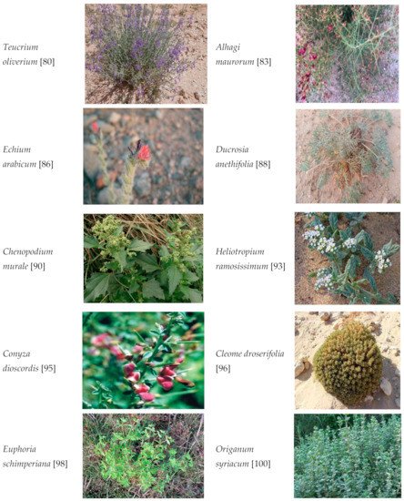
4. Which Are the Medicinal Plants of Saudi Arabia That Might Demonstrate Anti-Mutagenesis?

References
- Drakvik, E.; Altenburger, R.; Aoki, Y.; Backhaus, T.; Bahadori, T. Statement on advancing the assessment of chemical mixtures and their risks for human health and the environment. Environ. Int. 2020, 134, 105267.
- Munnia, A.; Giese, R.W.; Polvani, S.; Galli, A.; Cellai, F.; Peluso, M.E. Bulky DNA Adducts, Tobacco Smoking, Genetic Susceptibility, and Lung Cancer Risk. Adv. Clin. Chem. 2017, 81, 231–277.
- Matthew, D.W.; Fradet-Turcotte, A. Virus DNA Replication and the Host DNA Damage Response. Ann. Rev. Virol. 2018, 5, 141–164.
- Cellai, F.; Capacci, F.; Sgarrella, C.; Poli, C.; Arena, L.; Tofani, L.; Giese, R.W.; Peluso, M.A. Cross-Sectional Study on 3-(2-Deoxy-d-Erythro-Pentafuranosyl) Pyrimido
- Anderson, G.P.; Bozinovski, S. Acquired somatic mutations in the molecular pathogenesis of COPD. Trends Pharmacol. Sci. 2003, 24, 71–76.
- Baer, C.F.; Miyamoto, M.M.; Denver, D.R. Mutation rate variation in multicellular eukaryotes: Causes and consequences. Natl. Rev. Genet. 2007, 8, 619–631.
- Bae, T.; Tomasini, L.; Mariani, J.; Zhou, B.; Roychowdhury, T.; Franjic, D. Different mutational rates and mechanisms in human cells at pregastrulation and neurogenesis. Science 2018, 359, 550–555.
- Arvanitis, D.A.; Flouris, G.A.; Spandidos, D.A. Genomic rearrangements on VCAM1, SELE, APEG1 and AIF1 loci in atherosclerosis. J. Cell. Mol. Med. 2005, 9, 153–159.
- Andreassi, M.G. Coronary atherosclerosis and somatic mutations: An overview of the contributive factors for oxidative DNA damage. Mutat. Res. 2003, 543, 67–86.
- Lubos, E.; Loscalzo, J.; Handy, D.E. Homocysteine and glutathione peroxidase-1. Antioxid. Redox Signal. 2007, 9, 1923–1940.
- Saez, G.T.; Estan-Capell, N. Antioxidant enzymes. In Encyclopaedia of Cancer; Schwab, M., Ed.; Springer: Berlin, Germany, 2014.
- Devasangayam, T.P.A.; Tilak, J.C.; Boloor, K.K.; Sane, K.S.; Ghaskadbi, S.S.; Lele, R.D. Free radicals and antioxidant in human health: Current status and future prospects. J. Assoc. Physicians India 2004, 52, 794–804.
- Orrell, R.W. Amyotrophic lateral sclerosis: Copper/zinc superoxide dismutase (SOD1) gene mutations. Neuromuscul. Disord. 2000, 10, 63–68.
- Furukawa, Y.; Torres, A.S.; O’Halloran, T.V. Oxygen-induced maturation of SOD1: A key role for disulfide formation by the copper chaperone CCS. Eur. Mol. Biol. Organ. J. 2004, 23, 2872–2881.
- Crystal, R.G. Oxidants and antioxidants. Am. J. Med. 1991, 91, 3S–10S.
- Tomas-Barberan, F.A.; Robins, R.J. Phytochemistry of Fruits and Vegetables; Calendon Press: New York, NY, USA, 1997.
- Abdel-Wahhab, M.A.; Ahmed, H.H. Protective effect of Korean Panax ginseng against chromium VI toxicity and free radicals generation in rats. J. Ginseng Res. 2004, 28, 11–17.
- Li, J.; Yu, H.; Wang, S. Natural products, an important resource for discovery of multitarget drugs and functional food for regulation of hepatic glucose metabolism. Drug Des. Dev. Ther. 2018, 12, 121–135.
- Edenharder, R.; Worf-Wandelburg, A.; Decker, M.; Platt, K.L. Antimutagenic effects and possible mechanisms of action of vitamins and related compounds against genotoxic heterocyclic amines from cooked food. Mutat. Res. 1999, 444, 235–248.
- de Oliveira, M.R. The neurotoxic effects of vitamin A and retinoids. Ann. Braz. Acad. Sci. 2015, 87, 1361–1373.
- Naziroglu, M.; Cay, M. Protective role of intraperitoneally administered vitamin E and selenium on the oxidative defense mechanisms in rats with diabetes induced by streptozotocin. Biol. Trace Elem. Res. 2011, 79, 149–159.
- Rabbani, S.I.; Devi, K.; Khanam, S. Role of Pioglitazone with Metformin or Glimepiride on Oxidative Stress-induced Nuclear Damage and Reproductive Toxicity in Diabetic Rats. Malays. J. Med. Sci. 2010, 17, 3–11.
- Alpsoy, L.; Yildirim, A.; Agar, G. The antioxidant effects of vitamin A, C, and E on aflatoxin B1-induced oxidative stress in human lymphocytes. Toxicol. Ind. Health 2009, 25, 121–127.
- Sarma, L.; Kesavan, P.C. Protective effect of vitamin C and E against gamma-ray induced chromosomal damage in mouse. Int. J. Radiat. Biol. 1993, 759, 339–345.
- Konapacka, M.; Widel, M.; Rzeszowska-Wolny, J. Modifying effect of vitamins C, E and beta-carotene against gamma-ray-induced DNA damage in mouse cells. Mutat. Res. 1998, 417, 85–91.
- Monache, F.D.; MacQuhae, M.M.; Monache, G.D.; Bettolo, G.B.M.; De Lima, R.A. Xanthones, xanthonolignoids and other constituents of the roots of Vismia guaramirangae. Phytochemistry 1983, 22, 227–232.
- Rabbani, S.I.; Devi, K.; Khanam, S.; Zahra, N. Citral, a component of lemongrass oil inhibits the clastogenic effect of nickel chloride in mouse micronucleus test system. Pak. J. Pharm. Sci. 2006, 19, 108–113.
- Mossa, S.; AI-Yahya, M.A.; AI-Meshal, I.A. Medicinal Plants of Saudi Arabia; King Fahad National Library Publication: Riyadh, Saudi Arabia, 2000; Volume 2, pp. 1–355.
- Alfarhan, A.H.; Chaudhary, S.A.; Thomas, J. Notes on the flora of Saudi Arabia. J. King Saud Univ. 1998, 10, 31–40.
- Abdallah, E.M.; El-Ghazali, G. Screening for antimicrobial activity of some plants from Saudi folk medicine. Glob. J. Res. Med. Plants Indig. Med. 2013, 2, 210–218.
- Al-Sodany, Y.M.; Bazaid, S.A.; Mosallam, H.A. Medicinal plants in Saudi Arabia: I. Sarrwat Mountains at Taif, KSA. Acad. J. Plant Sci. 2013, 6, 134–145.
- El-Ghazali, G.E.; Al-Khalifa, K.S.; Saleem, G.A.; Abdallah, E.M. Traditional medicinal plants indigenous to Al-Rass province, Saudi Arabia. J. Med. Plants Res. 2010, 4, 2680–2683.
- Al-asmari, A.; Manthiri, R.A.; Abdo, N.; AL-duaiji, F.A.; Khan, H.A. Saudi medicinal plants for the treatment of scorpion sting envenomation. Saudi J. Biol. Sci. 2017, 24, 1204–1211.


