Submitted Successfully!
To reward your contribution, here is a gift for you: A free trial for our video production service.
Thank you for your contribution! You can also upload a video entry or images related to this topic.
Version Summary Created by Modification Content Size Created at Operation
1 + 2070 word(s) 2070 2021-09-01 10:01:03 |
2 format correct Meta information modification 2070 2021-09-17 03:08:51 |

Video Upload Options

Do you have a full video?

Confirm

Are you sure to Delete?
Cite
If you have any further questions, please contact Encyclopedia Editorial Office.
Burgelman, M. Extracellular Vesicles and Sepsis. Encyclopedia. Available online: https://encyclopedia.pub/entry/14265 (accessed on 27 July 2024).
Burgelman M. Extracellular Vesicles and Sepsis. Encyclopedia. Available at: https://encyclopedia.pub/entry/14265. Accessed July 27, 2024.
Burgelman, Marlies. "Extracellular Vesicles and Sepsis" Encyclopedia, https://encyclopedia.pub/entry/14265 (accessed July 27, 2024).
Burgelman, M. (2021, September 16). Extracellular Vesicles and Sepsis. In Encyclopedia. https://encyclopedia.pub/entry/14265
Burgelman, Marlies. "Extracellular Vesicles and Sepsis." Encyclopedia. Web. 16 September, 2021.
Extracellular Vesicles and Sepsis
Edit

Sepsis is defined as a life-threatening organ dysfunction caused by a dysregulated host response to an infection. Several studies on mouse and patient sepsis samples have revealed that the level of extracellular vesicles (EVs) in the blood is altered compared to healthy controls, but the different functions of EVs during sepsis pathology are not yet completely understood. Sepsis EVs are described as modulators of inflammation, lymphocyte apoptosis, coagulation and organ dysfunction. Furthermore, EVs can influence clinical outcome and it is suggested that EVs can predict survival. Both detrimental and beneficial roles for EVs have been described in sepsis, depending on the EV cellular source and the disease phase during which the EVs are studied.

extracellular vesicles sepsis inflammation

1. Overview

Sepsis is defined as a life-threatening organ dysfunction caused by a dysregulated host response to an infection. Several studies on mouse and patient sepsis samples have revealed that the level of extracellular vesicles (EVs) in the blood is altered compared to healthy controls, but the different functions of EVs during sepsis pathology are not yet completely understood. Sepsis EVs are described as modulators of inflammation, lymphocyte apoptosis, coagulation and organ dysfunction. Furthermore, EVs can influence clinical outcome and it is suggested that EVs can predict survival. Both detrimental and beneficial roles for EVs have been described in sepsis, depending on the EV cellular source and the disease phase during which the EVs are studied. In this review, we summarize the current knowledge of EV sources and functions during sepsis pathology based on in vitro and mouse models, as well as patient samples. 

2. Systemic Inflammation and Sepsis

Sepsis is defined as a life-threatening organ dysfunction caused by a dysregulated host response to infection [1]. With a mortality rate of 41% in Europe and 28.3% in the US, sepsis is one of the leading causes of mortality in critically ill patients at the intensive care unit (ICU) [2]. The global number of incident sepsis cases in 2017 was 48.9 million, 11 million of which resulted in sepsis-related deaths, accounting for 19.7% of all global deaths [3].
In 1992, the American College of Chest Physicians/Society of Critical Care Medicine Consensus Conference Committee defined infection as a microbial phenomenon characterized by an inflammatory response to the presence of micro-organisms or the invasion of normally sterile host tissue by those organisms [4]. A normal host response to infection is a very complex but localized process, executed by the innate immune system [5]. Upon bacterial entry, innate immune cells recognize evolutionary conserved structures of the invader, known as pathogen-associated molecular patterns (PAMPs), via their pathogen recognition receptors (PRRs), including Toll Like Receptors (TLRs) [6]. Recognition of PAMPs by PRRs triggers the release of pro-inflammatory cytokines and chemokines, leading to upregulated expression of adhesion molecules on endothelial cells [5][6]. All these factors ensure the attraction and migration of leukocytes to the infection site and ultimately lead to bacterial clearing via phagocytosis [6]. The release of pro-inflammatory signals is tightly regulated and in balance with anti-inflammatory cytokine release to assure a controlled and localized immune reaction [6][7].
During sepsis, the immune system is severely compromised and unable to eradicate pathogens [8]. Unlike what happens during a normal host response to infection, the immune response is now expanded over the whole body instead of being localized to the infection site. Sepsis typically involves an imbalance of the pro- and anti-inflammatory components of the immune system, characterized by an initial phase of overproduction of pro-inflammatory cytokines and hyperactivation of the immune system, which is then followed by an exacerbated anti-inflammatory state leading to immunosuppression [8][9][10]. Next to the systemic reaction to infection by the immune system, sepsis is accompanied by hemodynamic and coagulation alterations and cellular injuries, leading to the development of multiple organ dysfunction (MOD) [10][11]. Moreover, the peripheral inflammation during sepsis is also communicated to the central nervous system (CNS), leading to the development of encephalopathy as a complication in sepsis patients [12]. The cellular, circulatory and metabolic irregularities in a septic patient can profoundly aggravate until the patient needs to be supported with vasopressors to maintain an arterial pressure of 65 mmHg and the serum lactate levels are greater than 2 mmol/L in the absence of hypovolemia [1]. This sepsis subset is defined as septic shock and associated with higher hospital mortality rate than sepsis alone, raising above 40% [1].
The events that are linked to infection-triggered inflammation show similarities with non-infectious, sterile inflammation reactions, which makes it difficult to distinguish between them in early stages of the disease [13]. The systemic inflammatory response to a variety of clinical but non-infectious insults is called the systemic inflammation response syndrome (SIRS) [4]. These clinical insults typically imply trauma, burns or pancreatitis. Sepsis was therefore first defined as the systemic response to infection, thereby linked to SIRS [4]. However, as a result of new knowledge about the pathophysiology of sepsis, definitions of sepsis and septic shock have been reviewed several times. For an overview on the sepsis definitions, we refer to other reviews and the official reports of the international conferences where these definitions were defined [1][4][13][14][15]. When reading this review, keep in mind that both former and new definitions will be applied in studies depending on the time of publication. For this review, we will generally implement the term sepsis to avoid confusion and because most studies reviewed here are applying this term.
Over the past decades, a variety of murine models for sepsis were designed. In general, they can be divided into three categories: injection of a PRR agent, injection of live pathogens or surgical impairment of barrier tissue. One example of a mouse model for sepsis is the lipopolysaccharide (LPS) endotoxemia model (Figure 1A). Here, the mice are treated with LPS, which is a substance of the outer membrane of Gram-negative bacteria, as PRR agent. Although this model is frequently used in sepsis studies, its validity as a true sepsis model is debated due to the absence of an actual infection. On the other hand, the cecal ligation and puncture (CLP) model is established by a surgery in which the cecum is punctured and ligated. In this model, feces is leaking into the abdominal cavity, introducing peritonitis (Figure 1B). Therefore, the CLP model is frequently referred to as one of the golden standard rodent models for sepsis. None of the existing models can perfectly mimic all the human clinical features of sepsis, which implies that research in more than one model is most suitable. For an overview of the different existing sepsis models regarding the model characteristics, benefits and limitations, we refer to other recent reviews about commonly used sepsis models [16][17][18].
Figure 1. Schematic representation of two frequently used sepsis models. (A) The lipopolysaccharide (LPS) endotoxemia model implies injection (mostly intraperitoneally) of the PRR agent LPS. (B) The cecal ligation and puncture (CLP) model is established by puncturing and ligating the cecum, by which feces can enter the peritoneal cavity. In both models, one of the disease symptoms is hypothermia, which can be monitored by measuring body temperature. Figure created with BioRender.com.

3. Extracellular Vesicles (EVs)

EVs are nano-sized membrane vesicles which are secreted by a variety of cell types. As they can travel to distant tissues and transfer their cargo to cells, they comprise an important mechanism of intercellular communication. Based on biogenesis, EVs can be classified as exosomes, microvesicles or apoptotic bodies. Exosomes (~40–100 nm) are secreted via fusion of multivesicular bodies with the plasma membrane whereas microvesicles (~200–1000 nm) originate by direct membrane budding. Apoptotic bodies are large (500–3000 nm) and formed by random blebbing of the plasma membrane induced by cell death. EVs carry information in forms of proteins, genetic material, lipids and metabolites which they can transfer to other cells, influencing various physiological and pathological functions and explaining their involvement in both health and disease [19][20].
To justify the application of the term EV, several requirements regarding EV characterization and function should be satisfied as described by the Minimal Information for Studies of EVs (MISEV) guidelines [21]. These characterization guidelines demand that the EV preparation itself as well as the source of the EVs is properly quantified. Global quantification of EV source and EV preparation should include a description of the volume of fluid, and/or cell number, and/or tissue mass used to isolate EVs, as well as the quantification of EV amount per volume of initial fluid or per number of producing cells/mass of tissue by means of two methods such as assessing protein amount, particle number and lipid amount. Additionally, the presence of at least three protein markers needs to be verified. Among these, analysis of at least one transmembrane or glycosylphosphatidylinositol (GPI)-anchored protein associated with the plasma membrane and/or endosomes (general or cell-/tissue-specific), one cytosolic or periplasmic protein marker and one non-EV co-isolated structure is required to prove the presence and purity of the EV preparations. The presence of these pan-EV markers ensures their lipid bilayer character which encloses intracellular materials. It is important to stress that there are no markers that can distinguish between the EV subtypes. To claim the involvement of small EVs, an additional set of protein markers should be evaluated, specifically the proteins that are situated in/on intracellular compartments of eukaryotic secreting cells other than the plasma membrane and endosomes. These compartments include the Golgi apparatus, mitochondria, autophagosomes, peroxisomes and the endoplasmic reticulum. These transmembrane, lipid-bound and soluble proteins associated with these intracellular compartments are normally not enriched in smaller EVs (<200 nm diameter) [21].
When all guidelines regarding function and characterization are satisfied, it is justified to claim the presence of EVs and/or to categorize the EV into a certain EV subtype.

4. Conclusions

Currently, EVs in sepsis are associated with inflammation, apoptosis, bacterial clearance, coagulation and organ damage. Moreover, several studies suggest a link between EVs, clinical outcome and survival prediction. However, it is difficult to decipher whether EVs are detrimental or beneficial players in sepsis disease.
Sepsis is a condition which is characterized by its high heterogeneity. This is evident on different levels, including the infection source, individual patient heterogeneity, the plethora of pathways involving several processes (e.g., inflammation, apoptosis and coagulation) within sepsis pathology and the different pro- and anti-inflammatory phases during sepsis disease. It is therefore not surprising that EVs are found to have both pro- and anti-inflammatory or pro- and anti-apoptotic effects. The observed effect is strongly dependent on the cellular status of the EV source, which in its turn depends on the phase of sepsis disease. For example, an EV with pro-inflammatory content can be beneficial when produced during the immunosuppressive state; however, it will have detrimental effects within the hyperinflammatory phase. It is not clear yet which EV source is the most important during sepsis. Here, coherent data are still lacking, but overall, most studies indicate higher levels of leukocyte-, endothelial cell-, platelet- and RBC-derived EVs [22][23][24][25][26][27][28][29][30][31][32]. Conducted research focusing on unravelling EV cell sources in sepsis is greatly depending on flow cytometry studies, which in our opinion are valuable but also have their disadvantages such as the detection size limit and the great focus on PS-positive EVs. Moreover, a standardized EV isolation method and complete quality control as required by the guidelines in the field is often lacking. This represents a common pitfall in EV research which often impedes drawing unambiguous conclusions across studies. In future research, new emerging technologies such as Exoview/ExoFlex, ImageStream and Meso Scale Discovery (MSD) assays can be applied to further complement current knowledge about EV sources in sepsis. Regarding research focusing on biological effects of EVs, most studies have been focusing on the effects of neutrophil-, monocyte/macrophage- and endothelial cell-derived EVs in sepsis. In this review, both pro- and anti-inflammatory effects are described for macrophage- [33][34][35][36][37] and neutrophil-derived EVs [38][39]. Moreover, neutrophil-derived EVs are described as anti-bacterial effectors [29][38][39] and monocyte-derived EVs are linked with tissue factor (TF)-EV production and coagulation [25][40][41]. On the other hand, effects of endothelial-derived EVs are investigated in the context of organ damage [32][42] and coagulation [31][43][44]. To get more insight into the role of specific EV sources, transgenic mouse models in which EV production by the cells of interest is dampened would be of high value. In transgenic mice of interest, genes that are linked with EV biogenesis pathways (e.g., Alix and nSMase2) could be blocked, assuming this will result in reduced EV production or the release of EVs with an altered content by the target cells of interest. In this way, this approach will allow validation of whether withdrawal of certain EV subtypes has significant effects on survival, organ damage and inflammation during sepsis.

References

  1. Singer, M.; Deutschman, C.S.; Seymour, C.W.; Shankar-Hari, M.; Annane, D.; Bauer, M.; Bellomo, R.; Bernard, G.R.; Chiche, J.D.; Coopersmith, C.M.; et al. The third international consensus definitions for sepsis and septic shock (sepsis-3). JAMA 2016, 315, 801–810.
  2. Levy, M.M.; Artigas, A.; Phillips, G.S.; Rhodes, A.; Beale, R.; Osborn, T.; Vincent, J.L.; Townsend, S.; Lemeshow, S.; Dellinger, R.P. Outcomes of the surviving sepsis campaign in intensive care units in the USA and Europe: A prospective cohort study. Lancet Infect. Dis. 2012, 12, 919–924.
  3. Rudd, K.E.; Johnson, S.C.; Agesa, K.M.; Shackelford, K.A.; Tsoi, D.; Kievlan, D.R.; Colombara, D.V.; Ikuta, K.S.; Kissoon, N.; Finfer, S.; et al. Global, regional, and national sepsis incidence and mortality, 1990–2017: Analysis for the global burden of disease study. Lancet 2020, 395, 200–211.
  4. American College of Chest Physicians/Society of Critical Care Medicine Consensus Conference: Definitions for sepsis and organ failure and guidelines for the use of innovative therapies in sepsis. Crit. Care Med. 1992, 20, 864–874.
  5. Tosi, M.F. Innate immune responses to infection. J. Allergy Clin. Immunol. 2005, 116, 241–249.
  6. Mogensen, T.H. Pathogen recognition and inflammatory signaling in innate immune defenses. Clin. Microbiol. Rev. 2009, 22, 240–273.
  7. Cicchese, J.M.; Evans, S.; Hult, C.; Joslyn, L.R.; Wessler, T.; Millar, J.A.; Marino, S.; Cilfone, N.A.; Mattila, J.T.; Linderman, J.J.; et al. Dynamic balance of pro- and anti-inflammatory signals controls disease and limits pathology. Immunol. Rev. 2018, 285, 147–167.
  8. Hotchkiss, R.S.; Karl, I.E. The pathophysiology and treatment of sepsis. N. Engl. J. Med. 2003, 348, 138–150.
  9. Oberholzer, A.; Oberholzer, C.; Moldawer, L.L. Sepsis syndromes: Understanding the role of innate and acquired immunity. Shock 2001, 16, 83–96.
  10. Gyawali, B.; Ramakrishna, K.; Dhamoon, A.S. Sepsis: The evolution in definition, pathophysiology, and management. SAGE Open Med. 2019, 7, 2050312119835043.
  11. Caraballo, C.; Jaimes, F. Organ dysfunction in sepsis: An ominous trajectory from infection to death. Yale J. Biol. Med. 2019, 92, 629–640.
  12. Chaudhry, N.; Duggal, A.K. Sepsis associated encephalopathy. Adv. Med. 2014, 2014, 762320.
  13. Gul, F.; Arslantas, M.K.; Cinel, I.; Kumar, A. Changing definitions of sepsis. Turk. J. Anaesthesiol. Reanim. 2017, 45, 129–138.
  14. Levy, M.M.; Fink, M.P.; Marshall, J.C.; Abraham, E.; Angus, D.; Cook, D.; Cohen, J.; Opal, S.M.; Vincent, J.L.; Ramsay, G. 2001 SCCM/ESICM/ACCP/ATS/SIS international sepsis definitions conference. Intensive Care Med. 2003, 29, 530–538.
  15. Balk, R.A. Systemic inflammatory response syndrome (SIRS): Where did it come from and is it still relevant today? Virulence 2014, 5, 20–26.
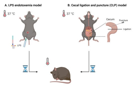
  16. Korneev, K.V. Mouse Models of Sepsis and Septic Shock. Mol. Biol. 2019, 53, 799–814.
  17. Lewis, A.J.; Seymour, C.W.; Rosengart, M.R. Current murine models of sepsis. Surg Infect. 2016, 17, 385–393.
  18. Dejager, L.; Pinheiro, I.; Dejonckheere, E.; Libert, C. Cecal ligation and puncture: The gold standard model for polymicrobial sepsis? Trends Microbiol. 2011, 19, 198–208.
  19. Andaloussi, S.E.; Mager, I.; Breakefield, X.O.; Wood, M.J. Extracellular vesicles: Biology and emerging therapeutic opportunities. Nat. Rev. Drug Discov. 2013, 12, 347–357.
  20. van Niel, G.; D’Angelo, G.; Raposo, G. Shedding light on the cell biology of extracellular vesicles. Nat. Rev. Mol. Cell Biol. 2018, 19, 213–228.
  21. Thery, C.; Witwer, K.W.; Aikawa, E.; Alcaraz, M.J.; Anderson, J.D.; Andriantsitohaina, R.; Antoniou, A.; Arab, T.; Archer, F.; Atkin-Smith, G.K.; et al. Minimal information for studies of extracellular vesicles 2018 (MISEV2018): A position statement of the International Society for Extracellular Vesicles and update of the MISEV2014 guidelines. J. Extracell Vesicles 2018, 7, 1535750.
  22. Zhang, Y.; Meng, H.; Ma, R.; He, Z.; Wu, X.; Cao, M.; Yao, Z.; Zhao, L.; Li, T.; Deng, R.; et al. Circulating microparticles, blood cells, and endothelium induce procoagulant activity in sepsis through phosphatidylserine exposure. Shock 2016, 45, 299–307.
  23. Mostefai, H.A.; Meziani, F.; Mastronardi, M.L.; Agouni, A.; Heymes, C.; Sargentini, C.; Asfar, P.; Martinez, M.C.; Andriantsitohaina, R. Circulating microparticles from patients with septic shock exert protective role in vascular function. Am. J. Respir. Crit. Care Med. 2008, 178, 1148–1155.
  24. Fendl, B.; Weiss, R.; Eichhorn, T.; Linsberger, I.; Afonyushkin, T.; Puhm, F.; Binder, C.J.; Fischer, M.B.; Weber, V. Extracellular vesicles are associated with C-reactive protein in sepsis. Sci. Rep. 2021, 11, 6996.
  25. Nieuwland, R.; Berckmans, R.J.; McGregor, S.; Boing, A.N.; Romijn, F.P.; Westendorp, R.G.; Hack, C.E.; Sturk, A. Cellular origin and procoagulant properties of microparticles in meningococcal sepsis. Blood 2000, 95, 930–935.
  26. Ogura, H.; Kawasaki, T.; Tanaka, H.; Koh, T.; Tanaka, R.; Ozeki, Y.; Hosotsubo, H.; Kuwagata, Y.; Shimazu, T.; Sugimoto, H. Activated platelets enhance microparticle formation and platelet-leukocyte interaction in severe trauma and sepsis. J. Trauma 2001, 50, 801–809.
  27. Joop, K.; Berckmans, R.J.; Nieuwland, R.; Berkhout, J.; Romijn, F.P.; Hack, C.E.; Sturk, A. Microparticles from patients with multiple organ dysfunction syndrome and sepsis support coagulation through multiple mechanisms. Thromb. Haemost. 2001, 85, 810–820.
  28. Lashin, H.M.S.; Nadkarni, S.; Oggero, S.; Jones, H.R.; Knight, J.C.; Hinds, C.J.; Perretti, M. Microvesicle subsets in sepsis due to community acquired pneumonia compared to faecal peritonitis. Shock 2018, 49, 393–401.
  29. Timar, C.I.; Lorincz, A.M.; Csepanyi-Komi, R.; Valyi-Nagy, A.; Nagy, G.; Buzas, E.I.; Ivanyi, Z.; Kittel, A.; Powell, D.W.; McLeish, K.R.; et al. Antibacterial effect of microvesicles released from human neutrophilic granulocytes. Blood 2013, 121, 510–518.
  30. Lehner, G.F.; Harler, U.; Haller, V.M.; Feistritzer, C.; Hasslacher, J.; Dunzendorfer, S.; Bellmann, R.; Joannidis, M. Characterization of microvesicles in septic shock using high-sensitivity flow cytometry. Shock 2016, 46, 373–381.
  31. Matsumoto, H.; Yamakawa, K.; Ogura, H.; Koh, T.; Matsumoto, N.; Shimazu, T. Enhanced expression of cell-specific surface antigens on endothelial microparticles in sepsis-induced disseminated intravascular coagulation. Shock 2015, 43, 443–449.
  32. Soriano, A.O.; Jy, W.; Chirinos, J.A.; Valdivia, M.A.; Velasquez, H.S.; Jimenez, J.J.; Horstman, L.L.; Kett, D.H.; Schein, R.M.; Ahn, Y.S. Levels of endothelial and platelet microparticles and their interactions with leukocytes negatively correlate with organ dysfunction and predict mortality in severe sepsis. Crit. Care Med. 2005, 33, 2540–2546.
  33. Essandoh, K.; Yang, L.; Wang, X.; Huang, W.; Qin, D.; Hao, J.; Wang, Y.; Zingarelli, B.; Peng, T.; Fan, G.C. Blockade of exosome generation with GW4869 dampens the sepsis-induced inflammation and cardiac dysfunction. Biochim. Biophys. Acta 2015, 1852, 2362–2371.
  34. Nair, R.R.; Mazza, D.; Brambilla, F.; Gorzanelli, A.; Agresti, A.; Bianchi, M.E. LPS-challenged macrophages release microvesicles coated with histones. Front. Immunol. 2018, 9, 1463.
  35. Anand, P.K.; Anand, E.; Bleck, C.K.; Anes, E.; Griffiths, G. Exosomal Hsp70 induces a pro-inflammatory response to foreign particles including mycobacteria. PLoS ONE 2010, 5, e10136.
  36. Wisler, J.R.; Singh, K.; McCarty, A.R.; Abouhashem, A.S.E.; Christman, J.W.; Sen, C.K. Proteomic pathway analysis of monocyte-derived exosomes during surgical sepsis identifies immunoregulatory functions. Surg Infect. 2020, 21, 101–111.
  37. Alarcon-Vila, C.; Baroja-Mazo, A.; de Torre-Minguela, C.; Martinez, C.M.; Martinez-Garcia, J.J.; Martinez-Banaclocha, H.; Garcia-Palenciano, C.; Pelegrin, P. CD14 release induced by P2X7 receptor restricts inflammation and increases survival during sepsis. Elife 2020, 9, e60849.
  38. Youn, Y.J.; Shrestha, S.; Lee, Y.B.; Kim, J.K.; Lee, J.H.; Hur, K.; Mali, N.M.; Nam, S.W.; Kim, S.H.; Lee, S.; et al. Neutrophil-derived trail is a proinflammatory subtype of neutrophil-derived extracellular vesicles. Theranostics 2021, 11, 2770–2787.
  39. Dalli, J.; Norling, L.V.; Montero-Melendez, T.; Federici Canova, D.; Lashin, H.; Pavlov, A.M.; Sukhorukov, G.B.; Hinds, C.J.; Perretti, M. Microparticle alpha-2-macroglobulin enhances pro-resolving responses and promotes survival in sepsis. EMBO Mol. Med. 2014, 6, 27–42.
  40. Janiszewski, M.; Do Carmo, A.O.; Pedro, M.A.; Silva, E.; Knobel, E.; Laurindo, F.R. Platelet-derived exosomes of septic individuals possess proapoptotic NAD(P)H oxidase activity: A novel vascular redox pathway. Crit. Care Med. 2004, 32, 818–825.
  41. Matsumoto, H.; Yamakawa, K.; Ogura, H.; Koh, T.; Matsumoto, N.; Shimazu, T. Clinical significance of tissue factor and CD13 double-positive microparticles in sirs patients with trauma and severe sepsis. Shock 2017, 47, 409–415.
  42. Liu, Y.; Zhang, R.; Qu, H.; Wu, J.; Li, L.; Tang, Y. Endothelial microparticles activate endothelial cells to facilitate the inflammatory response. Mol. Med. Rep. 2017, 15, 1291–1296.
  43. Guervilly, C.; Lacroix, R.; Forel, J.M.; Roch, A.; Camoin-Jau, L.; Papazian, L.; Dignat-George, F. High levels of circulating leukocyte microparticles are associated with better outcome in acute respiratory distress syndrome. Crit. Care 2011, 15, R31.
  44. Delabranche, X.; Quenot, J.P.; Lavigne, T.; Mercier, E.; Francois, B.; Severac, F.; Grunebaum, L.; Mehdi, M.; Zobairi, F.; Toti, F.; et al. Early detection of disseminated intravascular coagulation during septic shock: A multicenter prospective study. Crit. Care Med. 2016, 44, e930–e939.
More
Information
Subjects: Immunology
Contributor MDPI registered users' name will be linked to their SciProfiles pages. To register with us, please refer to https://encyclopedia.pub/register :
View Times: 440
Revisions: 2 times (View History)
Update Date: 17 Sep 2021
1000/1000
Video Production Service