Submitted Successfully!
To reward your contribution, here is a gift for you: A free trial for our video production service.
Thank you for your contribution! You can also upload a video entry or images related to this topic.
Version Summary Created by Modification Content Size Created at Operation
1 + 2991 word(s) 2991 2021-08-06 11:41:47 |
2 format correct Meta information modification 2991 2021-09-16 03:21:32 |

Video Upload Options

Do you have a full video?

Confirm

Are you sure to Delete?
Cite
If you have any further questions, please contact Encyclopedia Editorial Office.
Shoeb, E. VNPs for Anti-Cancer Therapy. Encyclopedia. Available online: https://encyclopedia.pub/entry/14131 (accessed on 28 March 2024).
Shoeb E. VNPs for Anti-Cancer Therapy. Encyclopedia. Available at: https://encyclopedia.pub/entry/14131. Accessed March 28, 2024.
Shoeb, Erum. "VNPs for Anti-Cancer Therapy" Encyclopedia, https://encyclopedia.pub/entry/14131 (accessed March 28, 2024).
Shoeb, E. (2021, September 13). VNPs for Anti-Cancer Therapy. In Encyclopedia. https://encyclopedia.pub/entry/14131
Shoeb, Erum. "VNPs for Anti-Cancer Therapy." Encyclopedia. Web. 13 September, 2021.
VNPs for Anti-Cancer Therapy
Edit

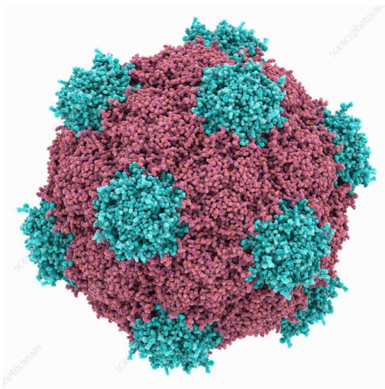
Naturally occurring viral nanomaterials have gained popularity owing to their biocompatible and biodegradable nature. Plant virus nanoparticles (VNPs) can be used as nanocarriers for a number of biomedical applications. Plant VNPs are inexpensive to produce, safe to administer and efficacious as treatments. Features which distinguish plant viruses from synthetic nanocarriers include stability, flexibility, diversity in shape and size for use in drug delivery and the nontoxic nature of plant viruses in humans. Cancer is one of the most common death causing disease worldwide and it is characterized by uncontrolled rapid cell division and differentiation. VNPs are an ideal choice to apply for cancer treatment owing to the enhanced permeation and retention (EPR) potential of cancer cells for these nanoparticles, whereas VNPs cannot penetrate through healthy tissues due to tightly packed endothelial cells.

nanoparticles plant virus-like particles therapeutics imaging cancer

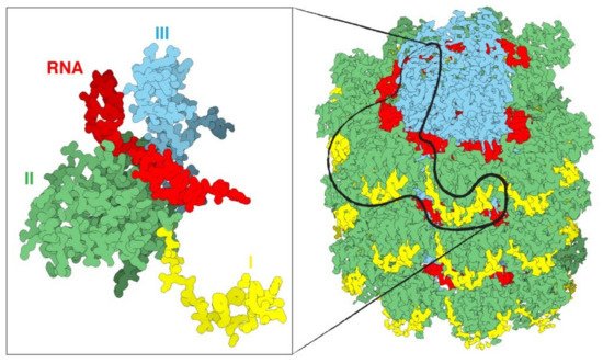
1. Plant Virus Architecture

Virus size data that is found in different databases has been obtained by transmission electron microscopy (TEM), and virus sizes and architecture differs significantly [1]. A method of nanoparticle tracking analysis (NTA) can be used to determine the size and concentration of viruses and VLPs. At present, this technique has been used to study icosahedral VLPs of Cowpea mosaic virus (CPMV) (Figure 1) [2], spherical VLPs of structurally modified Tobacco mosaic virus (TMV) [3], and VLPs of filamentous Potato virus X (PVX) [4]. Nanoparticle tracking analysis has also been reported to compare the intensity of coated and native helical TMV [5][6].
Figure 1. Cowpea mosaic virus structure, Protein subunits in red and blue. Source: fineartamerica.com (accessed on 11 May 2021).
Viruses are composed of outer protein shells that encapsulate the genomic material. The multiple copies of coat proteins that form the virus outer shell of viruses are collectively known as the capsid [7]. Primarily, the capsid occurs in different shapes and sizes and is meant to protect the genomic material to keep viruses safe under extreme environments [8]. Animal viruses are used as delivery vehicles for gene therapy, but the use of these viruses as nanocarriers can potentially create safety issues. Alternatively, plant viruses have not displayed toxicity and thus have received more attention for applications in nanomedicine [9]. The most well studied plant viruses are Tobacco mosaic virus (TMV), Cowpea mosaic virus (CPMV), and Potato virus X (PVX) (Figure 2) [10]. The immense diversity with respect to the shape and size of plant viruses enables them to be tailored for specific applications. For instance, rod shaped viruses are more suitable for penetrating into tumors [11]. The structural integrity of viruses remains intact even when surface properties have been altered through chemical and genetic modification; this allows control over targeting ligands, drugs and contrast agents for imaging [12]. The most interesting properties which give these plant based nanoparticles edge over synthetic nanomaterials include bulk production via host infection and available inexpensive large scale purification methods [13].
Figure 2. An overview of a portion of the PVX virus (right). The three domains of the protein are shin in yellow, green and cyan, the RNA in red. The magnification on the left displays only one single CP with a fragment of RNA. Source: https://naturemicrobiologycommunity.nature.com/posts/61805-a-detailed-view-of-a-filamentous-virus-potato-virus-x (accessed on 11 May 2021).
Researchers have the choice of selecting the most appropriate system for any given application owing to the diversity of plant virus shape and size. A wide range of nanomedical applications have been demonstrated using the capsids of icosahedral viruses [14]. Rod shaped viruses with a high aspect ratio have superior penetration into tumor tissue [11][15]. Various chemical and genetic approaches are reported to control the virus surface properties without affecting structural integrity, and that allow control on the attachment sites of drug molecules or contrast agents on the virus surface [12]. Plant-virus capsid pores are also reported to be employed to encapsulate small therapeutic molecules [16].

2. Biomedical Applications of Plant Virus Nanoparticles

Magnetic resonance imaging (MRI) is a promising technology for the diagnosis of disease due to its high resolution and deep contrast, however, virus-based nanoparticles have been explored to overcome the drawback of low sensitivity [17]. TMV is reported as a carrier to deliver high payloads of MRI contrast imaging agents to sites of disease [18] and fluorescent dyes for biosensing and bioimaging [19]. This inert nature together with biological compatibility and multi-valency makes plant viruses suitable carriers of in vivo imaging agents. TMV rods have been conjugated to ‘BF3’, a multi-photon absorbing fluorophore which allowed mouse brain image over an extended duration without crossing the blood–brain barrier [20]. Ultra-high-field magnetic resonance imaging (UHFMRI) advances the diagnostic accuracy of MRI scans depending on better contrast agents for better resolution and signal-to-noise ratio. A bimodal contrast agent has been prepared to target integrin α2β1by loading the internal cavity of TMV nanoparticles with the complex of dysprosium (Dy3+) and the near-infrared fluorescence (NIRF) dye Cy7.5, and externally conjugated with an Asp–Gly–Glu–Ala (DGEA) peptide through a linker polyethylene glycol. This nanoparticle (Dy–Cy7.5–TMV–DGEA) was not only stable but also biocompatible with a low cytotoxicity. It achieved a high resolution when targeted to PC-3 prostate cancer cells [21].
Noninvasive imaging systems that are for early diagnosis of cancer, such as magnetic resonance imaging (MRI) and computed tomography (CT), have some limitations [22][23]. Nanotechnology offers an accurate state-of-the-art solution for imaging systems [22][23]. The capacity for multifunctionality and multivalency makes plant nanoparticle platforms an ideal choice for theranostic applications [24]. Nanoparticles are capable of precise molecular imaging to achieve accurate cancer diagnosis and therapy [25]. Delivery of imaging probes through nanostructures can improve the chances of early-stage cancer diagnosis through the use of multiple modalities to improve resolution, sensitivity, penetration, time, cost and on the top of all clinical relevance compared to the single imaging modalities [26]. Drug conjugated nanoparticles administered intravenously target tumors, via the process of enhanced permeability and retention (EPR) effect, depending on the type of tumor [27].
The use of nanoparticles (NPs) as drug delivery carriers for the treatment of infectious and chronic diseases including cancer are advantageous when compared with naked drugs [28]. The most promising nanoparticle systems have been adopted from naturally occurring plant viruses. Plant viruses are ideal for drug delivery as they are safe, non-infectious and nontoxic to humans [24]. Cancer cells exhibit specific antigens on the surface of tumor cells which can be identified and targeted by plant-virus based nanoparticles, thus providing a clinical application of diagnosis and therapeutics for cancer. The most promising nano-scale systems have been adopted from naturally occurring plant viruses such as Tobacco mosaic virus (TMV), Cowpea mosaic virus (CPMV), Potato virus X (PVX) and many more. Currently, these new strategies are only applied in small scale production. As these approaches undergo further development, we will witness a spectrum of possible applications in the fields of medicine and biomedical engineering.
VLPs mimic the native conformation of viruses, propagate their innate immunogenicity and promote safety due to the lack of their genetic material, and therefore form an attractive platform for vaccine development [29][30]. Furthermore, plant viruses are incapable of replication within mammalian cells, thus ensuring an additional level of safety during administration into tumors. VLPs serve as ideal vehicles for both prophylaxis (vaccine design) and therapy against cancer. VLPs are highly immunogenic and are readily phagocytosed by the antigen presenting cells (APCs), which in turn elicit antigen processing and display of pathogenic epitopes on their surfaces. As the VLPs are composed of multiple copies of their respective capsid proteins, they present repetitive multivalent scaffolds which aid in antigen presentation. Therefore, the VLPs prove to be perfect platforms for delivery and presentation of antigenic epitopes, resulting in induction of more a robust immune response compared to those of their soluble counterparts. As the tumor microenvironment poses the challenge of self-antigen tolerance, VLPs are preferrable platforms for delivery and display of self-antigens as well as otherwise weakly immunogenic antigens. The inherent multivalency and geometric arrangement of proteins on the VLP surface promote immune recognition via PAMPs (pathogen-associated molecular pattern receptors), most commonly Toll-like Receptors (TLRs) [31]. These properties, in addition to their diminutive size, enable the VLPs to deliver vaccines to the draining lymph nodes in addition to promoting APC interactions. Furthermore, many plant viral VLPs possess inherent adjuvant properties dispensing with the requirement of additional adjuvants to stimulate immune activity. Some of the highly immunogenic VLPs elicit innate immune activity, which in turn instigate adaptive immunity in tumor micro-environments [32]. Plant viral VLPs are nontoxic, inherently stable, and capable of being mass-produced as well as being modified with antigens and drugs, therefore provide an attractive option for eliciting anti-tumor immunity.
VLPs have been attached to TAAs (tumor associated antigens) using chemical conjugation, genetic fusion and enzyme-mediated ligation techniques. Among these various strategies, the most common method has been chemical conjugation. For example, the human epidermal growth factor receptor 2 (HER2) epitope, when conjugated to the icosahedral CPMV, was successfully delivered to the lymphatic system with enhanced uptake and activation of APCs that led to an augmented anti-HER2 immune response. The CPMV HER2 candidate vaccine slowed tumor progression and metastasis in mouse models, enhancing survival [33]. Importantly, CPMV-HER2 stimulated a predominantly Th1 immune response while Sesbania mosaic virus-HER2 and CCMV-HER2 induced mostly a Th2 response in mouse models, thus proving that the nature of the epitope carrier itself plays an essential role in regulating the Th1/Th2 bias. This could be due to differences in epitope display on the surface of the VNPs as well as the capsid. In another study, when PVX was conjugated to a recombinant idiotypic (Id) TAA displayed a 7-times greater anti-Id IgG response compared to that elicited by Id alone in a mouse B-cell lymphoma model (BCL1) [34]. It was determined that TLR7 was crucial for the recognition of the viral RNA, in addition, cytokines such as IL-12 and IFN-α were induced.
Cancer vaccines against carbohydrate antigens associated with tumors (TACAs) could be useful for diminishing tumor progression. Nevertheless, carbohydrates are weakly immunogenic and therefore, plant viruses used as carriers of these molecules could enhance the immune response to TACAs. CPMV-TACA conjugates targeting the Tn antigen (GalNAc-α-O-Ser/Thr) [35] were demonstrated to induce enhanced IgG titers, implicating heightened T-cell mediated immunity and antibody isotype switching in mouse models. IgG binding to the Tn antigens were observed in experiments wherein mice sera were added to breast cancer cell lines. In another subsequent study, TMV was used as an equivalent system to display Tn antigens [36] in which conjugation to Tyr 139 of TMV generated robust immune responses. Finally, nucleic acid vaccines have become recognized as next-generation vaccines against cancers, and VLPs are currently being investigated as potential carriers. Nanocarriers such as plant VLPs could be used to protect RNA-based vaccines, thereby extending the half-lives and biodistribution of these therapeutics.
The tumor microenvironment poses a great challenge to immune clearance by virtue of being immunosuppressive and favoring immune escape of the tumors through the inhibition of anti-tumor T-cells [30][37]. CPMV VLP nanoparticles were shown to decrease tumor growth in murine models of lung melanomas, ovarian, colon and breast tumors [38][39][40]. Mechanistically, CPMV has been shown to reprogram the tumor microenvironment by recruitment of natural killer cells and neutrophils, while enabling the transition of M2 to M1 anti-tumor macrophages. This innate immune cell population subsequently combats the tumor leading to cell lysis while allowing the antigen presenting cells to process and display the tumor associated antigens resulting in strong T-cell activation and systemic immunity against the tumors. CPMV VNPs have also been formulated as slow-release aggregates along with polyamidoamine generation 4 dendrimers (CPMV-G4) [41], where they were shown to be effective in combating ovarian cancer in murine models, even when provided as a single dosage. Besides CPMV, TMV and PapMV have also been shown to have anti-tumor potential, although CPMV proved to be more effectual.
Plant virus VLPs have also been used as combination therapies to augment their immune efficacy. The PVX-DOX (doxorubicin) [42] combination was shown to be highly effective in stimulating cytokine/chemokine levels while prolonging the survival of mice in melanoma models when compared to that obtained through the administration of either PVX or DOX alone. Moreover, the strong chemotherapeutic cyclophosphamide, when used in combination with CPMV VNPs, profoundly elicited tumor cell death, releasing extracellular TAAs and stimulating immune cell invasion in addition to augmenting TAA recognition and antigen presentation [38] in mouse tumor models. CPMV VNPs have also been administered as combinations with CD47-blocking antibodies [43] which proved to have synergistic effects in combating tumor growth in murine ovarian tumor models, where it activated phagocytes. The anti-CD47 antibodies suppressed antiphagocytic signals, leading to stimulation of the adaptive immune response. Similar synergistic effects were observed when CPMV VNPs were used in combination with the anti-programmed cell death-1 checkpoint inhibitor. In addition to this, CPMV has been used successfully in promoting anti-tumor effects, when combined with radiation therapy. In this instance, CPMV was shown to enhance the recruitment of APCs, which in turn targeted the extracellular TAAs and phagocytosed them to induce a prolonged effectual immune response [44] in mice and canine models.
Presently, clinical treatment for cancer is primarily addressed by chemotherapy [21]. Nevertheless, the high recurrence and resistance rates as well as the fast clearance of anti-cancer drugs and non-targeted drug delivery necessitate the administration of maximum tolerable doses of drugs for enabling cancer therapy. This may lead to increased toxicity and diminished clinical pertinence. Therefore, drug delivery technologies that enable intracellular and targeted conveyance while promoting active drug accumulation in tumors, in concert with the limiting of dose requirements, could alleviate these concerns and augment treatment outcomes.
Plant virus VLPs have several attractive features that make them appropriate for targeted administration of therapeutic molecules. The anti-cancer drug doxorubicin (DOX), has been successfully delivered using VNPs and VLPs. Specifically, red clover necrotic mosaic virus (RCNMV) VLPs have been used to both encapsulate DOX [45] as well as bind DOX on its surface. This enabled the simultaneous quick release of the externally-bound DOX and the slow release of the encapsulated DOX from within the VLP. Similarly, TMV- and PVX-derived VLPs and VNPs have been successfully used to deliver DOX [46], which proved to be highly efficacious. In this context, molecules with high aspect ratio have proven to be of great use in effective drug delivery, of which helical plant viruses that form tubular and filamentous structures are good representative examples. VNPs have shown great promise as their cargo-RNA functions as a ruler establishing the length of the virus particle and simple adsorption of DOX on their surface was shown to be effective for reducing tumor growth [47][48].
Plant virus VNPs have been used for targeted administration of platinum-based drugs against cancer. This is important, as 50% of chemotherapy treatments involve the use of these platinum-derived drugs. TMV has been demonstrated to efficiently deliver Cisplatin [49] and Phenanthriplatin [50], both of which are platinum-based drugs. The drugs were loaded into the TMV VNP cavity using charge-driven interactions or by synthesizing stable covalent adducts. Such a TMV-based drug delivery system was proven to enable superior, targeted cytotoxicity as well as increased ease of uptake by cancer cells in in vitro systems using HepG2 and MCF-7 cancer cell lines [51].
Another anti-cancer drug, mitoxanthrone (MTO), is a topoisomerase II inhibitor and has been shown to be encapsulated by TMV [52] and CPMV [53] VNPs exhibited superior tumor-reduction in mouse cancer models, while precluding severe cardiac outcomes that sometimes accompany direct delivery of MTO. Yet another anti-neoplastic and antimitotic drug, valine-citrulline monomethyl auristatin E (vcMMAE), was bound to the exterior of TMV VNPs which targeted non-Hodgkin’s lymphoma. Internalization into endolysosomal compartments was reported [54], most likely accompanied by the protease-mediated release of the drug. This system was efficient in terms of cytotoxicity towards the in vitro Karpas 299 non-Hodgkin’s lymphoma cell line with an IC50 of 250 nM.
Viruses are inherently natural gene-delivery vehicles. Plant viral VLPs in particular, by virtue of being devoid of their own genetic materials, easily encapsidate nucleic acids. This makes them highly useful in delivering genes and therapeutic nucleic acids. It is reported that (Gene Therapy Clinical Trials Worldwide, 2019) 67% of clinical trials involving gene therapy use VLPs and viruses as vectors for transmission of genetic materials. Tian et al., (2018) demonstrated that transacting activation transduction (TAT) peptide conjugated to the external surface of TMV augmented internalization along with increased ability to escape endo/lysosomal compartments [55]. Most of these VLPS exhibited uptake by dendritic cells and macrophages and proved to be highly immunogenic. Thus, therapeutic nucleic acids can be easily delivered to immune cells during cancer treatments. The CpG oligodeoxynucleotide (ODN) was loaded onto CCMV VLPs which greatly increased uptake by TAM (tumor associated macrophages), not by cancer cells. Intra-tumoral delivery of CpG-loaded CCMV VLPs demonstrated significant inhibition of tumor growth in mouse models [38].
In comparison to nucleic acids, proteins and polypeptides are more difficult and complex to encapsulate, thus limiting their delivery into cancer cells. This could be resolved by fusing the polypeptide to the VLP capsid protein by chemical or genetic means. This could direct cargo localization into the interior of the VLPs during the synthesis and assembly of the capsid in vivo [56]. Viral capsid protein could spontaneously self-assemble around the polypeptide in the presence or absence of a targeting element in vitro [57]. Several drug-activating enzymes encapsulated within VLPs enable therapeutic functions [58]. Amongst these, the most important are the cytochrome P450 family of enzymes which catalyze into active forms of their precursors, namely the chemotherapeutic pro-drugs. This enables better tumor-targeting, enhancing their half-life within the tumors via better permeability while reducing side effects [59][60]. In this context, the CCMV has been shown to encapsulate cytochrome CYPBM3 with demonstrated activation effects on the pro-drugs resveratrol and tamoxifen [59]. Additionally, these proteins can be displayed on the VLP surfaces for therapeutic or targeting purposes.
Helical viruses such as PVX [61] and TMV are ideal vehicles for drug delivery by virtue of their shape and high aspect ratio that enables augmented cellular interactions with their conjugated cargo. PVX displaying TNF related apoptosis inducing ligand (TRAIL) was used to promote the recruitment and activation of death receptors in vitro in HCC-38 primary ductal carcinoma, BT-549 ductal carcinoma and the MDA-MB-231 breast cancer cell lines [42][61]. In vivo mouse models also demonstrated that the PVX-TRAIL formulation potently inhibited tumor growth.

References

  1. Nikitin, N.; Trifonova, E.; Evtushenko, E.; Kirpichnikov, M.; Atabekov, J.; Karpova, O. Comparative Study of Non-Enveloped Icosahedral Viruses Size. PLoS ONE 2015, 10, e0142415.
  2. Aljabali, A.A.; Shah, S.N.; Evans-Gowing, R.; Lomonossoff, G.P.; Evans, D.J. Chemically-coupled-peptide-promoted virus nanoparticle templated mineralization. Integr. Biol. (Camb.) 2011, 3, 119–125.
  3. Nikitin, N.; Trifonova, E.; Karpova, O.; Atabekov, J. Examination of biologically active nanocomplexes by nanoparticle tracking analysis. Microsc. Microanal. 2013, 19, 808–813.
  4. Petrova, E.K.; Nikitin, N.A.; Trifonova, E.A.; Protopopova, A.D.; Karpova, O.V.; Atabekov, J.G. The 5′-proximal region of Potato virus X RNA involves the potential cap-dependent “conformational element” for encapsidation. Biochimie 2015, 115, 116–119.
  5. Zhou, J.C.; Soto, C.M.; Chen, M.S.; Bruckman, M.A.; Moore, M.H.; Barry, E.; Ratna, B.R.; Pehrsson, P.E.; Spies, B.R.; Confer, T.S. Biotemplating rod-like viruses for the synthesis of copper nanorods and nanowires. J. Nanobiotechnology 2012, 10, 18.
  6. Steinmetz, N.F.; Shah, S.N.; Barclay, J.E.; Rallapalli, G.; Lomonossoff, G.P.; Evans, D.J. Virus-templated silica nanoparticles. Small 2009, 5, 813–816.
  7. Liu, Z.; Qiao, J.; Niu, Z.; Wang, Q. Natural supramolecular building blocks: From virus coat proteins to viral nanoparticles. Chem. Soc. Rev. 2012, 41, 6178–6194.
  8. Pokorski, J.K.; Steinmetz, N.F. The art of engineering viral nanoparticles. Mol. Pharm. 2011, 8, 29–43.
  9. Kaiser, C.R.; Flenniken, M.L.; Gillitzer, E.; Harmsen, A.L.; Harmsen, A.G.; Jutila, M.A.; Douglas, T.; Young, M.J. Biodistribution studies of protein cage nanoparticles demonstrate broad tissue distribution and rapid clearance in vivo. Int. J. Nanomed. 2007, 2, 715–733.
  10. Flynn, C.E.; Lee, S.-W.; Peelle, B.R.; Belcher, A.M. Viruses as vehicles for growth, organization and assembly of materials11The Golden Jubilee Issue—Selected topics in Materials Science and Engineering: Past, Present and Future, edited by S. Suresh. Acta Mater. 2003, 51, 5867–5880.
  11. Wen, A.M.; Wang, Y.; Jiang, K.; Hsu, G.C.; Gao, H.; Lee, K.L.; Yang, A.C.; Yu, X.; Simon, D.I.; Steinmetz, N.F. Shaping bio-inspired nanotechnologies to target thrombosis for dual optical-magnetic resonance imaging. J. Mater. Chem. B 2015, 3, 6037–6045.
  12. Rong, J.; Niu, Z.; Lee, L.A.; Wang, Q. Self-assembly of viral particles. Curr. Opin. Colloid Interface Sci. 2011, 16, 441–450.
  13. Sokullu, E.; Soleymani Abyaneh, H.; Gauthier, M.A. Plant/Bacterial Virus-Based Drug Discovery, Drug Delivery, and Therapeutics. Pharmaceutics 2019, 11, 211.
  14. Narayanan, K.; Han, S.S. Icosahedral plant viral nanoparticles—Bioinspired synthesis of nanomaterials/nanostructures. Adv. Colloid Interface Sci. 2017, 248.
  15. Shukla, S.; Ablack, A.L.; Wen, A.M.; Lee, K.L.; Lewis, J.D.; Steinmetz, N.F. Increased tumor homing and tissue penetration of the filamentous plant viral nanoparticle Potato virus X. Mol. Pharm. 2013, 10, 33–42.
  16. Zeng, Q.; Wen, H.; Wen, Q.; Chen, X.; Wang, Y.; Xuan, W.; Liang, J.; Wan, S. Cucumber mosaic virus as drug delivery vehicle for doxorubicin. Biomaterials 2013, 34, 4632–4642.
  17. Pokorski, J.K.; Breitenkamp, K.; Liepold, L.O.; Qazi, S.; Finn, M.G. Functional virus-based polymer-protein nanoparticles by atom transfer radical polymerization. J. Am. Chem. Soc. 2011, 133, 9242–9245.
  18. Bruckman, M.A.; Hern, S.; Jiang, K.; Flask, C.A.; Yu, X.; Steinmetz, N.F. Tobacco mosaic virus rods and spheres as supramolecular high-relaxivity MRI contrast agents. J. Mater. Chem. B 2013, 1, 1482–1490.
  19. Wen, J.; Xu, Y.; Li, H.; Lu, A.; Sun, S. Recent applications of carbon nanomaterials in fluorescence biosensing and bioimaging. Chem. Commun. 2015, 51, 11346–11358.
  20. Niehl, A.; Appaix, F.; Bosca San Jose, S.; van der Sanden, B.; Nicoud, J.-F.; Bolze, F.; Heinlein, M. Fluorescent Tobacco mosaic virus-Derived Bio-Nanoparticles for Intravital Two-Photon Imaging. Front. Plant Sci. 2016, 6.
  21. Hu, H.; Zhang, Y.; Shukla, S.; Gu, Y.; Yu, X.; Steinmetz, N.F. Dysprosium-Modified Tobacco Mosaic Virus Nanoparticles for Ultra-High-Field Magnetic Resonance and Near-Infrared Fluorescence Imaging of Prostate Cancer. ACS Nano 2017, 11, 9249–9258.
  22. Li, R.; Wu, W.; Liu, Q.; Wu, P.; Xie, L.; Zhu, Z.; Yang, M.; Qian, X.; Ding, Y.; Yu, L.; et al. Intelligently targeted drug delivery and enhanced antitumor effect by gelatinase-responsive nanoparticles. PLoS ONE 2013, 8, e69643.
  23. Li, R.; Xie, L.; Zhu, Z.; Liu, Q.; Hu, Y.; Jiang, X.; Yu, L.; Qian, X.; Guo, W.; Ding, Y.; et al. Reversion of pH-induced physiological drug resistance: A novel function of copolymeric nanoparticles. PLoS ONE 2011, 6, e24172.
  24. Beatty, P.H.; Lewis, J.D. Cowpea mosaic virus nanoparticles for cancer imaging and therapy. Adv. Drug Deliv. Rev. 2019, 145, 130–144.
  25. Ma, Y.-Y.; Jin, K.-T.; Wang, S.-B.; Wang, H.-J.; Tong, X.-M.; Huang, D.-S.; Mou, X.-Z. Molecular Imaging of Cancer with Nanoparticle-Based Theranostic Probes. Contrast Media Mol. Imaging 2017, 2017, 1026270.
  26. Key, J.; Leary, J.F. Nanoparticles for multimodal in vivo imaging in nanomedicine. Int. J. Nanomed. 2014, 9, 711–726.
  27. Hansen, A.E.; Petersen, A.L.; Henriksen, J.R.; Boerresen, B.; Rasmussen, P.; Elema, D.R.; Rosenschöld, P.M.A.; Kristensen, A.T.; Kjær, A.; Andresen, T.L. Positron Emission Tomography Based Elucidation of the Enhanced Permeability and Retention Effect in Dogs with Cancer Using Copper-64 Liposomes. ACS Nano 2015, 9, 6985–6995.
  28. Shoeb, E.; Hefferon, K. Future of cancer immunotherapy using plant virus-based nanoparticles. Future Sci. OA 2019, 5, FSO401.
  29. Chroboczek, J.; Szurgot, I.; Szolajska, E. Virus-like particles as vaccine. Acta. Biochim. Pol. 2014, 61, 531–539.
  30. Chung, H.-J.; Islam, S.; Rahman, M.; Hong, S.-T. Neuroprotective function of Omi to α-synuclein-induced neurotoxicity. Neurobiol. Dis. 2020, 136, 104706.
  31. Bachmann, M.F.; Jennings, G.T. Vaccine delivery: A matter of size, geometry, kinetics and molecular patterns. Nat. Rev. Immunol. 2010, 10, 787–796.
  32. Lebel, C.; Treit, S.; Beaulieu, C. Diffusion MRI of typical white matter development from childhood to adulthood. NMR Bio-Medicine 2017, in press.
  33. Shukla, A.K.; Spurrier, J.; Kuzina, I.; Giniger, E. Hyperactive Innate Immunity Causes Degeneration of Dopamine Neurons upon Altering Activity of Cdk5. Cell Rep. 2019, 26, 131–144.
  34. Jobsri, J.; Allen, A.; Rajagopal, D.; Shipton, M.; Kanyuka, K.; Lomonossoff, G.P.; Ottensmeier, C.; Diebold, S.S.; Stevenson, F.; Savelyeva, N. Plant Virus Particles Carrying Tumour Antigen Activate TLR7 and Induce High Levels of Protective Antibody. PLoS ONE 2015, 10, e0118096.
  35. Miermont, A.; Barnhill, H.; Strable, E.; Lu, X.; Wall, K.A.; Wang, Q.; Finn, M.G.; Huang, X. Cowpea Mosaic Virus Capsid: A Promising Carrier for the Development of Carbohydrate Based Antitumor Vaccines. Chem. A Eur. J. 2008, 14, 4939–4947.
  36. Yin, Z.; Nguyen, H.G.; Chowdhury, S.; Bentley, P.; Bruckman, M.A.; Miermont, A.; Gildersleeve, J.C.; Wang, Q.; Huang, X. Tobacco mosaic virus as a new carrier for tumor associated carbohydrate antigens. Bioconjug. Chem. 2012, 23, 1694–1703.
  37. Gajewski, T.F.; Schreiber, H.; Fu, Y.-X. Innate and adaptive immune cells in the tumor microenvironment. Nat. Immunol. 2013, 14, 1014–1022.
  38. Shukla, S.; Wang, C.; Beiss, V.; Cai, H.; Washington, T.; Murray, A.A.; Gong, X.; Zhao, Z.; Masarapu, H.; Zlotnick, A.; et al. The unique potency of Cowpea mosaic virus (CPMV) in situ cancer vaccine. Biomater. Sci. 2020, 8, 5489–5503.
  39. Lizotte, P.; Wen, A.M.; Sheen, M.R.; Fields, J.; Rojanasopondist, P.; Steinmetz, N.F.; Fiering, S. In situ vaccination with cowpea mosaic virus nanoparticles suppresses metastatic cancer. Nat. Nanotechnol. 2016, 11, 295–303.
  40. Kerstetter-Fogle, A.; Shukla, S.; Wang, C.; Beiss, V.; Harris, P.L.R.; Sloan, A.E.; Steinmetz, N.F.; Fogle, K. Wang Plant Virus-Like Particle In Situ Vaccine for Intracranial Glioma Immunotherapy. Cancers 2019, 11, 515.
  41. Czapar, A.E.; Tiu, B.D.B.; Veliz, F.A.; Pokorski, J.K.; Steinmetz, N.F. Slow-Release Formulation of Cowpea Mosaic Virus for In Situ Vaccine Delivery to Treat Ovarian Cancer. Adv. Sci. 2018, 5, 1700991.
  42. Le, D.H.T.; Lee, K.L.; Shukla, S.; Commandeur, U.; Steinmetz, N.F. Potato virus X, a filamentous plant viral nanoparticle for doxorubicin delivery in cancer therapy. Nanoscale 2017, 9, 2348–2357.
  43. Wang, C.; Steinmetz, N.F. CD47 Blockade and Cowpea Mosaic Virus Nanoparticle In Situ Vaccination Triggers Phagocytosis and Tumor Killing. Adv. Heal. Mater. 2019, 8, e1801288.
  44. Masarapu, H.; Patel, B.K.; Chariou, P.L.; Hu, H.; Gulati, N.M.; Carpenter, B.L.; Ghiladi, R.A.; Shukla, S.; Steinmetz, N.F. Physalis Mottle Virus-Like Particles as Nanocarriers for Imaging Reagents and Drugs. Biomacromolecules 2017, 18, 4141–4153.
  45. Cao, Y.; Ruan, Y.; Shen, T.; Huang, X.; Li, M.; Yu, W.; Zhu, Y.; Man, Y.; Wang, S.; Li, J. AstragalusPolysaccharide Suppresses Doxorubicin-Induced Cardiotoxicity by Regulating the PI3k/Akt and p38MAPK Pathways. Oxidative Med. Cell. Longev. 2014, 2014, 1–12.
  46. Finbloom, J.A.; Aanei, I.L.; Bernard, J.M.; Klass, S.H.; Elledge, S.K.; Han, K.; Ozawa, T.; Nicolaides, T.P.; Berger, M.S.; Francis, M.B. Evaluation of Three Morphologically Distinct Virus-Like Particles as Nanocarriers for Convection-Enhanced Drug Delivery to Glioblastoma. Nanomaterials 2018, 8, 1007.
  47. Pitek, A.S.; Jameson, S.A.; Veliz, F.A.; Shukla, S.; Steinmetz, N.F. Serum albumin ‘camouflage’ of plant virus based nanoparticles prevents their antibody recognition and enhances pharmacokinetics. Biomaterials 2016, 89, 89–97.
  48. Bruckman, M.A.; Czapar, A.E.; VanMeter, A.; Randolph, L.N.; Steinmetz, N.F. Tobacco mosaic virus-based protein nanoparticles and nanorods for chemotherapy delivery targeting breast cancer. J. Control. Release 2016, 231, 103–113.
  49. Franke, C.E.; Czapar, A.E.; Patel, R.; Steinmetz, N.F. Tobacco Mosaic Virus-Delivered Cisplatin Restores Efficacy in Platinum-Resistant Ovarian Cancer Cells. Mol. Pharm. 2017, 15, 2922–2931.
  50. Vernekar, A.; Berger, G.; Czapar, A.E.; Veliz, F.A.; Wang, D.I.; Steinmetz, N.F.; Lippard, S.J. Speciation of Phenanthriplatin and Its Analogs in the Core of Tobacco Mosaic Virus. J. Am. Chem. Soc. 2018, 140, 4279–4287.
  51. Liu, X.; Wu, F.; Tian, Y.; Wu, M.; Zhou, Q.; Jiang, S.; Niu, Z. Size Dependent Cellular Uptake of Rod-like Bionanoparticles with Different Aspect Ratios. Sci. Rep. 2016, 6, 24567.
  52. Lin, R.D.; Steinmetz, N.F. Tobacco mosaic virus delivery of mitoxantrone for cancer therapy. Nanoscale 2018, 10, 16307–16313.
  53. Lam, P.; Lin, R.D.; Steinmetz, N.F. Delivery of mitoxantrone using a plant virus-based nanoparticle for the treatment of glioblastomas. J. Mater. Chem. B 2018, 6, 5888–5895.
  54. Kernan, K.F.; A Carcillo, J. Hyperferritinemia and inflammation. Int. Immunol. 2017, 29, 401–409.
  55. Tian, Y.; Zhou, M.; Shi, H.; Gao, S.; Xie, G.; Zhu, M.; Wu, M.; Chen, J.; Niu, Z. Integration of Cell-Penetrating Peptides with Rod-like Bionanoparticles: Virus-Inspired Gene-Silencing Technology. Nano Lett. 2018, 18, 5453–5460.
  56. Medeiros, S.D.O.; Martins, A.N.; Dias, C.G.A.; Tanuri, A.; Brindeiro, R.D.M. Natural transmission of feline immunodeficiency virus from infected queen to kitten. Virol. J. 2012, 9, 99.
  57. Comellas-Aragonès, M.; Engelkamp, H.; Claessen, V.I.; Sommerdijk, N.A.J.M.; Rowan, A.E.; Christianen, P.C.M.; Maan, J.C.; Verduin, B.J.M.; Cornelissen, J.J.L.M.; Nolte, R.J.M. A virus-based single-enzyme nanoreactor. Nat. Nanotechnol. 2007, 2, 635–639.
  58. Koyani, C.N.; Kolesnik, E.; Woelkart, G.; Shrestha, N.; Scheruebel, S.; Trummer, C.; Zorn-Pauly, K.; Hammer, A.; Lang, P.; Reicher, H.; et al. Dipeptidyl peptidase-4 independent cardiac dysfunction links saxagliptin to heart failure. Biochem. Pharmacol. 2017, 145, 64–80.
  59. Sánchez, Y.; Segura, V.; Marín-Béjar, O.; Athie, A.; Marchese, F.; González, J.; Bujanda, L.; Guo, S.; Matheu, A.; Huarte, M. Genome-wide analysis of the human p53 transcriptional network unveils a lncRNA tumour suppressor signature. Nat. Commun. 2014, 5, 5812.
  60. Strable, E.; Finn, M.G. Chemical Modification of Viruses and Virus-Like Particles. Curr. Top. Microbiol. Immunol. 2009, 327, 1–21.
  61. Röder, J.; Dickmeis, C.; Fischer, R.; Commandeur, U. Systemic Infection of Nicotiana benthamiana with Potato virus X Nanoparticles Presenting a Fluorescent iLOV Polypeptide Fused Directly to the Coat Protein. BioMed Res. Int. 2018, 2018, 1–12.
More
Information
Contributor MDPI registered users' name will be linked to their SciProfiles pages. To register with us, please refer to https://encyclopedia.pub/register :
View Times: 553
Revisions: 2 times (View History)
Update Date: 16 Sep 2021
1000/1000