Submitted Successfully!
To reward your contribution, here is a gift for you: A free trial for our video production service.
Thank you for your contribution! You can also upload a video entry or images related to this topic.
Version Summary Created by Modification Content Size Created at Operation
1 + 2624 word(s) 2624 2021-07-26 05:48:13 |
2 format change Meta information modification 2624 2021-07-29 04:23:23 |

Video Upload Options

Do you have a full video?

Confirm

Are you sure to Delete?
Cite
If you have any further questions, please contact Encyclopedia Editorial Office.
Gonzalez-Cabo, P. Cofilin and Neurodegeneration. Encyclopedia. Available online: https://encyclopedia.pub/entry/12543 (accessed on 27 July 2024).
Gonzalez-Cabo P. Cofilin and Neurodegeneration. Encyclopedia. Available at: https://encyclopedia.pub/entry/12543. Accessed July 27, 2024.
Gonzalez-Cabo, Pilar. "Cofilin and Neurodegeneration" Encyclopedia, https://encyclopedia.pub/entry/12543 (accessed July 27, 2024).
Gonzalez-Cabo, P. (2021, July 28). Cofilin and Neurodegeneration. In Encyclopedia. https://encyclopedia.pub/entry/12543
Gonzalez-Cabo, Pilar. "Cofilin and Neurodegeneration." Encyclopedia. Web. 28 July, 2021.
Cofilin and Neurodegeneration
Edit

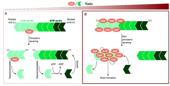
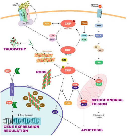
Cofilin is an actin-binding protein that plays a major role in the regulation of actin dynamics, an essential cellular process. This protein has emerged as a crucial molecule for functions of the nervous system including motility and guidance of the neuronal growth cone, dendritic spine organization, axonal branching, and synaptic signalling. Recently, other important functions in cell biology such as apoptosis or the control of mitochondrial function have been attributed to cofilin. Moreover, novel mechanisms of cofilin function regulation have also been described. The activity of cofilin is controlled by complex regulatory mechanisms, with phosphorylation being the most important, since the addition of a phosphate group to cofilin renders it inactive. Due to its participation in a wide variety of key processes in the cell, cofilin has been related to a great variety of pathologies, among which neurodegenerative diseases have attracted great interest.

cofilin neurodegenerative diseases cofilin–actin rods apoptosis mitochondrial fission microtubule instability

1. Introduction

In 1980, Bamburg et al. identified a protein that acted as an actin disassembly factor in chicken brain extracts [1]. Proteins with similar functions were isolated from different organisms and tissues during the following years [2] and were grouped under the ADF/cofilin family, which includes actin-depolymerizing factor (ADF, also known as destrin); cofilin-1, the major ubiquitous isoform in non-muscle tissues; and cofilin-2, the major isoform in differentiated muscle. In this review, we will focus on cofilin-1 (hereinafter called cofilin).
Actin microfilaments (F-actin) are linear polymers composed of globular actin monomers (G-actin), which are polarized with a barbed end, where the addition of available actin monomers bound to ATP occurs, and a pointed end, where actin bound to ADP is released [3]. The addition and release of actin monomers allows actin microfilaments to be a dynamic structure capable of responding to stimuli. These actin dynamics are possible thanks to accessory proteins such as the Arp2/3 complex and formin, involved in actin nucleation, a process of the formation of a complex composed of actin monomers from which an actin filament can elongate; profilin, involved in microfilament elongation; or ADF/cofilin, involved in actin disassembly, although its function has been shown to vary depending on cofilin concentration relative to actin [4], as Figure 1 shows. At low cofilin/actin ratios, cofilin binds to the ADP-actin region of the F-actin, and it severs filaments in a persistent way creating new barbed and pointed ends. Then, cofilin is released from binding to an ADP-actin subunit and both can be recycled. At the same time, the pieces of F-actin generated by severing can nucleate filament growth or enhance depolymerization if ATP-actin is limited [5]. At higher cofilin/actin ratios, many cofilin subunits bind to ADP-actin, induce conformational changes in the microfilament modifying its twist and sever it in a rapid, but not persistent, way as most cofilin is sequestered and bound to actin. Cofilin saturates the fragments, which are stabilized, although they can be depolymerized to generate monomers or may be used to nucleate growth [4]. Under ATP-deficient conditions, stabilized actin bound to cofilin can form rods, slowing the actin dynamics and associated ATP hydrolysis. This mechanism allows the preservation of ATP, but if it persists, the rods can cause neurite degeneration (reviewed below) [6]. When even higher cofilin/actin ratios occur, cofilin can bind actin monomers and nucleate new filaments promoting polymerization [7].
Figure 1. Function of cofilin in actin dynamics according to the cofilin/actin ratio. Cofilin has a pointed end, where actin subunits can be released from the microfilament, and a barbed end, where actin subunits can be added. (A) At low cofilin/actin ratios, cofilin binds to ADP-actin of filaments and severs it, creating new pointed and barbed ends. Then, cofilin binds to the generated fragments and induces depolymerization. Both cofilin and the released actin subunits can be recycled. (B) At higher cofilin/actin ratios, many cofilin subunits bind to ADP-actin, induce conformational changes in the microfilament, and sever it. However, severing is not persistent because cofilin is sequestered and bound to the microfilament. The generated F-actin fragments bound to cofilin are stabilized and can form rods under ATP-deficient conditions.
However, cofilin has been recently related to cellular processes other than actin microfilament disassembly. For example, cofilin could take part in rod formation, apoptosis induction, mitochondrial dynamics, microtubule instability, or even in the regulation of the gene expression [8][9][10]. Alterations in these processes could trigger pathological conditions such as neurodegenerative diseases. Neurons are cells susceptible to cytoskeleton disturbances that can lead to deficiencies in axonal transport. In addition, neurons present a high energetic demand, which, to be satisfied, requires that mitochondrial dynamics works properly [11][12][13]. However, not only neurons are involved in the correct functioning of the nervous system. The role of glial cells is essential by providing support to neurons in relevant processes including synapses, neuronal plasticity, brain fluid transport mediated by the glymphatic system, and the inflammatory response [14][15][16][17]. As in neurons, cofilin could cause alterations in glial functions or morphology changes and, consequently, lead to neurodegenerative diseases [18]. In this regard, great advances have been made to discover how cofilin and its regulatory mechanisms may be involved in neurodegenerative diseases such as Alzheimer’s disease (AD), Huntington’s disease (HD), or Parkinson’s disease (PD) [19][20][21]. In this review, we analyse the main mechanisms through which cofilin, and its regulation could be taking part in the pathology of different neurodegenerative diseases (Figure 2).
Figure 2. Role and regulation of cofilin in neurodegeneration. Cofilin function depends on the phosphorylation of its Ser3 residue, among others. When Ser3 is dephosphorylated by CIN and SSH1, cofilin enters an active state that can be enhanced by Aβ oligomer accumulation. On the other hand, LIMK and TESK can phosphorylate, and thus, inactivate cofilin. Parkin can interfere with cofilin phosphorylation by blocking LIMK function. Under oxidative stress conditions, active and oxidized cofilin can be translocated into the mitochondria, allowing it to take part in mitochondrial fission. This process is mediated by both active cofilin and the DRP1 protein and can be induced by activation of the ROCK pathway. Mitochondrial fission, in turn, triggers the release of the cytochrome C, leading to apoptosis. Apoptosis can also begin with the cofilin/p53 pathway, although inactive cofilin can prevent it activating PLD1. Moreover, active and oxidized cofilin can form cofilin–actin rods. The rods have been found to colocalize in the nucleus with huntingtin. Cofilin can also enter the nucleus together with actin through importin-9. Once in the nucleus, cofilin and actin can regulate gene expression. Finally, cofilin competes with tau for microtubule binding, displacing tau and promoting tauopathies. Aβ, amyloid β; APP, amyloid precursor protein; CIN, chronophin; COF, cofilin; Drp1, dynamin-related protein 1; HTT, huntingtin; LIMK, LIM-domain containing kinase; PLD1, phospholipase D1; PP1/PP2, protein phosphatase 1 and 2; PRKN, parkin; SSH1, slingshot; TESK, testis-specific kinase.

2. Cofilin Regulation and Its Implication in Neurodegenerative Diseases

Cofilin can undergo post-translational modifications that regulate its function and, among these, phosphorylation stands out. Phospho-regulation of cofilin is mediated by signalling pathways in response to extracellular signals. This signal transduction is very complex, and numerous studies have been carried out to characterize it [22]. Cofilin is inactive when it is phosphorylated on its Ser3 residue by LIM-domain containing kinases 1 and 2 (LIMK1 y LIMK2) and testis-specific kinases 1 and 2 (TESK1 y TESK2). LIMK1 is in turn activated by the small GTPase Rho and its downstream Rho-associated protein kinase (ROCK) [23], and TESK1 is activated downstream of an integrin signal. Both contribute to stabilize the actin cytoskeleton. The phosphatases chronophin (CIN) and slingshot (SSH1) remove the phosphate of Ser3, activating cofilin and accelerating the dynamics of the actin cytoskeleton [24]. However, in addition, the regulation of cofilin has to be spatiotemporally controlled to obtain a precise actin cytoskeleton reorganization. While cofilin is expressed in almost all tissues and cells, distinctly different expression patterns of LIMKs, TESKs, and SSHs exist [22].
Moreover, other regulatory mechanisms of cofilin are known. Dephosphorylation by CIN and SSH is enhanced when ATP levels are low and reactive oxygen species (ROS) high [25][26], demonstrating how oxidative stress plays a role in the regulation of cofilin activity. Moreover, cofilin function is also directly regulated by the oxidation of its cysteine residues [27]. Oxidized cofilin can induce the formation of an intramolecular disulfide bridge in vivo and the loss of its Ser3 phosphorylation site. This activated and oxidized cofilin is responsible for inducing the apoptosis cascade through its translocation to the mitochondria [28]. In addition, cofilin oxidation can induce not only intramolecular but also intermolecular disulfide bonds between cofilin molecules. In these cases, cofilin–actin rod formation is enhanced, thus preventing the cell from entering apoptosis [29]. Apart from phosphorylation and oxidation, other regulatory mechanisms have been proposed. For example, phosphatidylinositol 4,5-bisphosphate (PIP2) is a membrane phospholipid that inhibits cofilin activity by competing with actin at the binding site [30]. High-pH conditions could also regulate the activity of cofilin by increasing it [31]. Cofilin phosphorylation at its Y68 residue induces cofilin ubiquitination and degradation through the proteasome, adding another regulation mechanism [32].
According to current evidence, it seems clear that cofilin is involved in neurodegenerative processes, as we will describe in the following paragraphs, and more specifically, the regulation of cofilin function has also been associated with different disease states. However, much remains to be known about the complex regulatory mechanisms of this protein and how they are affected in different neurodegenerative diseases.
Mutations in the Parkin 2 (PARK) gene are the main cause of autosomal recessive parkinsonism, a type of early onset familial PD, and also seem to play a role in sporadic PD. Parkin is a ubiquitin E3 ligase that ubiquitinates dysfunctional proteins for their degradation in the proteasome. The loss of parkin activity leads to the accumulation of parkin substrates. These have been proposed as a cause for toxicity and neurodegeneration through mechanisms that are not completely understood [33]. Previous studies had already shown that parkin was associated with actin filaments by colocalizing with them, so it was suggested that parkin could have a role in actin stabilization [34]. According to this finding, Lim et al. observed that parkin interacts with and ubiquitinates LIMK1 in the human dopaminergic neuroblastoma cell line BE(2)-M17 but not in the human embryonic kidney-derived cell line HEK293, indicating a tissue-specific regulation. As a result, since LIMK1 phosphorylates cofilin, LIMK1 ubiquitination by PARK2 reduces the level of inactive cofilin [35]. Overall, parkin modulates the level of phosphocofilin by negatively regulating LIMK1 activity in a cell-type-dependent way, which can help understand how cofilin contributes to familial PD.
Friedreich’s ataxia (FRDA) is an inherited peripheral neuropathy characterized by an early loss of neurons in the dorsal root ganglia, among other clinical symptoms caused by frataxin deficiency. Cytoskeletal abnormalities have been proposed to contribute to dying-back neurodegeneration in FRDA [24]. In fact, at the molecular level, the F-actin:G-actin ratio has been shown to be increased in sensory neurons of FRDA mice, suggesting an alteration of the normal turnover of actin filaments. This disturbance could be the cause of the changes observed in the morphology of the growth cones of FRDA mouse neurons [36]. The authors observed a hyperactivation of cofilin in the dorsal nerve roots of the FRDA mouse model compared to controls, which can be partially explained by the increased levels of CIN [36]. Altogether, the dysregulation of cofilin might explain the reduced neurite growth and alterations of cytoskeleton, suggesting a role in FRDA neuropathy.
Regulation of active cofilin could also be involved in AD. A significant reduction of total SSH1 phosphatase in AD brains could be the cause of the increased cofilin inactivation observed in human samples. γ-Secretase is an enzyme that makes the second cleavage to amyloid precursor protein (APP) to give rise to the amyloid-β (Aβ) peptides, whose pathological accumulation is related to AD. This enzyme has been discovered to takes part in promoting actin/cofilin-related pathology. In this sense, γ-secretase inhibitors promote cofilin activation by increasing SSH1 levels in mouse primary cortical neurons [37]. However, other authors point to the excessive cofilin activation, and not the opposite, as the cause of AD pathogenesis. For example, Kim et al. suggest that the binding of Aβ oligomers to the leukocyte immunoglobulin-like receptor B2 (LilrB2) present in human brain enhances cofilin signalling, which leads to synapse elimination [38].
The contribution of active cofilin to AD pathogenesis does not seem to be limited only to its levels since its location also has an influence. Cofilin is translocated to the spine upon long-term potentiation induction and promotes the assembly of F-actin, essential for spine expansion and to potentiate synaptic transmission. This activity-dependent plasticity phenomenon is altered in the hippocampus of AD patients, with cofilin having a potential role in this defect since it is aberrantly localized in neuron spines [39]. The authors showed how downregulation of cyclase-associated protein 2 (CAP2), by synergizing with cofilin to accelerate depolymerization of the pointed end of actin filaments, is able to control cofilin synaptic availability in long-term potentiation processes that, as it is known, are extremely important for stabilizing memory.
It seems clear that the regulation of cofilin is crucial for the activity and function of the protein, so that alterations in the cofilin regulation mechanisms trigger pathological processes such as neurodegenerative diseases.

3. New Functions for Cofilin in Neurodegenerative Diseases

Chua et al. have shown that after staurosporine-induced apoptosis, active cofilin can translocate from the cytoplasm to the mitochondria, leading to the opening of the mitochondrial permeability transition pore and the release of cytochrome C, which is the first step in the apoptotic cell death cascade [28]. This suggests that cofilin has an important function during the initiation phase of apoptosis. In this regard, the translocation of cofilin to mitochondria has been also linked to mitochondrial fission, a process in which cofilin would participate regulating the function of the dynamin-related protein 1 (Drp1) [40]. Moreover, cofilin could also be translocated to the nucleus due to its nuclear localization sequence [41]. In this context, cofilin has been suggested to participate in actin import to the nucleus, where actin can regulate gene expression [42]. Even inactive cofilin, a form that was thought to have no function, has been shown to have stimulatory effects on phospholipase D1 (PLD1), an enzyme involved in a wide variety of cellular responses, including calcium mobilization, superoxide production, endocytosis, vesicle trafficking, etc. [43].
All these processes, highly related to neurodegeneration, point towards cofilin as a key protein involved in many neurological diseases, but its role in the pathophysiology of these diseases still remains to be elucidated.

4. Future Perspectives

Cofilin has a main role in actin dynamics and, therefore, in cytoskeletal homeostasis. However, current evidence on its new functions seems to show that cofilin also contributes to degenerative processes through the formation of cofilin–actin rods that impair axonal transport and promotion of neuronal cell death, or by changes in mitochondrial dynamics and in the endoplasmic reticulum–mitochondria (ER–mitochondria) connection and communication. However, much remains to be known about cofilin, starting with its complex regulatory mechanisms. This is a key piece to understand how the modulation of cofilin activity and levels could be a critical point in neurodegenerative diseases to modify its natural history. At this point, the question arises as to whether cofilin could be used as a neurodegenerative progression biomarker. In this sense, cofilin 2 expression was demonstrated to be significantly increased in the serum of Alzheimer’s disease patients, and it performed well as a diagnostic and non-invasive biomarker with high sensitivity and specificity [44].
Thus, the biggest challenge is to determine whether cofilin is a potential target for neurodegenerative diseases, which can open new possibilities for drug development with the purpose, for example, of preventing mitochondrial alteration and axonopathy in neurodegenerative diseases. Another therapeutic approach could be to design drugs that target cofilin-regulatory proteins since many of the pathological processes related to cofilin are due to alterations in its regulation. Anyway, the recent advances in the use of high-throughput screening and computer-aided drug design can help in the search of treatments for neurodegenerative diseases [45].

References

  1. Bamburg, J.R.; Harris, H.E.; Weeds, A.G. Partial purification and characterization of an actin depolymerizing factor from brain. FEBS Lett. 1980, 121, 178–182.
  2. Bamburg, J.R. Proteins of the ADF/Cofilin Family: Essential regulators of actin dynamics. Annu. Rev. Cell Dev. Biol. 1999, 15, 185–230.
  3. Blanchoin, L.; Boujemaa-Paterski, R.; Sykes, C.; Plastino, J. Actin dynamics, architecture and mechanics in cell motility. Physiol. Rev. 2014, 94, 235–263.
  4. Bamburg, J.R.; Bernstein, B.W. Roles of ADF/cofilin in actin polymerization and beyond. F1000 Biol. Rep. 2010, 2, 62.
  5. Wioland, H.; Guichard, B.; Senju, Y.; Myram, S.; Lappalainen, P.; Jégou, A.; Romet-Lemonne, G. ADF/Cofilin accelerates actin dynamics by severing filaments and promoting their depolymerization at both ends. Curr. Biol. 2017, 27, 1956–1967.e7.
  6. Bernstein, B.W.; Chen, H.; Boyle, J.A.; Bamburg, J.R. Formation of actin-ADF/cofilin rods transiently retards decline of mitochondrial potential and ATP in stressed neurons. Am. J. Physiol. Cell Physiol. 2006, 291, C828–C839.
  7. Andrianantoandro, E.; Pollard, T.D. Mechanism of actin filament turnover by severing and nucleation at different concentrations of ADF/Cofilin. Mol. Cell 2006, 24, 13–23.
  8. Kanellos, G.; Frame, M.C. Cellular functions of the ADF/cofilin family at a glance. J. Cell Sci. 2016, 129, 3211–3218.
  9. Li, G.-B.; Zhang, H.-W.; Fu, R.-Q.; Hu, X.-Y.; Liu, L.; Li, Y.-N.; Liu, Y.-X.; Liu, X.; Hu, J.-J.; Deng, Q.; et al. Mitochondrial fission and mitophagy depend on cofilin-mediated actin depolymerization activity at the mitochondrial fission site. Oncogene 2018, 37, 1485–1502.
  10. Woo, J.-A.A.; Liu, T.; Fang, C.C.; Cazzaro, S.; Kee, T.; LePochat, P.; Yrigoin, K.; Penn, C.; Zhao, X.; Wang, X.; et al. Activated cofilin exacerbates tau pathology by impairing tau-mediated microtubule dynamics. Commun. Biol. 2019, 2, 112.
  11. Itoh, K.; Nakamura, K.; Iijima, M.; Sesaki, H. Mitochondrial dynamics in neurodegeneration. Trends Cell Biol. 2013, 23, 64–71.
  12. Gibson, R.M. Does apoptosis have a role in neurodegeneration? BMJ 2001, 322, 1539–1540.
  13. McMurray, C.T. Neurodegeneration: Diseases of the cytoskeleton? Cell Death Differ. 2000, 7, 861–865.
  14. Mestre, H.; Hablitz, L.M.; Xavier, A.L.R.; Feng, W.; Zou, W.; Pu, T.; Monai, H.; Murlidharan, G.; Castellanos Rivera, R.M.; Simon, M.J.; et al. Aquaporin-4-dependent glymphatic solute transport in the rodent brain. eLife 2018, 7, e40070.
  15. Bordone, M.P.; Salman, M.M.; Titus, H.E.; Amini, E.; Andersen, J.V.; Chakraborti, B.; Diuba, A.V.; Dubouskaya, T.G.; Ehrke, E.; Espindola de Freitas, A.; et al. The energetic brain—A review from students to students. J. Neurochem. 2019, 151, 139–165.
  16. Verkhratsky, A.; Nedergaard, M. Physiology of astroglia. Physiol. Rev. 2018, 98, 239–389.
  17. Araque, A.; Parpura, V.; Sanzgiri, R.P.; Haydon, P.G. Tripartite synapses: Glia, the unacknowledged partner. Trends Neurosci. 1999, 22, 208–215.
  18. Alsegiani, A.; Shah, Z. The role of cofilin in age-related neuroinflammation. Neural Regen. Res. 2020, 15, 1451–1459.
  19. Kang, D.E.; Woo, J.A. Cofilin, a master node regulating cytoskeletal pathogenesis in Alzheimer’s Disease. J. Alzheimer’s Dis. 2019, 72, S131–S144.
  20. Zhang, Q.; Hu, C.; Huang, J.; Liu, W.; Lai, W.; Leng, F.; Tang, Q.; Liu, Y.; Wang, Q.; Zhou, M.; et al. ROCK1 induces dopaminergic nerve cell apoptosis via the activation of Drp1-mediated aberrant mitochondrial fission in Parkinson’s Disease. Exp. Mol. Med. 2019, 51, 1–13.
  21. Munsie, L.; Caron, N.; Atwal, R.S.; Marsden, I.; Wild, E.J.; Bamburg, J.R.; Tabrizi, S.J.; Truant, R. Mutant huntingtin causes defective actin remodeling during stress: Defining a new role for transglutaminase 2 in neurodegenerative disease. Hum. Mol. Genet. 2011, 20, 1937–1951.
  22. Ohashi, K. Roles of cofilin in development and its mechanisms of regulation. Dev. Growth Differ. 2015, 57, 275–290.
  23. Ohashi, K.; Nagata, K.; Maekawa, M.; Ishizaki, T.; Narumiya, S.; Mizuno, K. Rho-associated Kinase ROCK Activates LIM-kinase 1 by Phosphorylation at Threonine 508 within the Activation Loop. J. Biol. Chem. 2000, 275, 3577–3582.
  24. Van Troys, M.; Huyck, L.; Leyman, S.; Dhaese, S.; Vandekerkhove, J.; Ampe, C. Ins and outs of ADF/cofilin activity and regulation. Eur. J. Cell Biol. 2008, 87, 649–667.
  25. Huang, T.Y.; Minamide, L.S.; Bamburg, J.R.; Bokoch, G.M. Chronophin mediates an ATP-sensing mechanism for cofilin dephosphorylation and neuronal cofilin-actin rod formation. Dev. Cell 2008, 15, 691–703.
  26. Kim, J.-S.; Huang, T.Y.; Bokoch, G.M. Reactive oxygen species regulate a slingshot-cofilin activation pathway. Mol. Biol. Cell 2009, 20, 2650–2660.
  27. Cameron, J.M.; Gabrielsen, M.; Chim, Y.H.; Munro, J.; McGhee, E.J.; Sumpton, D.; Eaton, P.; Anderson, K.I.; Yin, H.; Olson, M.F. Polarized cell motility induces hydrogen peroxide to inhibit cofilin via cysteine oxidation. Curr. Biol. 2015, 25, 1520–1525.
  28. Chua, B.T.; Volbracht, C.; Tan, K.O.; Li, R.; Yu, V.C.; Li, P. Mitochondrial translocation of cofilin is an early step in apoptosis induction. Nat. Cell Biol. 2003, 5, 1083–1089.
  29. Bernstein, B.W.; Shaw, A.E.; Minamide, L.S.; Pak, C.W.; Bamburg, J.R. Incorporation of cofilin into rods depends on disulfide intermolecular bonds: Implications for actin regulation and neurodegenerative disease. J. Neurosci. 2012, 32, 6670–6681.
  30. Zhao, H.; Hakala, M.; Lappalainen, P. ADF/cofilin binds phosphoinositides in a multivalent manner to act as a PIP(2)-density sensor. Biophys. J. 2010, 98, 2327–2336.
  31. Yeoh, S.; Pope, B.; Mannherz, H.G.; Weeds, A. Determining the differences in actin binding by human ADF and cofilin. J. Mol. Biol. 2002, 315, 911–925.
  32. Yoo, Y.; Ho, H.J.; Wang, C.; Guan, J.-L. Tyrosine phosphorylation of cofilin at Y68 by v-Src leads to its degradation through ubiquitin–proteasome pathway. Oncogene 2010, 29, 263–272.
  33. Dawson, T.M.; Dawson, V.L. The role of parkin in familial and sporadic Parkinson’s disease. Mov. Disord. 2010, 25 (Suppl. 1), S32–S39.
  34. Huynh, D.P.; Scoles, D.R.; Ho, T.H.; Del Bigio, M.R.; Pulst, S.-M. Parkin is associated with actin filaments in neuronal and nonneural cells. Ann. Neurol. 2000, 48, 737–744.
  35. Lim, M.K.; Kawamura, T.; Ohsawa, Y.; Ohtsubo, M.; Asakawa, S.; Takayanagi, A.; Shimizu, N. Parkin interacts with LIM Kinase 1 and reduces its cofilin-phosphorylation activity via ubiquitination. Exp. Cell Res. 2007, 313, 2858–2874.
  36. Muñoz-Lasso, D.C.; Mollá, B.; Calap-Quintana, P.; García-Giménez, J.L.; Pallardo, F.V.; Palau, F.; Gonzalez-Cabo, P. Cofilin dysregulation alters actin turnover in frataxin-deficient neurons. Sci. Rep. 2020, 10, 5207.
  37. Barone, E.; Mosser, S.; Fraering, P.C. Inactivation of brain Cofilin-1 by age, Alzheimer’s disease and γ-secretase. Biochim. Biophys. Acta Mol. Basis Dis. 2014, 1842, 2500–2509.
  38. Kim, T.; Vidal, G.S.; Djurisic, M.; William, C.M.; Birnbaum, M.E.; Garcia, K.C.; Hyman, B.T.; Shatz, C.J. Human LilrB2 is a β-amyloid receptor and its murine homolog PirB regulates synaptic plasticity in an Alzheimer’s model. Science 2013, 341, 1399–1404.
  39. Pelucchi, S.; Vandermeulen, L.; Pizzamiglio, L.; Aksan, B.; Yan, J.; Konietzny, A.; Bonomi, E.; Borroni, B.; Padovani, A.; Rust, M.B.; et al. Cyclase-associated protein 2 dimerization regulates cofilin in synaptic plasticity and Alzheimer’s disease. Brain Commun. 2020, 2.
  40. Rehklau, K.; Hoffmann, L.; Gurniak, C.B.; Ott, M.; Witke, W.; Scorrano, L.; Culmsee, C.; Rust, M.B. Cofilin1-dependent actin dynamics control DRP1-mediated mitochondrial fission. Cell Death Dis. 2017, 8, e3063.
  41. Kanellos, G.; Zhou, J.; Patel, H.; Ridgway, R.A.; Huels, D.; Gurniak, C.B.; Sandilands, E.; Carragher, N.O.; Sansom, O.J.; Witke, W.; et al. ADF and cofilin1 control actin stress fibers, nuclear Integrity and cell survival. Cell Rep. 2015, 13, 1949–1964.
  42. Dopie, J.; Skarp, K.-P.; Kaisa Rajakylä, E.; Tanhuanpää, K.; Vartiainen, M.K. Active maintenance of nuclear actin by importin 9 supports transcription. Proc. Natl. Acad. Sci. USA 2012, 109, E544–E552.
  43. Han, L.; Stope, M.B.; de Jesús, M.L.; Oude Weernink, P.A.; Urban, M.; Wieland, T.; Rosskopf, D.; Mizuno, K.; Jakobs, K.H.; Schmidt, M. Direct stimulation of receptor-controlled phospholipase D1 by phospho-cofilin. EMBO J. 2007, 26, 4189–4202.
  44. Sun, Y.; Liang, L.; Dong, M.; Li, C.; Liu, Z.; Gao, H. Cofilin 2 in serum as a novel biomarker for Alzheimer’s disease in Han Chinese. Front. Aging Neurosci. 2019.
  45. Aldewachi, H.; Al-Zidan, R.N.; Conner, M.T.; Salman, M.M. High-Throughput screening platforms in the discovery of novel drugs for neurodegenerative diseases. Bioengineering 2021, 8, 30.
More
Information
Subjects: Clinical Neurology
Contributor MDPI registered users' name will be linked to their SciProfiles pages. To register with us, please refer to https://encyclopedia.pub/register :
View Times: 639
Revisions: 2 times (View History)
Update Date: 29 Jul 2021
1000/1000
Video Production Service