Submitted Successfully!
To reward your contribution, here is a gift for you: A free trial for our video production service.
Thank you for your contribution! You can also upload a video entry or images related to this topic.
Version Summary Created by Modification Content Size Created at Operation
1 + 1533 word(s) 1533 2021-06-24 10:40:44 |
2 Reference formatted Meta information modification 1533 2021-06-24 12:30:46 | |
3 Content change based on AB's suggestion + 694 word(s) 2227 2021-06-30 05:20:31 |

Video Upload Options

Do you have a full video?

Confirm

Are you sure to Delete?
Cite
If you have any further questions, please contact Encyclopedia Editorial Office.
Tsai, C.; Cheng, N. Glyoxal-Crosslinked Chitosan/Gelatin Hydrogel. Encyclopedia. Available online: https://encyclopedia.pub/entry/11250 (accessed on 18 April 2024).
Tsai C, Cheng N. Glyoxal-Crosslinked Chitosan/Gelatin Hydrogel. Encyclopedia. Available at: https://encyclopedia.pub/entry/11250. Accessed April 18, 2024.
Tsai, Ching-Cheng, Naichen Cheng. "Glyoxal-Crosslinked Chitosan/Gelatin Hydrogel" Encyclopedia, https://encyclopedia.pub/entry/11250 (accessed April 18, 2024).
Tsai, C., & Cheng, N. (2021, June 24). Glyoxal-Crosslinked Chitosan/Gelatin Hydrogel. In Encyclopedia. https://encyclopedia.pub/entry/11250
Tsai, Ching-Cheng and Naichen Cheng. "Glyoxal-Crosslinked Chitosan/Gelatin Hydrogel." Encyclopedia. Web. 24 June, 2021.
Glyoxal-Crosslinked Chitosan/Gelatin Hydrogel
Edit

The clinical application of human platelet lysate (HPL) holds promise for tissue regeneration, and the development of an efficient vehicle for its delivery is desired. Chitosan-based hydrogels are potential candidates, but they often exhibit weak mechanical properties. In this study, a chitosan/gelatin (CS-GE) hydrogel crosslinked by glyoxal was fabricated for sustained release of HPL. After an ideal glyoxal concentration was determined, we further characterized the crosslinked CS-GE hydrogels encapsulated with different amounts of HPL. The HPL-incorporated hydrogel was shown to significantly promote the proliferation of Hs68 cells and the migration of HUVECs. Moreover, the release pattern of transforming growth factor-β1 (TGF-β1) and platelet-derived growth factor-BB (PDGF-BB) from hydrogel was examined in vitro, demonstrating a sustained release profile of the growth factors. Finally, the chick chorioallantoic membrane assay revealed that HPL encapsulation in the hydrogel significantly stimulated angiogenesis in ovo. These results demonstrate the great potential of the crosslinked CS-GE hydrogel to serve as an effective delivery system for HPL to promote tissue regeneration. 

human platelet lysate chitosan gelatin glyoxal hydrogel angiogenesis tissue regeneration

1. Introduction

Abundant growth factors and cytokines are stored in platelet granules, and they can be naturally released by thrombin activation and clotting or artificially released by freeze/thaw-mediated platelet lysis, sonication or chemical treatment [1]. Clinically, these platelet concentrates have been widely applied in a variety of fields, such as the treatment of symptomatic oral lichen planus [2], lateral epicondylitis [3] and medication-related osteonecrosis of the jaws [4]. Although the efficacy of platelet concentrates in promoting wound healing and tissue regeneration is at the center of a recent academic debate, different formats of platelet concentrates have been prepared to test their regenerative effects.
Human platelet lysate (HPL), a biological material derived from platelet concentrates, can be manufactured as a cost-effective and standardized cell culture supplement [5][6]. HPL is known to contain abundant mitogenic growth factors, including vascular endothelial growth factor (VEGF), basic fibroblast growth factor (bFGF), epidermal growth factor (EGF), platelet-derived growth factors (PDGF) and transforming growth factor (TGF) [7][8]. These growth factors not only exert a positive effect on angiogenesis and post-ischemic vascular remodeling but also play a critical role in tissue inflammation and regeneration [9][10][11]. Therefore, platelet lysates have demonstrated the advantages of replacing fetal bovine serum (FBS) for culturing various types of cells, including stem cells, fibroblasts and endothelial cells [1][12]. The use of HPL in cell culture is desired for clinical use by circumventing the risks of xeno-immunization against bovine antigens and the transmission of zoonotic diseases [13][14]. Moreover, the effect of HPL on directly promoting angiogenesis and tissue regeneration has been demonstrated [15][16], so HPL holds great promise for treating various tissue defects.
The development of efficient biomaterials to assist HPL delivery for tissue regeneration remains a challenge. To facilitate a sustained release of its contents in the defect site, HPL could be blended with biomaterials, adsorbed onto scaffolds or used as a gel to mediate neo-angiogenesis [17][18][19]. However, the currently available options are not ideal, and a novel hydrogel system is desired to allow sustained HPL delivery. Hydrogel is a web of crosslinked polymer filmaments soaked by an aqueous solution. It is usually soft and flexible with good fluid-retaining capability and biocompatibility, so it has been widely applied in various biomedical fields as a kind of drug delivery system [20][21]. Chitosan-based thermosensitive hydrogels, fabricated with a chitosan/β-glycerophosphate (β-GP) system, have been successfully applied in drug release studies [22], and they were further applied to deliver cells for tissue engineering purposes [23][24]. β-GP acts as a proton receiver from the positively charged chitosan at elevated temperature, thereby inducing chitosan gelation [25]. Moreover, our previous study demonstrated superior biocompatibility when gelatin was blended into this chitosan/β-GP system [26].
When gelatin and chitosan are mixed for hydrogel fabrication, they form poly-electrolytic complexes in different gelated states, and the structure mimics the natural components of the extracellular matrix [27][28]. However, the mechanical property of the composite hydrogel is still not ideal. To achieve better mechanical strength and a controlled release pattern, glyoxal was selected as a crosslinker to reinforce the hydrogel in this study [29][30]. Glyoxal is the smallest dialdehyde with high water solubility, and it exhibits lower cytotoxicity when compared to most chemical crosslinkers [29][30][31]. It was expected to crosslink the available amine groups on chitosan and gelatin chains to form the network [32], rendering improved mechanical properties of the chitosan/gelatin (CS-GE) hydrogel. In this study, we aimed to fabricate a CS-GE hydrogel crosslinked by glyoxal for the sustained release of HPL. An ideal concentration of glyoxal was selected, resulting in negligible cytotoxicity with a reasonable level of mechanical strength. These characteristics can facilitate efficient HPL delivery from the crosslinked CS-GE hydrogel to promote angiogenesis and regeneration in damaged tissues.

2. Results

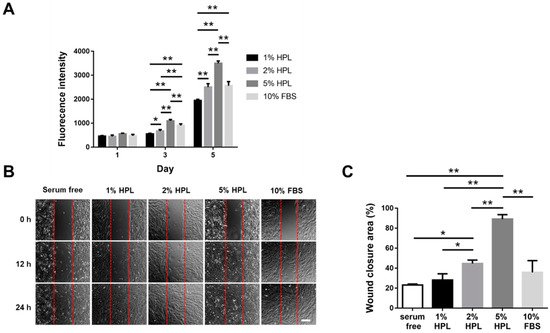
2.1. HPL Enhanced the Proliferation and Migration of Hs68 Fibroblasts

In the result of the Alamar Blue assay, a higher concentration of HPL in the medium resulted in higher fluorescence intensity, suggesting a higher proliferative activity. The 5% HPL group even demonstrated a significantly higher value of fluorescence intensity on day 5 than the 10% FBS group (3498.0 ± 96.0 vs. 2560.0 ± 175.0, p < 0.01; Figure 1A). Moreover, fibroblast migration was assessed to determine whether HPL can accelerate the process of wound healing. After Hs68 cells were cultured in different conditions for 12 h, the images showed that the cell coverage area in the experimental group, 5% HPL, was higher than that in the other experimental groups. At 24 h, the gap was nearly filled by the migrant cells in the 5% HPL group, while some cell-free zones could still be observed in other groups (Figure 1B). ImageJ software was used to quantify the wound closure area at the time point of 12 h, revealing a significantly higher wound closure area in the 5% HPL group relative to the 10% FBS group (89.2% ± 7.6% vs. 35.8% ± 20.3%, p < 0.01; Figure 1C).
Figure 1. The influence of different concentrations of HPL on the proliferation and migration of Hs68 fibroblasts. (A) Alamar Blue assay revealed the proliferative activity of Hs68 fibroblasts incubated in different concentrations of HPL or 10% FBS. (B) The representative images of in vitro cell migration at 0, 12 and 24 h (scale bar = 200 μm). (C) Quantification of the wound closure percentage of Hs68 fibroblasts at 12 h. Serum-free was used as the control group. (* p < 0.05, ** p < 0.01).

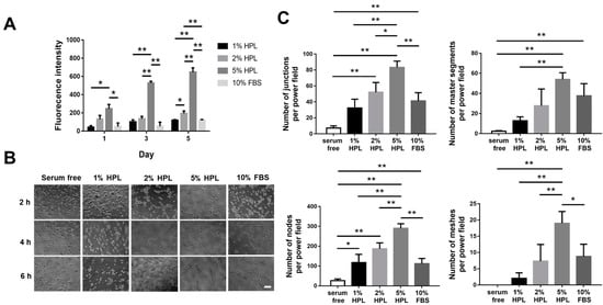
2.2. HPL Enhanced the Proliferation and Tube Formation of Endothelial Cells

In the proliferation assay of human umbilical vein endothelial cells (HUVECs), the 5% HPL group on day 5 displayed a significantly higher proliferative activity than the other groups (fluorescence intensity: 5% HPL: 647.7 ± 46.8 vs. 1% HPL: 119.0 ± 8.5, 2% HPL: 194.7 ± 26.5 and 10% FBS: 118.7 ± 8.5, p < 0.01, respectively; Figure 2A). Furthermore, we examined the tube formation of HUVECs under different culture conditions at different time points (Figure 2B). ImageJ software was used to quantify the cell images at 4 h. The 5% HPL group exhibited significantly more tube-like structures than the 10% FBS group (83.3 ± 7.8 vs. 41.3 ± 10.2 for junctions, p < 0.01; 290.0 ± 23.1 vs. 111.8 ± 26.4 for nodes, p < 0.01; 19.0 ± 3.6 vs. 17.4 ± 2.9 for meshes, p < 0.05; Figure 2C).
Figure 2. The influence of different concentrations of HPL on the proliferation and tube formation of HUVECs. (A) Alamar Blue assay revealed the proliferative activity of HUVECs incubated in different concentrations of HPL or 10% FBS. (B) The representative images of endothelial cell tube formation under different conditions at 2, 4 and 6 h (scale bar = 100 μm). (C) The number of junctions, master segments, nodes and meshes at 4 h was compared among different groups. Serum-free was used as the control group. (* p < 0.05, ** p < 0.01).

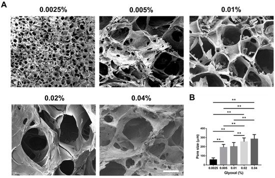
2.3. Scanning Electron Micrographs and Pore Size Measurement

Scanning electron microscope (SEM) images showed the morphology of lyophilized hydrogel crosslinked with different concentrations of glyoxal, and all groups presented with high porous structures (Figure 3A). For the pore size measurement, using a higher concentration of glyoxal resulted in a larger pore size of the hydrogel. The 0.0025% glyoxal group possessed a significantly smaller pore size than the other groups (0.0025% glyoxal: 57.8 ± 19.7 μm vs. 0.005% glyoxal: 193.9 ± 31.3 μm, 0.01% glyoxal: 202.4 ± 43.2 μm, 0.02% glyoxal: 254.8 ± 41.8 μm and 0.04% glyoxal: 285.2 ± 47.6 μm, p < 0.01, respectively; Figure 3B).
Figure 3. The porosity of CS-GE hydrogels crosslinked with different glyoxal concentrations. (A) The representative scanning electron micrographs of lyophilized CS-GE hydrogels crosslinked with different concentrations of glyoxal (scale bar = 200 μm). (B) Pore size measurement revealed significant differences among different CS-GE hydrogels. (** p < 0.01).

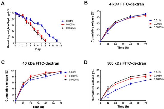
2.4. Enzymatic Degradation Assay

Although a higher glyoxal concentration for hydrogel crosslinking resulted in stronger mechanical properties, the shorter gelation time (less than 20 s) and higher viscosity may render it difficult to apply or inject. Hence, we chose the 0.0025%, 0.005% and 0.01% glyoxal groups to further conduct the hydrogel degradation and drug release experiments.
To evaluate the degradation process of the CS-GE hydrogels under collagenase treatment, the remaining weight of hydrogels was measured. The 0.0025% and 0.005% glyoxal-crosslinked hydrogels were degraded within 8 days, and it took 12 days to fully degrade the 0.01% glyoxal-crosslinked hydrogel (Figure 4A).
Figure 4. The degradation of CS-GE hydrogels and the release pattern of encapsulated FITC-dextrans. (A) In vitro degradation test of CS-GE hydrogel crosslinked with different concentrations of glyoxal. The release percentage of (B) 4 kDa, (C) 40 kDa and (D) 500 kDa FITC-dextrans from CS-GE hydrogels crosslinked with different glyoxal concentrations.

3. Current Insights

HPL contains lots of well-known platelet growth factors, which have been considered to play an essential role in cell-mediated angiogenesis and skin fibroblast behavior [33][34]. Previous publications have shown that HPL can be utilized for the restoration of corneal lesions [35], diabetic ulcers [36] and other soft tissue pathologies [37]. Apart from the soft tissue, HPL can be successfully applied for the local treatment of osteoarthritis, which is attributed to the growth factors released from platelet granules after intra-articular activation [38]. Mesenchymal stem cells can also be combined with HPL for bone regeneration [39]. However, HPL-based treatment may be limited because of the poor retention of HPL in the defect sites. In the present study, HPL-incorporated CS-GE hydrogel was successfully fabricated, and its mechanical property was improved by crosslinking with glyoxal. The HPL-incorporated CS-GE hydrogel not only significantly promoted the proliferative activity of Hs68 fibroblasts and the migration of HUVECs in vitro but also enhanced angiogenesis in the in ovo CAM model. Hence, the novel crosslinked hydrogel system allows sustained release of HPL-derived growth factors to promote tissue regeneration.
Chitosan-based hydrogels are often combined with β-GP to constitute a thermally responsive hydrogel system. Due to the electrostatic attractions between the chitosan protonated amine groups (-NH3+) and negatively charged phosphate molecules of β-GP (-HPO4 or -PO42−), the hydrogel system composed of chitosan, gelatin and β-GP can stay in a liquid state without aggregation at low temperature [40][41]. By adding β-GP as a weak base, the pH of the solution is escalated close to physiological pH so that the available -NH2 on the chitosan and gelatin chains may increase [42]. Therefore, the more available reaction sites can be bound by adding glyoxal as a crosslinker to form a robust network structure. Consequently, a higher glyoxal concentration led to greater mechanical strength and a shorter gelation time (less than ~70 s) in this study.
Recently, glyoxal has been used as an alternative dialdehyde crosslinker in various biomedical applications [42][43]. Although a higher amount of glyoxal was reported to trigger oxidative stress, thereby inducing cytotoxicity [44], we demonstrated no significant cytotoxicity if the concentration of glyoxal was less than 0.01% in cell culture. Compared to other chemical crosslinkers for CS-GE hydrogels, glyoxal exhibits several advantages for its application in tissue engineering and drug delivery. Genipin-crosslinked chitosan-based hydrogels were shown to have good biocompatibility and low cytotoxicity, but the gelation time was at least 1 h, rendering it not suitable for clinical use [45][46]. Moreover, an anti-angiogenesis effect of genipin was reported to decrease the rate of wound healing [47]. Glutaraldehyde can crosslink chitosan chains to form hydrogels within 1 h; however, it is considered toxic for the respiratory tract, eyes and skin [48].
Our previous study manifested blending gelatin in the chitosan-based hydrogel, resulting in a more porous architecture after in vivo application due to the degradation of gelatin [26]. This porous structure can ensure that more growth factors are released rather than stuck in the hydrogel. By modifying this design in the present study, the chemical crosslinker, glyoxal, was used to increase the mechanical strength of the hydrogel, rendering a more sustained release of the encapsulated HPL. Lim et al. fabricated HPL-incorporated hydroxybutyl chitosan hydrogels, which displayed a cumulative release of more than 80% of PDGF-BB and TGF-β1 on day 7 during in vitro degradation [49]. Compared to their result, the cumulative releases of PDGF-BB and TGF-β1 in our experiment were less than 80%, demonstrating a more sustained release of HPL.
In this study, a significantly higher cell proliferative activity was noted as the HPL concentration increased in the medium. In line with the previous publications suggesting that HPL is more suitable than FBS as culture supplements [50][51], our results revealed that 5% HPL was significantly more effective than 10% FBS to promote Hs68 fibroblast proliferation and migration. Li et al. have elucidated the mechanism of human dermal fibroblast migration driven by PDGF-BB, indicating the important role of PDGF in regulating the fibroblast behavior [52]. HPL exhibits abundant PDGF-BB, indicating the great potential of HPL in fibroblast-driven soft tissue regeneration, such as wound healing.
Tissue regeneration normally needs the formation of vessels to transfer essential materials including oxygen and nutrition. Therefore, capillary angiogenesis within the damaged tissue is critical during the tissue repair process. Angiogenesis is a coordinated process, involving endothelial cellular proliferation, migration and association in tubular structures [53]. In this work, HUVECs cultured in the medium containing 5% HPL revealed significantly more tube-like structures relative to the 10% FBS group, and the HPL-incorporated hydrogel significantly induced the migration of HUVECs. Moreover, the CAM assay was employed for further evaluation because it can be applied to study in ovo both angiogenesis and anti-angiogenesis in response to different materials [54].

References

  1. Burnouf, T.; Strunk, D.; Koh, M.B.; Schallmoser, K. Human platelet lysate: Replacing fetal bovine serum as a gold standard for human cell propagation? Biomaterials 2016, 76, 371–387.
  2. Bennardo, F.; Liborio, F.; Barone, S.; Antonelli, A.; Buffone, C.; Fortunato, L.; Giudice, A. Efficacy of platelet-rich fibrin compared with triamcinolone acetonide as injective therapy in the treatment of symptomatic oral lichen planus: A pilot study. Clin. Oral Investig. 2021, 25, 3747–3755.
  3. Mi, B.; Liu, G.; Zhou, W.; Lv, H.; Liu, Y.; Wu, Q.; Liu, J. Platelet rich plasma versus steroid on lateral epicondylitis: Meta-analysis of randomized clinical trials. Physician Sportsmed. 2017, 45, 97–104.
  4. Bennardo, F.; Bennardo, L.; Del Duca, E.; Patruno, C.; Fortunato, L.; Giudice, A.; Nisticò, S.P. Autologous platelet-rich fibrin injections in the management of facial cutaneous sinus tracts secondary to medication-related osteonecrosis of the jaw. Dermatol. Ther. 2020, 33, e13334.
  5. Hemeda, H.; Giebel, B.; Wagner, W. Evaluation of human platelet lysate versus fetal bovine serum for culture of mesenchymal stromal cells. Cytotherapy 2014, 16, 170–180.
  6. Shanskii, Y.D.; Sergeeva, N.; Sviridova, I.; Kirakozov, M.; Kirsanova, V.; Akhmedova, S.; Antokhin, A.; Chissov, V. Human platelet lysate as a promising growth-stimulating additive for culturing of stem cells and other cell types. Bull. Exp. Biol. Med. 2013, 156, 146–151.
  7. Chen, M.-S.; Wang, T.-J.; Lin, H.-C.; Burnouf, T. Four types of human platelet lysate, including one virally inactivated by solvent-detergent, can be used to propagate Wharton jelly mesenchymal stromal cells. New Biotechnol. 2019, 49, 151–160.
  8. Chen, L.W.; Huang, C.-J.; Tu, W.-H.; Lu, C.-J.; Sun, Y.-C.; Lin, S.-Y.; Chen, W.-L. The corneal epitheliotrophic abilities of lyophilized powder form human platelet lysates. PLoS ONE 2018, 13, e0194345.
  9. Brill, A.; Dashevsky, O.; Rivo, J.; Gozal, Y.; Varon, D. Platelet-derived microparticles induce angiogenesis and stimulate post-ischemic revascularization. Cardiovasc. Res. 2005, 67, 30–38.
  10. Varon, D.; Hayon, Y.; Dashevsky, O.; Shai, E. Involvement of platelet derived microparticles in tumor metastasis and tissue regeneration. Thromb. Res. 2012, 130, S98–S99.
  11. Boilard, E.; Nigrovic, P.A.; Larabee, K.; Watts, G.F.; Coblyn, J.S.; Weinblatt, M.E.; Massarotti, E.M.; Remold-O’Donnell, E.; Farndale, R.W.; Ware, J. Platelets amplify inflammation in arthritis via collagen-dependent microparticle production. Science 2010, 327, 580–583.
  12. Cheng, N.-C.; Tu, Y.-K.; Lee, N.-H.; Young, T.-H. Influence of Human Platelet Lysate on Extracellular Matrix Deposition and Cellular Characteristics in Adipose-Derived Stem Cell Sheets. Front. Cell Dev. Biol. 2020, 8, 1122.
  13. Schallmoser, K.; Strunk, D. Generation of a pool of human platelet lysate and efficient use in cell culture. In Basic Cell Culture Protocols; Springer: Berlin/Heidelberg, Germany, 2013; pp. 349–362.
  14. Barro, L.; Nebie, O.; Chen, M.-S.; Wu, Y.-W.; Koh, M.B.; Knutson, F.; Watanabe, N.; Takahara, M.; Burnouf, T. Nanofiltration of growth media supplemented with human platelet lysates for pathogen-safe xeno-free expansion of mesenchymal stromal cells. Cytotherapy 2020, 22, 458–472.
  15. Hayon, Y.; Dashevsky, O.; Shai, E.; Varon, D.; Leker, R.R. Platelet lysates stimulate angiogenesis, neurogenesis and neuroprotection after stroke. Thromb. Haemost. 2013, 110, 323–330.
  16. Oliveira, S.M.; Pirraco, R.P.; Marques, A.P.; Santo, V.E.; Gomes, M.E.; Reis, R.L.; Mano, J.F. Platelet lysate-based pro-angiogenic nanocoatings. Acta Biomater. 2016, 32, 129–137.
  17. Babo, P.S.; Pires, R.L.; Santos, L.; Franco, A.; Rodrigues, F.; Leonor, I.; Reis, R.L.; Gomes, M.E. Platelet lysate-loaded photocrosslinkable hyaluronic acid hydrogels for periodontal endogenous regenerative technology. ACS Biomater. Sci. Eng. 2017, 3, 1359–1369.
  18. Robinson, S.T.; Douglas, A.M.; Chadid, T.; Kuo, K.; Rajabalan, A.; Li, H.; Copland, I.B.; Barker, T.H.; Galipeau, J.; Brewster, L.P. A novel platelet lysate hydrogel for endothelial cell and mesenchymal stem cell-directed neovascularization. Acta Biomater. 2016, 36, 86–98.
  19. Wang, C.; Xiao, R.; Cao, Y.-L.; Yin, H.-Y. Evaluation of human platelet lysate and dimethyl sulfoxide as cryoprotectants for the cryopreservation of human adipose-derived stem cells. Biochem. Biophys. Res. Commun. 2017, 491, 198–203.
  20. Ruel-Gariepy, E.; Leroux, J.-C. In situ-forming hydrogels—Review of temperature-sensitive systems. Eur. J. Pharm. Biopharm. 2004, 58, 409–426.
  21. Ta, H.T.; Dass, C.R.; Dunstan, D.E. Injectable chitosan hydrogels for localised cancer therapy. J. Control Release 2008, 126, 205–216.
  22. Viyoch, J.; Sudedmark, T.; Srema, W.; Suwongkrua, W. Development of hydrogel patch for controlled release of alpha-hydroxy acid contained in tamarind fruit pulp extract. Int. J. Cosmet. Sci. 2005, 27, 89–99.
  23. Richardson, S.M.; Hughes, N.; Hunt, J.A.; Freemont, A.J.; Hoyland, J.A. Human mesenchymal stem cell differentiation to NP-like cells in chitosan–glycerophosphate hydrogels. Biomaterials 2008, 29, 85–93.
  24. Roughley, P.; Hoemann, C.; DesRosiers, E.; Mwale, F.; Antoniou, J.; Alini, M. The potential of chitosan-based gels containing intervertebral disc cells for nucleus pulposus supplementation. Biomaterials 2006, 27, 388–396.
  25. Ahmadi, R.; de Bruijn, J.D. Biocompatibility and gelation of chitosan–glycerol phosphate hydrogels. J. Biomed. Mater. Res. Part A 2008, 86, 824–832.
  26. Cheng, N.-C.; Lin, W.-J.; Ling, T.-Y.; Young, T.-H. Sustained release of adipose-derived stem cells by thermosensitive chitosan/gelatin hydrogel for therapeutic angiogenesis. Acta Biomater. 2017, 51, 258–267.
  27. Kim, S.; Nimni, M.E.; Yang, Z.; Han, B. Chitosan/gelatin–based films crosslinked by proanthocyanidin. J. Biomed. Mater. Res. Part B 2005, 75, 442–450.
  28. Cheng, N.C.; Chang, H.H.; Tu, Y.K.; Young, T.H. Efficient transfer of human adipose-derived stem cells by chitosan/gelatin blend films. J. Biomed. Mater. Res. Part B 2012, 100, 1369–1377.
  29. Dessì, M.; Borzacchiello, A.; Mohamed, T.H.; Abdel-Fattah, W.I.; Ambrosio, L. Novel biomimetic thermosensitive β-tricalcium phosphate/chitosan-based hydrogels for bone tissue engineering. J. Biomed. Mater. Res. Part A 2013, 101, 2984–2993.
  30. Wang, L.; Stegemann, J.P. Glyoxal crosslinking of cell-seeded chitosan/collagen hydrogels for bone regeneration. Acta Biomater. 2011, 7, 2410–2417.
  31. Hoemann, C.D.; Chenite, A.; Sun, J.; Hurtig, M.; Serreqi, A.; Lu, Z.; Rossomacha, E.; Buschmann, M.D. Cytocompatible gel formation of chitosan-glycerol phosphate solutions supplemented with hydroxyl ethyl cellulose is due to the presence of glyoxal. J. Biomed. Mater. Res. A 2007, 83, 521–529.
  32. Jawad, A.H.; Norrahma, S.S.A.; Hameed, B.; Ismail, K. Chitosan-glyoxal film as a superior adsorbent for two structurally different reactive and acid dyes: Adsorption and mechanism study. Int. J. Biol. Macromol. 2019, 135, 569–581.
  33. Boccardo, S.; Gaudiello, E.; Melly, L.; Cerino, G.; Ricci, D.; Martin, I.; Eckstein, F.; Banfi, A.; Marsano, A. Engineered mesenchymal cell-based patches as controlled VEGF delivery systems to induce extrinsic angiogenesis. Acta Biomater. 2016, 42, 127–135.
  34. Ranzato, E.; Mazzucco, L.; Patrone, M.; Burlando, B. Platelet lysate promotes in vitro wound scratch closure of human dermal fibroblasts: Different roles of cell calcium, P38, ERK and PI3K/AKT. J. Cell. Mol. Med. 2009, 13, 2030–2038.
  35. Griffith, G.L.; Holt, A.W.; Eriksson, E.; Johnson, A.J.; McDaniel, J.S. Human platelet lysate delivered via an ocular wound chamber for the treatment of corneal epithelial injuries. Exp. Eye Res. 2021, 206, 108493.
  36. Jafar, H.; Hasan, M.; Al-Hattab, D.; Saleh, M.; Ameereh, L.A.; Khraisha, S.; Younes, N.; Awidi, A. Platelet lysate promotes the healing of long-standing diabetic foot ulcers: A report of two cases and in vitro study. Heliyon 2020, 6, e03929.
  37. Rozman, P.; Bolta, Z. Use of platelet growth factors in treating wounds and soft-tissue injuries. Acta Dermatovenerol. Alp. Panon. Et Adriat. 2007, 16, 156.
  38. Al-Ajlouni, J.; Awidi, A.; Samara, O.; Al-Najar, M.; Tarwanah, E.; Saleh, M.; Awidi, M.; Hassan, F.A.; Samih, M.; Bener, A. Safety and efficacy of autologous intra-articular platelet lysates in early and intermediate knee osteoarthrosis in humans: A prospective open-label study. Clin. J. Sport Med. 2015, 25, 524–528.
  39. Altaie, A.; Owston, H.; Jones, E. Use of platelet lysate for bone regeneration—Are we ready for clinical translation? World J. Stem Cells 2016, 8, 47.
  40. G Tahrir, F.; Ganji, F.; M Ahooyi, T. Injectable thermosensitive chitosan/glycerophosphate-based hydrogels for tissue engineering and drug delivery applications: A review. Recent Pat. Drug Deliv. Formul. 2015, 9, 107–120.
  41. Kim, S.; Nishimoto, S.K.; Bumgardner, J.D.; Haggard, W.O.; Gaber, M.W.; Yang, Y. A chitosan/β-glycerophosphate thermo-sensitive gel for the delivery of ellagic acid for the treatment of brain cancer. Biomaterials 2010, 31, 4157–4166.
  42. Jarry, C.; Chaput, C.; Chenite, A.; Renaud, M.A.; Buschmann, M.; Leroux, J.C. Effects of steam sterilization on thermogelling chitosan-based gels. J. Biomed. Mater. Res. 2001, 58, 127–135.
  43. Vaz, C.M.; De Graaf, L.A.; Reis, R.L.; Cunha, A.M. In vitro degradation behaviour of biodegradable soy plastics: Effects of crosslinking with glyoxal and thermal treatment. Polym. Degrad. Stab. 2003, 81, 65–74.
  44. Shangari, N.; O’Brien, P.J. The cytotoxic mechanism of glyoxal involves oxidative stress. Biochem. Pharmacol. 2004, 68, 1433–1442.
  45. Jin, J.; Song, M.; Hourston, D. Novel chitosan-based films cross-linked by genipin with improved physical properties. Biomacromolecules 2004, 5, 162–168.
  46. Chiono, V.; Pulieri, E.; Vozzi, G.; Ciardelli, G.; Ahluwalia, A.; Giusti, P. Genipin-crosslinked chitosan/gelatin blends for biomedical applications. J. Mater. Sci. Mater. Med. 2008, 19, 889–898.
  47. Koo, H.-J.; Song, Y.S.; Kim, H.-J.; Lee, Y.-H.; Hong, S.-M.; Kim, S.-J.; Kim, B.-C.; Jin, C.; Lim, C.-J.; Park, E.-H. Antiinflammatory effects of genipin, an active principle of gardenia. Eur. J. Pharmacol. 2004, 495, 201–208.
  48. Gough, J.E.; Scotchford, C.A.; Downes, S. Cytotoxicity of glutaraldehyde crosslinked collagen/poly (vinyl alcohol) films is by the mechanism of apoptosis. J. Biomed. Mater. Res. 2002, 61, 121–130.
  49. Lim, T.; Tang, Q.; Zhu, Z.; Wei, X.; Zhang, C. Sustained release of human platelet lysate growth factors by thermosensitive hydroxybutyl chitosan hydrogel promotes skin wound healing in rats. J. Biomed. Mater. Res. Part. A 2020, 108, 2111–2122.
  50. Schallmoser, K.; Bartmann, C.; Rohde, E.; Reinisch, A.; Kashofer, K.; Stadelmeyer, E.; Drexler, C.; Lanzer, G.; Linkesch, W.; Strunk, D. Human platelet lysate can replace fetal bovine serum for clinical-scale expansion of functional mesenchymal stromal cells. Transfusion 2007, 47, 1436–1446.
  51. Mojica-Henshaw, M.P.; Morris, J.; Kelley, L.; Pierce, J.; Boyer, M.; Reems, J.-A. Serum-converted platelet lysate can substitute for fetal bovine serum in human mesenchymal stromal cell cultures. Cytotherapy 2013, 15, 1458–1468.
  52. Li, W.; Fan, J.; Chen, M.; Guan, S.; Sawcer, D.; Bokoch, G.M.; Woodley, D.T. Mechanism of human dermal fibroblast migration driven by type I collagen and platelet-derived growth factor-BB. Mol. Biol. Cell 2004, 15, 294–309.
  53. Gonzalez, A.C.; Costa, T.F.; Andrade, Z.D.; Medrado, A.R. Wound healing—A literature review. An. Bras. Dermatol. 2016, 91, 614–620.
  54. Ribatti, D.; Annese, T.; Tamma, R. The use of the chick embryo CAM assay in the study of angiogenic activiy of biomaterials. Microvasc. Res. 2020, 131, 104026.
More
Information
Contributors MDPI registered users' name will be linked to their SciProfiles pages. To register with us, please refer to https://encyclopedia.pub/register : ,
View Times: 657
Revisions: 3 times (View History)
Update Date: 23 Nov 2021
1000/1000