 +1 credit
| Version | Summary | Created by | Modification | Content Size | Created at | Operation |
|---|---|---|---|---|---|---|
| 1 | Gaskon Ibarretxe | + 1925 word(s) | 1925 | 2021-04-08 09:50:48 | | | |
| 2 | Bruce Ren | -21 word(s) | 1904 | 2021-04-15 03:26:19 | | |
Video Upload Options
Human dental pulp stem cells (hDPSCs) are some of the most promising stem cell types for regenerative therapies given their high ability differentiate to neural and vascular lineage cells, their growth in animal serum-free media, their secretion of neuroprotective factors and extracellular vesicles, their high resistance to hypoxia/ischemia, their immunomodulatory properties, and their wide range of possibilities to be used in autologous grafts.
1. Introduction: Neural and Mesenchymal Stem Cells, and Neuroregenerative Cell Therapies
Neuroregenerative therapies have always been a priority for health research in developed countries due the overwhelming social, economic and dependency burdens suffered by both the affected patients and their close relatives [1][2][3]. The nervous system in humans possesses a very limited capacity of self-repair in the event of injury. This is the reason why nerve lesions caused by trauma or neurodegenerative diseases often result in highly disabling irreversible conditions and chronic dependency [4][5][6]. Unlike other cells of the body, dead or damaged neurons cannot be easily replaced. Neurogenesis takes place in the developing brain, but it declines in the adulthood. Moreover, neuroinflammation and gliosis following neural trauma or disease make the tissue refractory to the rooting and establishment of new neural connections [7][8][9][10].
Neurogenesis is driven by specific multipotent stem cells known as neural stem cells (NSCs), which give rise to both neurons and glial cells. Embryonic NSCs are the first stone paving the way to brain (re)generation and impairments in their correct function are associated to several types of cortical malformations, with dramatic outcomes on the life of an individual [11]. Although the neurogenic capacity is significantly decreased in the adult mammalian brain, two regions bear NSCs during the entire lifetime of different species. The lateral ventricles and the hippocampus harbor adult NSCs capable of triggering a staggered process that ends up in the integration of a newborn neuron into the adult neuronal circuitry [12][13][14]. In humans, adult newborn neurons have been detected in the lateral ventricles, yet in highly infrequent basis [15][16][17]. In the hippocampus, controversial results over the existence of adult neurogenesis have been recently reported and the topic remains currently under hot debate [16][18]. Regardless, there is agreement about the presence of these neurogenic niches during the first years of life in young infants [16][17][19][20], a time period when NSC dysfunctions might play an important role in the development and chronification of neurodegenerative diseases [21]. Indeed, studies in rodents have shown that adult NSCs undergo changes when facing neurodegenerative challenges, including morphological and functional abnormalities that lead to disruption of neurogenesis and contribute to the detrimental tissue environment in these regions [22][23][24]. The existence of NSCs that could react in pathological conditions has been also suggested in other areas, like the cerebral cortex and spinal cord [25][26]. The amygdala, although with rare adult neurogenic events in normal conditions, has also been postulated to bear quiescent NSCs that could get activated upon peripheric lesions, at least in primates [27].
The scarce numbers of adult NSCs or their aberrant alterations in neurodegenerative diseases suppose the lack of a reliable endogenous mechanism to replenish neurons in the event of their loss. Not in vain, stem cells offer the potential to reduce deleterious signaling and improve traumatic lesions [28][29], and also to slowdown the progression of devastating neurodegenerative diseases such as Huntington’s (HD) [30][31], Parkinson’s (PD) [32][33] or Alzheimer’s disease (AD) [34][35][36]. The idea of using stem cells to treat neurodegenerative diseases was proposed very long ago, obtaining valuable and abundant data using fetal human tissue [31][37][38] or induced pluripotent stem cells (IPSCs) [39][40]. However, these methods raise both safety and ethical concerns that are still under intense debate [41][42][43][44][45][46]. The main practical problems are the security, the very low yields of extraction, and the troublesome conditions of intervention on premature infants to harvest human NSCs [34]. Stem cells from the spinal cord of 8-week fetuses have been tested in clinical trials for chronic spinal cord injury [47][48]. However, it is unlikely that these strategies will ever reach a widespread implementation, due to the scarcity of embryo donors and the associated ethical issues. IPSCs have been proposed to overcome ethical concerns about the use of human embryos. IPSCs can be very efficently differentiated to neurons and glial cells [49][50][51] and they have been proposed as a promising alternative for cell therapy in brain and spinal cord injury [39], as a tool to screen genetic bases of neurological diseases [52] or even as an approach to correct alterations in chronic neurodegenerative diseases [53]. However, a better understanding is still required to regulate the generation of specific neuronal and glial populations in a balanced and coordinated manner. Furthermore, the increased risk of cancer related to the use of IPSCs is regarded as a major drawback for autologous personalized neuroregenerative therapy [49].
In view of the limitations of endogenous NSCs and pluripotent stem cells, it is not surprising that the research community has turned its eyes to alternative sources of stem cells with neural regeneration capacity. Of all of them, the ones that seem the best positioned are mesenchymal stem cells (MSCs), which can give rise to all cell lineages of both proper and specialized connective tissues, including bone, cartilage, muscle and adipose cells, among others. MSCs can be extracted from different sources like the bone marrow, the adipose tissue and the umbilical cord [54]. Human dental pulp stem cells (hDPSCs) had also been traditionally included within MSCs, because they fulfill the standard criteria of plastic-adherent growth, multilineage differentiation and a characteristic molecular marker expression as defined by the presence of CD73, CD90 and CD105, which are required by International Society of Cell Therapy to classify a cell type as a MSC [55], in addition to other accessory markers like CD27, CD29, CD44, CD146, CD166, CD271 and STRO-1 [56][57]. This marker expression profile can be found in hDPSCs, as well as in MSCs from many other tissue sources [58]. On the contrary, MSCs and hDPSCs do not express CD45 (hematopoietic marker), CD14 (monocyte or macrophage marker), CD19 (B cell marker) or MHC-II (major histocompatibility complex II) surface molecules [56][57][59]. MSCs have raised substantial hopes for the clinical management of neural lesions, with very promising results [60].
2. DPSCs as Neural Crest Stem Cells. The postnatal DPSC Niche
Compared to other MSC sources, dental stem cells and DPSCs were discovered relatively late. It was not until the advent of the XXI century that the presence of stem cells in the postnatal human dental pulp was reported [61]. Later on, many other related MSCs with similar characteristics to DPSCs were discovered in other nearby dental tissues, like the periodontal ligament [62], the gingival mucosa [63], the apical papilla [64], the dental follicle [65] or the dental pulp of childhood deciduous teeth [66], among others. Over the last decades hDPSCs have remained as the most extensively studied type of dental stem cells, because of their ease of extraction, absence of ethical issues and relative abundance as biological waste from dental clinics.
It soon became apparent that hDPSCs possessed multilineage differentiation potential that exceeded that of conventional MSCs [59][67][68]. Contrary to other MSC sources, the dental pulp tissue is generated by the neural crest, a structure formed at the fusing borders of the neural tube during development. Cells of the neural crest undergo an epithelial-mesenchymal transition and acquire migratory ability, thus extensively colonizing other parts of the embryo, including the pharyngeal arches which are the precursors of craniomaxillofacial organs and tissues (Figure 1). These neural crest stem cells can subsequently commit to generate the diverse tissues of the oral cavity. Some neural crest stem cells differentiate to MSCs to generate oral neural crest-derived mesenchyme (i.e., ectomesenchyme) which will then give rise to the different oral connective tissues, cartilages, muscles and bones. However, these neural crest stem cells are also the precursors of the cranial peripheral nerve system [69]. Perhaps due to their shared origin, it is not uncommon to observe that hDPSCs express a varied repertoire of both neural progenitor and mature cell markers, even in normal standard (control) culture conditions [59][70][71][72]. Some of the neural markers that are most prominently expressed by hDPSC cultures include Neuroectodermal Stem Cell Intermediate filament marker (Nestin), β-3 tubulin (Tuj1), neurotrophin receptors, and neurofilaments [71][72]. As it can be expected from neural crest-related cells, hDPSC cultures also express neural crest markers like Snail, Slug, Sox10 and HNK1, and also pluripotency-related core factors like Oct4, Sox2 and Nanog [71]. Importantly, the expression of neural crest and pluripotency markers by hDPSCs and the corresponding stemness of these cells can be stimulated by the transient activation of specific signaling pathways, in the absence of any genetic modification [71][73]. Some of these treatments (e.g., Wnt/β-catenin signaling stimulation) have been shown to substantially modify the epigenetic and metabolic footprint of hDPSCs [74][75]. Of particular importance to cell therapy, hDPSCs have a great adaptability to adverse metabolic conditions [76], and can also secrete a large variety of neuroprotective and immunomodulatory factors (discussed in Section 6 of this manuscript) which make them a very attractive tool to promote neural regeneration.
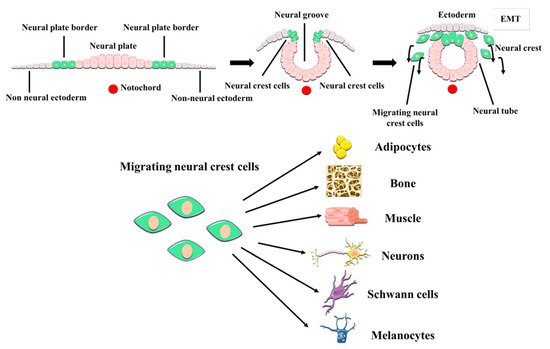
Figure 1. Embryonic origin and multilineage differentiation of human dental pulp stem cells (hDPSCs). hDPSCs derive from neural crest stem cells that generate craniomaxillofacial tissues, including the dental pulp. During development, neural crest cells undergo an epithelial-mesenchymal transition (EMT) and migrate out of the neural tube, to give rise to both mesenchymal and non-mesenchymal cell lineages of the oral cavity, like the neurons and glial cells of the craniofacial PNS. hDPSCs show many neural crest characteristics such as their expression of neural crest markers, and a higher differentiation potential to neural cell lineages than other MSCs.
The relationship between DPSCs and nerve tissue goes beyond a shared embryonic origin. The postnatal niches of DPSCs are the neurovascular bundles of the dental pulp, which are intricate associations of peripheral nerves and blood vessels which cross together through the apical foramen to irrigate and innervate the dental pulp tissue (Figure 2). The niche of DPSCs is thus extraordinarily rich in nerve fibers and blood vessels, in contrast to the surrounding loose connective tissue of the rest of the dental pulp [77]. Immunolabeling of STRO-1 expressing cells revealed that hDPSCs were located precisely within these perivascular niches [78]. Later on, lineage tracing experiments in a murine model revealed that stem cells of the dental pulp were neural crest marker-expressing cells associated with neurovascular bundles [79]. Thus, the same DPSC population could ultimately give rise to both non-mesenchymal (e.g., Schwann cells) and mesenchymal (e.g., odontoblasts) lineage-derived cells [79]. Probably because of their close association with neurovascular structures of the dental pulp, hDPSCs also have a very high capacity to generate vascular cells like endothelia and pericytes [80][81]. This higher ability to differentiate to vascular cells comes at the expense of a reduced capacity for commitment to other more conventional types of mesenchymal-related cell lineages, like chondrocyte differentiation [82]. Thus, according to this model, DPSCs would be located at a similar level to neural crest cells within the stem cell hierarchy, with a higher capacity to generate neural and vascular cells than conventional MSCs. Interestingly, DPSCs also exhibit natural niche homing characteristics when they are engrafted in vivo, as they tend to spontaneously migrate to nerves and vascular structures of the host organism after transplantation [80][83].
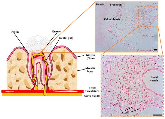
Figure 2. Cellular niche of hDPSCs in postnatal teeth. hDPSCs are harbored in neurovascular bundles of the dental pulp of mature teeth, containing a high concentration of nerve fibers and blood vessels. These neurovascular niches contain many myelinated axons (shown in cross-section) and a higher cellular density than in the rest of the dental pulp tissue. Scale bars: 50 µm.
References
- Yang, W.; Hamilton, J.L.; Kopil, C.; Beck, J.C.; Tanner, C.M.; Albin, R.L.; Ray Dorsey, E.; Dahodwala, N.; Cintina, I.; Hogan, P.; et al. Current and Projected Future Economic Burden of Parkinson’s Disease in the U.S. NPJ Park. Dis. 2020, 6, 1–9.
- Glidden, A.M.; Luebbe, E.A.; Elson, M.J.; Goldenthal, S.B.; Snyder, C.W.; Zizzi, C.E.; Dorsey, E.R.; Heatwole, C.R. Patient-Reported Impact of Symptoms in Huntington Disease: PRISM-HD. Neurology 2020, 94, e2045–e2053.
- Carnahan, J.L.; Judge, K.S.; Daggy, J.K.; Slaven, J.E.; Coleman, N.; Fortier, E.L.; Suelzer, C.; Fowler, N.R. Supporting Caregivers of Veterans with Alzheimer’s Disease and Traumatic Brain Injury: Study Protocol for a Randomized Controlled Trial. Trials 2020, 21, 340.
- Sivanandam, T.M.; Thakur, M.K. Traumatic Brain Injury: A Risk Factor for Alzheimer’s Disease. Neurosci. Biobehav. Rev. 2012, 36, 1376–1381.
- Smith, D.H.; Johnson, V.E.; Stewart, W. Chronic Neuropathologies of Single and Repetitive TBI: Substrates of Dementia? Nat. Rev. Neurol. 2013, 9, 211–221.
- Chiti, F.; Dobson, C.M. Protein Misfolding, Amyloid Formation, and Human Disease: A Summary of Progress Over the Last Decade. Annu. Rev. Biochem. 2017, 86, 27–68.
- Nieto-Sampedro, M. Neurite Outgrowth Inhibitors in Gliotic Tissue. Adv. Exp. Med. Biol. 1999, 468, 207–224.
- Bovolenta, P.; Fernaud-Espinosa, I.; Méndez-Otero, R.; Nieto-Sampedro, M. Neurite Outgrowth Inhibitor of Gliotic Brain Tissue. Mode of Action and Cellular Localization, Studied with Specific Monoclonal Antibodies. Eur. J. Neurosci. 1997, 9, 977–989.
- Arvidsson, A.; Collin, T.; Kirik, D.; Kokaia, Z.; Lindvall, O. Neuronal Replacement from Endogenous Precursors in the Adult Brain after Stroke. Nat. Med. 2002, 8, 963–970.
- Solleiro-Villavicencio, H.; Rivas-Arancibia, S. Effect of Chronic Oxidative Stress on Neuroinflammatory Response Mediated by CD4+T Cells in Neurodegenerative Diseases. Front. Cell. Neurosci. 2018, 12.
- Subramanian, L.; Calcagnotto, M.E.; Paredes, M.F. Cortical Malformations: Lessons in Human Brain Development. Front. Cell. Neurosci. 2020, 13, 576.
- Doetsch, F.; García-Verdugo, J.M.; Alvarez-Buylla, A. Cellular Composition and Three-Dimensional Organization of the Subventricular Germinal Zone in the Adult Mammalian Brain. J. Neurosci. 1997, 17, 5046–5061.
- Seri, B.; García-Verdugo, J.M.; McEwen, B.S.; Alvarez-Buylla, A. Astrocytes Give Rise to New Neurons in the Adult Mammalian Hippocampus. J. Neurosci. 2001, 21, 7153–7160.
- Van Praag, H.; Schinder, A.F.; Christie, B.R.; Toni, N.; Palmer, T.D.; Gage, F.H. Functional Neurogenesis in the Adult Hippocampus. Nature 2002, 415, 1030–1034.
- Wang, C.; You, Y.; Qi, D.; Zhou, X.; Wang, L.; Wei, S.; Zhang, Z.; Huang, W.; Liu, Z.; Liu, F.; et al. Human and Monkey Striatal Interneurons Are Derived from the Medial Ganglionic Eminence But Not from the Adult Subventricular Zone. J. Neurosci. 2014, 34, 10906–10923.
- Sorrells, S.F.; Paredes, M.F.; Cebrian-Silla, A.; Sandoval, K.; Qi, D.; Kelley, K.W.; James, D.; Mayer, S.; Chang, J.; Auguste, K.I.; et al. Human Hippocampal Neurogenesis Drops Sharply in Children to Undetectable Levels in Adults. Nature 2018, 555, 377–381.
- Sanai, N.; Nguyen, T.; Ihrie, R.A.; Mirzadeh, Z.; Tsai, H.-H.; Wong, M.; Gupta, N.; Berger, M.S.; Huang, E.; Garcia-Verdugo, J.-M.; et al. Corridors of Migrating Neurons in the Human Brain and Their Decline during Infancy. Nature 2011, 478, 382–386.
- Moreno-Jiménez, E.P.; Flor-García, M.; Terreros-Roncal, J.; Rábano, A.; Cafini, F.; Pallas-Bazarra, N.; Ávila, J.; Llorens-Martín, M. Adult Hippocampal Neurogenesis Is Abundant in Neurologically Healthy Subjects and Drops Sharply in Patients with Alzheimer’s Disease. Nat. Med. 2019, 25, 554–560.
- Cipriani, S.; Journiac, N.; Nardelli, J.; Verney, C.; Delezoide, A.-L.; Guimiot, F.; Gressens, P.; Adle-Biassette, H. Dynamic Expression Patterns of Progenitor and Neuron Layer Markers in the Developing Human Dentate Gyrus and Fimbria. Cereb. Cortex 2017.
- Knoth, R.; Singec, I.; Ditter, M.; Pantazis, G.; Capetian, P.; Meyer, R.P.; Horvat, V.; Volk, B.; Kempermann, G. Murine Features of Neurogenesis in the Human Hippocampus across the Lifespan from 0 to 100 Years. PLoS ONE 2010, 5, e8809.
- Blümcke, I.; Schewe, J.-C.; Normann, S.; Brüstle, O.; Schramm, J.; Elger, C.E.; Wiestler, O.D. Increase of Nestin-Immunoreactive Neural Precursor Cells in the Dentate Gyrus of Pediatric Patients with Early-Onset Temporal Lobe Epilepsy: Increased Hippocampal Neurogenesis in Human TLE. Hippocampus 2001, 11, 311–321.
- Sierra, A.; Martín-Suárez, S.; Valcárcel-Martín, R.; Pascual-Brazo, J.; Aelvoet, S.-A.; Abiega, O.; Deudero, J.J.; Brewster, A.L.; Bernales, I.; Anderson, A.E.; et al. Neuronal Hyperactivity Accelerates Depletion of Neural Stem Cells and Impairs Hippocampal Neurogenesis. Cell Stem Cell 2015, 16, 488–503.
- Jure, I.; De Nicola, A.F.; Encinas, J.M.; Labombarda, F. Spinal Cord Injury Leads to Hippocampal Glial Alterations and Neural Stem Cell Inactivation. Cell. Mol. Neurobiol. 2020.
- Faiz, M.; Sachewsky, N.; Gascón, S.; Bang, K.W.A.; Morshead, C.M.; Nagy, A. Adult Neural Stem Cells from the Subventricular Zone Give Rise to Reactive Astrocytes in the Cortex after Stroke. Cell Stem Cell 2015, 17, 624–634.
- Llorens-Bobadilla, E.; Chell, J.M.; Le Merre, P.; Wu, Y.; Zamboni, M.; Bergenstråhle, J.; Stenudd, M.; Sopova, E.; Lundeberg, J.; Shupliakov, O.; et al. A Latent Lineage Potential in Resident Neural Stem Cells Enables Spinal Cord Repair. Science 2020, 370.
- Zamboni, M.; Llorens-Bobadilla, E.; Magnusson, J.P.; Frisén, J. A Widespread Neurogenic Potential of Neocortical Astrocytes Is Induced by Injury. Cell Stem Cell 2020, 27, 605–617.e5.
- Chareyron, L.J.; Amaral, D.G.; Lavenex, P. Selective Lesion of the Hippocampus Increases the Differentiation of Immature Neurons in the Monkey Amygdala. Proc. Natl. Acad. Sci. USA 2016, 113, 14420–14425.
- Kawabori, M.; Weintraub, A.H.; Imai, H.; Zinkevych, L.; McAllister, P.; Steinberg, G.K.; Frishberg, B.M.; Yasuhara, T.; Chen, J.W.; Cramer, S.C.; et al. Cell Therapy for Chronic TBI: Interim Analysis of the Randomized Controlled STEMTRA Trial. Neurology 2021.
- Dewan, S.; Schimmel, S.; Borlongan, C.V. Treating Childhood Traumatic Brain Injury with Autologous Stem Cell Therapy. Expert Opin. Biol. Ther. 2018, 18, 515–524.
- Bachoud-Lévi, A.-C.; Massart, R.; Rosser, A. Cell Therapy in Huntington’s Disease: Taking Stock of Past Studies to Move the Field Forward. Stem Cells Dayt. Ohio 2021, 39, 144–155.
- Bachoud-Lévi, A.-C. From Open to Large-Scale Randomized Cell Transplantation Trials in Huntington’s Disease: Lessons from the Multicentric Intracerebral Grafting in Huntington’s Disease Trial (MIG-HD) and Previous Pilot Studies. Prog. Brain Res. 2017, 230, 227–261.
- Parmar, M. Towards Stem Cell Based Therapies for Parkinson’s Disease. Dev. Camb. Engl. 2018, 145.
- Kim, T.W.; Koo, S.Y.; Studer, L. Pluripotent Stem Cell Therapies for Parkinson Disease: Present Challenges and Future Opportunities. Front. Cell Dev. Biol. 2020, 8, 729.
- Fernández-Muñoz, B.; Rosell-Valle, C.; Ferrari, D.; Alba-Amador, J.; Montiel, M.Á.; Campos-Cuerva, R.; Lopez-Navas, L.; Muñoz-Escalona, M.; Martín-López, M.; Profico, D.C.; et al. Retrieval of Germinal Zone Neural Stem Cells from the Cerebrospinal Fluid of Premature Infants with Intraventricular Hemorrhage. Stem Cells Transl. Med. 2020, 9, 1085–1101.
- Si, Z.; Wang, X. Stem Cells Therapies in Alzheimer’s Disease: Applications for Disease Modeling. J. Pharmacol. Exp. Ther. 2021.
- Vasic, V.; Barth, K.; Schmidt, M.H.H. Neurodegeneration and Neuro-Regeneration-Alzheimer’s Disease and Stem Cell Therapy. Int. J. Mol. Sci. 2019, 20, 4272.
- Bachoud-Lévi, A.; Bourdet, C.; Brugières, P.; Nguyen, J.P.; Grandmougin, T.; Haddad, B.; Jény, R.; Bartolomeo, P.; Boissé, M.F.; Barba, G.D.; et al. Safety and Tolerability Assessment of Intrastriatal Neural Allografts in Five Patients with Huntington’s Disease. Exp. Neurol. 2000, 161, 194–202.
- Gaura, V.; Bachoud-Lévi, A.-C.; Ribeiro, M.-J.; Nguyen, J.-P.; Frouin, V.; Baudic, S.; Brugières, P.; Mangin, J.-F.; Boissé, M.-F.; Palfi, S.; et al. Striatal Neural Grafting Improves Cortical Metabolism in Huntington’s Disease Patients. Brain J. Neurol. 2004, 127, 65–72.
- Li, Y.; Shen, P.-P.; Wang, B. Induced Pluripotent Stem Cell Technology for Spinal Cord Injury: A Promising Alternative Therapy. Neural Regen. Res. 2021, 16, 1500–1509.
- Morizane, A. Cell therapy for Parkinson’s disease with induced pluripotent stem cells. Rinsho Shinkeigaku 2019, 59, 119–124.
- Lavazza, A.; Massimini, M. Cerebral Organoids: Ethical Issues and Consciousness Assessment. J. Med. Ethics 2018, 44, 606–610.
- Ferrari, D.; Gelati, M.; Profico, D.C.; Vescovi, A.L. Human Fetal Neural Stem Cells for Neurodegenerative Disease Treatment. Results Probl. Cell Differ. 2018, 66, 307–329.
- Shorr, A.F. Abortion and Fetal Tissue Research: Some Ethical Concerns. Fetal Diagn. Ther. 1994, 9, 196–203.
- Panikkar, B.; Smith, N.; Brown, P. Reflexive Research Ethics in Fetal Tissue Xenotransplantation Research. Account. Res. 2012, 19, 344–369.
- Yamaguchi, S.; Marumoto, T.; Nii, T.; Kawano, H.; Liao, J.; Nagai, Y.; Okada, M.; Takahashi, A.; Inoue, H.; Sasaki, E.; et al. Characterization of Common Marmoset Dysgerminoma-like Tumor Induced by the Lentiviral Expression of Reprogramming Factors. Cancer Sci 2014, 105, 402–408.
- Qiao, Y.; Agboola, O.S.; Hu, X.; Wu, Y.; Lei, L. Tumorigenic and Immunogenic Properties of Induced Pluripotent Stem Cells: A Promising Cancer Vaccine. Stem Cell Rev. Rep. 2020, 16, 1049–1061.
- Guo, X.; Johe, K.; Molnar, P.; Davis, H.; Hickman, J. Characterization of a Human Fetal Spinal Cord Stem Cell Line NSI-566RSC and Its Induction to Functional Motoneurons. J. Tissue Eng. Regen. Med. 2010, 4, 181–193.
- Curtis, E.; Martin, J.R.; Gabel, B.; Sidhu, N.; Rzesiewicz, T.K.; Mandeville, R.; Van Gorp, S.; Leerink, M.; Tadokoro, T.; Marsala, S.; et al. A First-in-Human, Phase I Study of Neural Stem Cell Transplantation for Chronic Spinal Cord Injury. Cell Stem Cell 2018, 22, 941–950.
- Ford, E.; Pearlman, J.; Ruan, T.; Manion, J.; Waller, M.; Neely, G.G.; Caron, L. Human Pluripotent Stem Cells-Based Therapies for Neurodegenerative Diseases: Current Status and Challenges. Cells 2020, 9, 2517.
- Sepehrimanesh, M.; Ding, B. Generation and Optimization of Highly Pure Motor Neurons from Human Induced Pluripotent Stem Cells via Lentiviral Delivery of Transcription Factors. Am. J. Physiol. Cell Physiol. 2020, 319, C771–C780.
- Klapper, S.D.; Garg, P.; Dagar, S.; Lenk, K.; Gottmann, K.; Nieweg, K. Astrocyte Lineage Cells Are Essential for Functional Neuronal Differentiation and Synapse Maturation in Human IPSC-Derived Neural Networks. Glia 2019, 67, 1893–1909.
- Toft, M. Advances in Genetic Diagnosis of Neurological Disorders. Acta Neurol. Scand. Suppl. 2014, 20–25.
- Fatima, A.; Gutiérrez-Garcia, R.; Vilchez, D. Induced Pluripotent Stem Cells from Huntington’s Disease Patients: A Promising Approach to Define and Correct Disease-Related Alterations. Neural Regen. Res. 2019, 14, 769–770.
- Dabrowska, S.; Andrzejewska, A.; Janowski, M.; Lukomska, B. Immunomodulatory and Regenerative Effects of Mesenchymal Stem Cells and Extracellular Vesicles: Therapeutic Outlook for Inflammatory and Degenerative Diseases. Front. Immunol. 2021, 11.
- Dominici, M.; Le Blanc, K.; Mueller, I.; Slaper-Cortenbach, I.; Marini, F.; Krause, D.; Deans, R.; Keating, A.; Prockop, D.; Horwitz, E. Minimal Criteria for Defining Multipotent Mesenchymal Stromal Cells. The International Society for Cellular Therapy Position Statement. Cytotherapy 2006, 8, 315–317.
- Anitua, E.; Troya, M.; Zalduendo, M. Progress in the Use of Dental Pulp Stem Cells in Regenerative Medicine. Cytotherapy 2018, 20, 479–498.
- Kawashima, N. Characterisation of Dental Pulp Stem Cells: A New Horizon for Tissue Regeneration? Arch. Oral Biol. 2012, 57, 1439–1458.
- Luo, L.; He, Y.; Wang, X.; Key, B.; Lee, B.H.; Li, H.; Ye, Q. Potential Roles of Dental Pulp Stem Cells in Neural Regeneration and Repair. Stem Cells Int. 2018, 2018, 1731289.
- Gronthos, S.; Brahim, J.; Li, W.; Fisher, L.W.; Cherman, N.; Boyde, A.; DenBesten, P.; Robey, P.G.; Shi, S. Stem Cell Properties of Human Dental Pulp Stem Cells. J. Dent. Res. 2002, 81, 531–535.
- Vaquero, J.; Zurita, M.; Rico, M.A.; Bonilla, C.; Aguayo, C.; Montilla, J.; Bustamante, S.; Carballido, J.; Marin, E.; Martinez, F.; et al. An Approach to Personalized Cell Therapy in Chronic Complete Paraplegia: The Puerta de Hierro Phase I/II Clinical Trial. Cytotherapy 2016, 18, 1025–1036.
- Gronthos, S.; Mankani, M.; Brahim, J.; Robey, P.G.; Shi, S. Postnatal Human Dental Pulp Stem Cells (DPSCs) in vitro and in Vivo. Proc. Natl. Acad. Sci. USA 2000, 97, 13625–13630.
- Seo, B.-M.; Miura, M.; Gronthos, S.; Bartold, P.M.; Batouli, S.; Brahim, J.; Young, M.; Robey, P.G.; Wang, C.-Y.; Shi, S. Investigation of Multipotent Postnatal Stem Cells from Human Periodontal Ligament. Lancet Lond. Engl. 2004, 364, 149–155.
- Zhang, Q.Z.; Nguyen, A.L.; Yu, W.H.; Le, A.D. Human Oral Mucosa and Gingiva: A Unique Reservoir for Mesenchymal Stem Cells. J. Dent. Res. 2012, 91, 1011–1018.
- Huang, G.T.-J.; Sonoyama, W.; Liu, Y.; Liu, H.; Wang, S.; Shi, S. The Hidden Treasure in Apical Papilla: The Potential Role in Pulp/Dentin Regeneration and Bioroot Engineering. J. Endod. 2008, 34, 645–651.
- Morsczeck, C.; Götz, W.; Schierholz, J.; Zeilhofer, F.; Kühn, U.; Möhl, C.; Sippel, C.; Hoffmann, K.H. Isolation of Precursor Cells (PCs) from Human Dental Follicle of Wisdom Teeth. Matrix Biol. J. Int. Soc. Matrix Biol. 2005, 24, 155–165.
- Miura, M.; Gronthos, S.; Zhao, M.; Lu, B.; Fisher, L.W.; Robey, P.G.; Shi, S. SHED: Stem Cells from Human Exfoliated Deciduous Teeth. Proc. Natl. Acad. Sci. USA 2003, 100, 5807–5812.
- Huang, G.T.-J.; Gronthos, S.; Shi, S. Mesenchymal Stem Cells Derived from Dental Tissues vs. Those from Other Sources: Their Biology and Role in Regenerative Medicine. J. Dent. Res. 2009, 88, 792–806.
- Janebodin, K.; Horst, O.V.; Ieronimakis, N.; Balasundaram, G.; Reesukumal, K.; Pratumvinit, B.; Reyes, M. Isolation and Characterization of Neural Crest-Derived Stem Cells from Dental Pulp of Neonatal Mice. PLoS ONE 2011, 6, e27526.
- Méndez-Maldonado, K.; Vega-López, G.A.; Aybar, M.J.; Velasco, I. Neurogenesis From Neural Crest Cells: Molecular Mechanisms in the Formation of Cranial Nerves and Ganglia. Front. Cell Dev. Biol. 2020, 8, 635.
- Ibarretxe, G.; Crende, O.; Aurrekoetxea, M.; García-Murga, V.; Etxaniz, J.; Unda, F. Neural Crest Stem Cells from Dental Tissues: A New Hope for Dental and Neural Regeneration. Stem Cells Int. 2012, 2012, 103503.
- Luzuriaga, J.; Pineda, J.R.; Irastorza, I.; Uribe-Etxebarria, V.; García-Gallastegui, P.; Encinas, J.M.; Chamero, P.; Unda, F.; Ibarretxe, G. BDNF and NT3 Reprogram Human Ectomesenchymal Dental Pulp Stem Cells to Neurogenic and Gliogenic Neural Crest Progenitors Cultured in Serum-Free Medium. Cell. Physiol. Biochem. Int. J. Exp. Cell. Physiol. Biochem. Pharmacol. 2019, 52, 1361–1380.
- Martens, W.; Wolfs, E.; Struys, T.; Politis, C.; Bronckaers, A.; Lambrichts, I. Expression Pattern of Basal Markers in Human Dental Pulp Stem Cells and Tissue. Cells Tissues Organs 2012, 196, 490–500.
- Uribe-Etxebarria, V.; Luzuriaga, J.; García-Gallastegui, P.; Agliano, A.; Unda, F.; Ibarretxe, G. Notch/Wnt Cross-Signalling Regulates Stemness of Dental Pulp Stem Cells through Expression of Neural Crest and Core Pluripotency Factors. Eur. Cell. Mater. 2017, 34, 249–270.
- Uribe-Etxebarria, V.; García-Gallastegui, P.; Pérez-Garrastachu, M.; Casado-Andrés, M.; Irastorza, I.; Unda, F.; Ibarretxe, G.; Subirán, N. Wnt-3a Induces Epigenetic Remodeling in Human Dental Pulp Stem Cells. Cells 2020, 9, 652.
- Uribe-Etxebarria, V.; Agliano, A.; Unda, F.; Ibarretxe, G. Wnt Signaling Reprograms Metabolism in Dental Pulp Stem Cells. J. Cell. Physiol. 2019, 234, 13068–13082.
- Mitsiadis, T.A.; Woloszyk, A. Odyssey of Human Dental Pulp Stem Cells and Their Remarkable Ability to Survive in Extremely Adverse Conditions. Front. Physiol. 2015, 6, 99.
- Nancy, A. Ten Cate’s Oral Histology, 9th ed.; Elsevier: Amsterdam, The Netherlands, 2017; ISBN 978-0-323-48524-1.
- Shi, S.; Gronthos, S. Perivascular Niche of Postnatal Mesenchymal Stem Cells in Human Bone Marrow and Dental Pulp. J. Bone Miner. Res. Off. J. Am. Soc. Bone Miner. Res. 2003, 18, 696–704.
- Kaukua, N.; Shahidi, M.K.; Konstantinidou, C.; Dyachuk, V.; Kaucka, M.; Furlan, A.; An, Z.; Wang, L.; Hultman, I.; Ahrlund-Richter, L.; et al. Glial Origin of Mesenchymal Stem Cells in a Tooth Model System. Nature 2014, 513, 551–554.
- Luzuriaga, J.; Pastor-Alonso, O.; Encinas, J.M.; Unda, F.; Ibarretxe, G.; Pineda, J.R. Human Dental Pulp Stem Cells Grown in Neurogenic Media Differentiate Into Endothelial Cells and Promote Neovasculogenesis in the Mouse Brain. Front. Physiol. 2019, 10, 347.
- Luzuriaga, J.; Irurzun, J.; Irastorza, I.; Unda, F.; Ibarretxe, G.; Pineda, J.R. Vasculogenesis from Human Dental Pulp Stem Cells Grown in Matrigel with Fully Defined Serum-Free Culture Media. Biomedicines 2020, 8, 483.
- Pisciotta, A.; Bertani, G.; Bertoni, L.; Di Tinco, R.; De Biasi, S.; Vallarola, A.; Pignatti, E.; Tupler, R.; Salvarani, C.; de Pol, A.; et al. Modulation of Cell Death and Promotion of Chondrogenic Differentiation by Fas/FasL in Human Dental Pulp Stem Cells (HDPSCs). Front. Cell Dev. Biol. 2020, 8, 279.
- Zordani, A.; Pisciotta, A.; Bertoni, L.; Bertani, G.; Vallarola, A.; Giuliani, D.; Puliatti, S.; Mecugni, D.; Bianchi, G.; de Pol, A.; et al. Regenerative Potential of Human Dental Pulp Stem Cells in the Treatment of Stress Urinary Incontinence: In Vitro and in Vivo Study. Cell Prolif. 2019, 52, e12675.


