Submitted Successfully!
To reward your contribution, here is a gift for you: A free trial for our video production service.
Thank you for your contribution! You can also upload a video entry or images related to this topic.
Version Summary Created by Modification Content Size Created at Operation
1 + 4707 word(s) 4707 2021-11-15 03:58:54 |
2 format correction Meta information modification 4707 2021-11-24 07:07:00 | |
3 format correction Meta information modification 4707 2021-11-24 07:07:49 |

Video Upload Options

Do you have a full video?

Confirm

Are you sure to Delete?
Cite
If you have any further questions, please contact Encyclopedia Editorial Office.
Won, J.Y. Role of Retinal Pigment Epithelium. Encyclopedia. Available online: https://encyclopedia.pub/entry/16322 (accessed on 24 April 2024).
Won JY. Role of Retinal Pigment Epithelium. Encyclopedia. Available at: https://encyclopedia.pub/entry/16322. Accessed April 24, 2024.
Won, Jae Yon. "Role of Retinal Pigment Epithelium" Encyclopedia, https://encyclopedia.pub/entry/16322 (accessed April 24, 2024).
Won, J.Y. (2021, November 24). Role of Retinal Pigment Epithelium. In Encyclopedia. https://encyclopedia.pub/entry/16322
Won, Jae Yon. "Role of Retinal Pigment Epithelium." Encyclopedia. Web. 24 November, 2021.
Role of Retinal Pigment Epithelium
Edit

The retinal pigment epithelium (RPE), situated upon Bruch’s membrane, plays multiple roles in the ocular system by interacting with photoreceptors and. Therefore, dysfunction of the RPE causes diseases related to vision loss, such as age-related macular degeneration (AMD). 

age-related macular degeneration retinal pigment epithelium

1. Introduction

Retinal pigment epithelium (RPE) cells form a monolayer on Bruch’s membrane and play various roles in the retina and choroid, maintaining homeostasis of the ocular system. The pigmented monolayer absorbs the entered light and alleviates oxidative stress. Tight junctions in the RPE control the molecular transportation by forming a blood-retinal-barrier. The RPE also removes molecular wastes from photoreceptors to support the visual cycle. Furthermore, the RPE secretes several types of growth factors and provides ocular immunity [1].
Due to their crucial role, dysfunction in the RPE causes diseases related to human vision, including age-related macular degeneration (AMD). AMD is the one of the most common causes of blindness in developed countries, especially in the elderly population, and the number of patients is expected to increase with an increase in population aging [2][3]. Moreover, the patients are expected to over 288 million by 2040 due to the lack of the therapies for dry AMD, the most prevalent form [4][5]. Unfortunately, the exact disease pathogenesis is still unknown. Several factors, including inherited genetic variations, oxidative stress, ethnicity, obesity, smoking, and hypertension, are reported as risk factors; however, aging is considered the most important one [6]. Despite the cause of AMD being unknown, the disease is affected by various human body systems including chronic low-grade inflammation, imbalance of the systemic immunity, and local ocular factors, such microglia, ganglion and Muller cells [7][8]. Among them, one of the major factors is the dysfunction of the RPE. In addition, changes in the RPE and its microenvironment are reported in AMD patients.

2. AMD

AMD is an age-related ocular dysfunction, which causes the central vision loss. It is classified as either dry or wet type. Wet or neovascular AMD is characterized by the invasion of blood vessel from choroid into the sub-retinal/-RPE space, which manifests as fluid release or hemorrhage (intraretinal, subretinal, or subretinal pigment epithelium), retinal pigment epithelium detachment, hard exudate, or subretinal fibrous scar tissue. These symptoms destruct the ocular system and lead to vision loss [9]. Depending on their stages, the disease also be classified into early, intermediate, or advanced AMD [10] (Figure 1). The Age-Related Eye Disease Study categorized disease based on several factors—density and size of drusen, location and area of RPE distruption, and choroidal neovascularization (CNV) [11]. Pigment irregularities in the retina and drusen and the accumulated extracellular debris between RPE and choroid are presented in early AMD. The drusen could be classified by size as small (smaller than 63 µm), intermediate (between 63 µm and 125 µm), and large (larger than 125 µm) [12]. Intermediate and large drusen were observed in intermediate AMD patients and indicate a higher risk of late or advanced AMD [11]. Half of the patients with extensive drusen progress to geographic atrophy and develop blindness or neovascularization within 5 years [13][14][15]. Unfortunately, these severe conditions do not have a cure. Anti-angiogenic factors have been tested in neovascular AMD patients; however, they only delay disease progression, and vision loss could occur depending on the initial retinal tissue state, such as disruption, scarring, and atrophy.
Figure 1. Multimodal image of age-related macular degeneration (AMD). Color fundus photograph and swept source optical coherence tomography (SS-OCT) images show the feature of early, intermediated AMD, neovascular AMD and geographic atrophy (a,b). Non-neovascular AMD (dry AMD): small and intermediate soft drusen. (c) Neovacular AMD (wet AMD): submacular hemorrhage, subretinal fluid and pigment epithelial detachement. (d) Geographic atrophy: retinal pigment epithelial pigment and photoreceptor atrophy at fovea.

3. Structure of the RPE

The RPE forms pigmented monolayer with hexagonal cells. Their size and shape are diverse depending on the features of retinal anatomy [16][17][18][19][20][21]. Their diameter ranges from 14 μm in the fovea to 60 μm in the peripheral retina. Their height ranges from 10–15 μm in the fovea, while it is 7.5 μm in the peripheral retina [22]. In addition, the fovea has a higher density of RPE cells than other regions [23]. The density of RPE cells in the peripheral retina decreases with age, however, the inward migration of peripheral RPE preserves density of RPE in fovea [24]. The RPE plays a multifunctional role to maintain retinal homeostasis. It interacts with overlaying photoreceptors by direct contact via microvilli on their apical surface [1]. Each RPE connects 30–40 photoreceptors, and the microvilli envelop the photoreceptor outer segments (POS) to facilitate molecular transportation [24]. Complex infoldings of the basal surface of the RPE also allow molecular transportation in the choroid.
The structure of RPE also reveals this apical-basal polarity. Most of the components, including nucleus and melano-lipofuscin granules, are located on the basal side; however, melanosomes are located on the apical side [19][25]. The directionality of molecular movements depends on ion pumps, polarly distributed, and channels on the apical and basal sides. For instance, for the maintenance of ionic homeostasis in the sub-retina, nutrients are transported to the sub-retina or molecular wastes are removed from photoreceptors.

4. Role of the RPE

4.1. Blood-Retinal-Barrier

The molecular control system called outer blood-retinal-barrier (oBRB) is formed via tight junction of RPE together with Bruch’s membrane [26][27]. The cells are connected via tight junction protein, such as ZO-1, with adjacent RPE cells to seal the interconnected regions. These junctions are crucial for oBRB formation, blocking the free movement of toxins, large molecules, blood-borne products, and even water. This barrier system makes intercellular molecular transportation 10-fold more efficient than pericellular molecular transportation [28]. However, retina-derived diffusible factors could damage the system. Among them, vascular endothelial growth factor (VEGF) is the most widely studied molecule; the studies reported the breaking down of the oBRB in diabetic edema and in in vitro experiments [29][30][31].

4.2. Protection from Oxidative Stress

The high metabolism rate induces oxidative stress in the retina. The RPE pigments absorb reflected and scattered light, which not only enhances the image quality but also protects the retina against oxidative damage due to increased local oxygen tension, high metabolism, continuous exposure to light, and the photo-oxidation of lipofuscin [19]. Especially, the melanin in the RPE reduces the photo-oxidation of lipofuscin by filtering the harmful light [32][33]. In addition, it removes reactive oxygen species (ROS) [34]. The density of melanin is higher in the center of the retina, with the highest density in the fovea. The intrinsic antioxidant property of the RPE can be attributed to the presence of enzymes, including superoxide dismutase (SOD), catalase, and cytochrome P450 monooxygenase, and non-enzymatic molecules, such as thiol, ascorbate, thioredoxin, β-carotene, and glutathione [35][36].

4.3. Transport of Nutrients, Wastes, and Water

4.3.1. Transport from Blood to Photoreceptors

The oBRB controls the molecular transportation in the ocular system. Nutrients, including fatty acids, ascorbate, and glucose, and fatty acids, are transported from the choroid to photoreceptors via transporters on the RPE membrane.
GLUT1 (glucose transporter 1) and GLUT3 transport glucose passively [37]. The fundamental glucose transportation is conducted by GLUT3; however, depending on the metabolic situation, GLUT1 is used for inducible glucose transportation. GLUT2 and GLUT5 are recently discovered glucose transporters found on cultured RPE cells [38]. Sodium-dependent transportation is used to transport ascorbic acid, a scavenger of superoxide radicals [39]. The transportation of fatty acids occurs in a concentra-tion-dependent manner [1]. Docosahexaenoic acid (DHA) is important for visual func-tion since it is the main component of the photoreceptor membrane and because the membrane is continuously consumed in the POS due to the clearance functions of RPE via phagocytosis [40]. Furthermore, DHA precursor is transformed into the anti-oxidant antioxidant neuroprotectin D1.

4.3.2. Transport from Subretinal Area to Blood

Several ions and water are transported from the subretinal area to the choroid. Metabolic activities of photoreceptors produce a large amount of water. Water movement from the vitreous humor induces pressure on the retina. Therefore, continuous removal of the water is required, which is facilitated by Müller cells in the inner retina and RPE in the subretinal region [41]. In the RPE, the transportation of water depends on Cl- and K+ movements, and the removal of water enhances the adhesion force between the retina and RPE [42][43]. RPE is classified as a tight epithelium and has a 10-fold higher resistance to paracellular transportation than transcellular transportation, making it nearly impossible for water to pass via boundaries of cells; water is mainly transported through transcellular pathways via aquaporin-1 [44][45][46].

4.4. Phagocytosis of POS

The phagocytosis of POS by RPE is essential for the maintenance of photoreceptor excitability, recycling the nutrients and preventing the photo-oxidation of damaged POS. The high light exposure induces the accumulation of photo-damaged proteins and lipids in photoreceptors. Therefore, the constant renewal of POS is required to maintain photoreceptor excitability [47]. Especially, free radicals and photo-damaged molecules are accumulated in the tips of the POS. RPE eliminates shed POS, containing molecular waste, via phagocytosis.

4.5. Production and Secretion of Growth Factors

The RPE also secretes various cytokines and growth factors to maintain homeostasis of the ocular system that provide structural stability to maintain the supply and circulation of nutrients and the survival of photoreceptors. These factors include pigment epithelium-derived growth factor (PEDF), VEGF, lens epithelium-derived growth factor (LEDGF), platelet-derived growth factor (PDGF), ciliary neurotrophic factor (CNTF), fibroblast growth factor (FGF), tissue inhibitor of metalloprotease (TIMP), insulin-like growth factor-1 (IGF-1), and members of the interleukin family [48][49][50][51][52][53][54]. PEDF is an anti-angiogenic factor secreted to the apical layer to maintain the fenestrated structure of the choriocapillaris. TGF-β regulates inflammation and extracellular matrix secretion. In addition, TGF-β and TIMP together regulate the turnover in the extracellular matrix. While PDGF regulates cell growth and healing, photoreceptors are protected by PEDF, CNTF, IGF-I, FGF, and LEDGF as neuroprotectant growth factors. VEGF is secreted from the basal side of the RPE and controls the permeability of the choriocapillaris. VEGF overexpression is key for choroidal neovascularization and is the main focus of wet AMD research [55].

4.6. Visual Cycle

The visual cycle is the conversion of the projected image data into electric signals and depends on the retinoid exchange between RPE and photoreceptors. The initial step of the cycle is light absorption by rhodopsin in the POS, composed of opsin, G-coupled receptors, and the chromophore 11-cis-retinal, and 11-cis-retinal is converted to all-trans-retinal [56]. The lack of cis-trans-isomerase in photoreceptors induces the metabolism of all-trans-retinal to all-trans-retinol. Then, the retinol is transported into RPE and re-isomerized to 11-cis-retinal via cis-trans-isomerase and re-transported into photoreceptors for subsequent visual cycles.

4.7. Immune Privilege

The RPE maintains immune privilege in the eye via the oBRB, immunosuppressive factors TGF β, interleukin 11, and interferon β, and complement proteins and regulators [57]. The oBRB forms a microenvironment that carefully regulates immune cell infiltration into the retina. In addition, Fas-ligand and Fas-expressing leukocytes induce apoptosis [58]. Furthermore, mass histocompatibility complex class I and II are expressed in the RPE and act as antigen-presenting cells in the ocular system [59]. Complement proteins and their related proteins, including complement 3 (C3), complement factor B (CFB), complement factor H (CFH), complement factor D (CFD), and complement factor I (CFI) are also synthesized in the RPE. In addition, the cells express complementary regulatory proteins such as membrane cofactor protein (MCP), decay accelerating factor, and CD59 on their membrane.

5. AMD Pathogenesis

While the exact pathogenesis of AMD is not fully understood, RPE dysfunction has a crucial role in both dry and wet AMD.

5.1. Complement Dysregulation in AMD

The complement system of innate immunity is essential for preventing inflammation. The eye is an immune-privileged organ, which can tolerate the introduction of antigens with its limited immune responses. The RPE is the primary driver source of complement activation in the retina. The constituents of the complement system are strictly regulated to small quantities in the eye [60]. This system could be activated via classical, mannose-binding lectin, and the alternative pathway. Among these systems, the AP is the major pathway related to AMD pathogenesis.
Inappropriate increases in complement activation are implicated in AMD pathogenesis [60][61][62][63]. Immunocytochemical analysis of drusen components and AMD lesions revealed a significant number of complement components, such as C3, C5, C9, complement factor F and H (CFF, CFH), and membrane attack complex (MAC) [64][65]. The AMD patients have elevated level of C3, C3d, Bb, and C5a [66].
The geographic atrophy (GA) is believed to occur usually because of drusen disturbing the transportation and removal of nutrients and wastes, respectively. This disturbance results in cell death in GA [61]. While the relationship between neovascular AMD and the impaired complement system is unknown, C3a, C5a, complement factor B, and MAC were shown to increase CNV lesions in a laser-induced CNV animal model by increasing the angiogenic factors secretion such as VEGF, TGF-β2, and β-FGF from the RPE [67][68].
The AMD is also affected via genetic variants of the complement system [69][70][71]. Genetic variations in CFB, C2, serpin peptidase inhibitor clade G member 1 (a complement component 1 inhibitor), and C3 increase the risk of AMD. The complement system may exacerbate the chronic local inflammation in AMD. C3a and C5a can stimulate the secretion of inflammatory cytokines including interleukin-1β, -6, -8, granulocyte-macrophage colony-stimulating factor, and MCP-1 from the RPE [72].
Oxidative stress could make the RPE more susceptible to complement-associated injury [73][74]. RPE cells under oxidative stress exhibit reduced expression of CD55 and CD59 and increased expression of CFH [75]. In human RPE cells, VEGF secretion is increased up to 100 times due to synergy between the complement cascade and oxidative stress [76].

5.2. Dysfunctional Mitochondria in RPE

Dysfunctional mitochondria in the RPE may be a critical cause of AMD pathogenesis [77][78][79]. Mitochondria mainly fulfill the demands of energy from cells by producing adenosine triphosphate (ATP) via oxidative phosphorylation, citric acid cycle, and β-oxidation. RPE also metabolizes fatty acids to synthesize β-hydroxybutyrate as an auxiliary source of energy [80]. RPE has abundant mitochondria to fulfill the energy needs for the outer retina [81].
Mitochondrial dysfunction leads to damaged respiration, which results in ROS accumulation [82]. Oxidative stress in mitochondria may aggravate the production of ROS leading to apoptosis. The study showed the mitochondrial-based AMD model via treatment of H2O2 to RPE for exhibiting mitochondrial DNA damage [83]. ROS overproduction could cause and result in significant mitochondrial DNA damage [84][85]. The intracellular ROS level is regulated by the antioxidant system, but an overwhelming ROS level leads to cell damage [86]. Although the mechanism of production of ROS in cells is not exactly clear, theories include cytochrome c interaction, damage to the SOD2 gene, and lipofuscin deposition [87][88][89].
SOD2, a primary antioxidant enzyme, protects the cell from damage due to oxidative stress by removing ROS [90]. In an SOD2-knockout mouse model, an increase in ROS level resulted in mitochondrial changes and RPE dysfunction [91]. ROS levels can increase due to interactions between the mitochondria and cytochrome c oxidase and the accumulations of lipofuscin in RPE cells [92][93].

5.3. Pathways of RPE Cell Death in AMD

The death of photoreceptors and RPE occurs through apoptosis in AMD. However, necrosis and pyroptosis pathways have also been reported [94][95]. In the subsequent sections, we have concisely reviewed necrosis, apoptosis, and pyroptosis, the cell death pathways, to understand the mechanisms of RPE degeneration in AMD.
Necrosis is uncontrolled cell death induced by hypoxia or inflammation. This pathway is activated by an increase in diverse pro-inflammatory proteins, such as nuclear factor-κB, leading to the destruction of the cell membrane. The cell contents spilled in the pericellular space cause inflammation and tissue damage. The binding of the tumor necrosis factor (TNF) ligand to death receptors of cellular membrane and its trimerization initiates the necrosis pathway. This pathway is mostly controlled by receptor-interacting protein kinases (RIPs). Necrosome formation occurs after autophosphorylation of RIPK1 and RIPK3 in the absence of caspase 8. The necrosome phosphorylates mixed lineage kinase domain-like and recruits phosphoglycerate mutase 5, which attaches to the mitochondrial membrane to stimulate dynamin-related protein 1, resulting in death of the cell [94]. The involvement of the necrosis pathway in RPE cell death has been studied previously [96][97]. In these studies, the typical characteristics of necrosis included depletion of ATP and aggregation of RIP3 in ARPE 19 cells treated with H2O2 or tertbutyl hydroperoxide to induce oxidative stress. RPE cell death due to oxidative stress was prevented when RIPK3 was not activated [96]. Furthermore, RPE cells exposed to oxidative stress presented morphological features similar to necrosis such as swelling of the cells and loss of cell membrane integrity [97].
Apoptosis is the process including, inhibition of growing and dividing, eventually resulting in controlled death without leakage of its contents into the nearby environment. It is also called programmed cell death. The activation of a chain of cysteine-aspartic proteases known as caspases initiates the apoptosis. There are two types of caspases: initiator caspases and executioner caspases [98]. The damage of cell activates the initiator caspases (caspases 8 and 9) to induce the activation of executioner caspases (caspases 3, 6, and 7). This process results in DNA and nuclear fragmentation, cytoskeleton destruction, and the formation of apoptotic bodies. Apoptosis can be initiated through the intrinsic and extrinsic pathways. The intrinsic pathway involved in the mitochondria depends on factors secreted from the mitochondria. Stressors such as hypoxia, toxins, radiation, ROS, and viruses activate the intrinsic pathway [98]. The cellular damage leads to severe damage in DNA, which results in the suppression of anti-apoptotic factors and secretion of proapoptotic factors, like Bcl-2-associated X protein (Bax). Under Bax stimulation, the cytochrome C is released into the cytoplasm. Then, the apoptosome is formed via binding of cytochrome c with apoptotic protease activating factor 1 (Apaf1). The apoptosome then induce the activation of pro-caspase-9 and caspase-9. Among them, the caspase-9 induces the apoptosis by activating caspase-3 [98][99]. After binding of the TNF ligand to death receptors, the extrinsic pathway is activated. The death-inducing signaling complex is formed via activation of TNF-receptor 1-associated death domain and Fas-associated death domain. Then, the caspase-8 is activated to induce caspase-3 based apoptosis. The involvement of apoptosis in RPE degeneration is well known. Active caspase-3 was found in the RPE cells of a patient with GA [100]. Furthermore, Ho et al. reported that because c-Jun N-terminal kinases and p38 mitogen-activated protein kinase are necessary for Bax translocation to mitochondria, inhibitors of these suppressed the transposition of Bax to the mitochondria in oxidative stress [101]. In a recent study, exposure of RPE cells to the NOD-like receptor family pyrin domain containing 3 (NLRP3) inflammasome, which initiates inflammatory cell death, activated the apoptosis and pyroptosis pathways [102].
Pyroptosis is the process of programmed cell death with collateral damage through inflammation. This pathway can be activated by caspase 1 either independently or dependently. In a caspase 1-dependent pathway, inflammasomes play an important role. Inflammasomes are large multiprotein complexes formed in response to pathogen-associated molecular patterns and damage-associated molecular patterns. NOD-like receptor family proteins such as NLPR1, NLPR3, and NLPR4 of inflammasomes activate caspase-1 with ASC as the adapter protein. Caspase-1 then promotes the cleavage of the pro-pyroptotic factor gasdermin D, producing an N-terminal fragment that induces cell death. In case of the caspase-1-independent pathway, caspase-4/5 of human and caspase-11 of mouse promote the cleavage of gasdermin D to activate pyroptosis [95].
NLRP3 inflammasomes have been reported in GA and CNV lesions [103]. The proteolytic cleavage of caspase-3 (apoptotic pathway) and gasdermin D (pyroptotic pathway) was found in the RPE and choroid tissues of rats treated by intravitreal amyloid-beta injections [104]. Exposure of primary RPE cells to oxidative stress primed inflammasome formation, and the mechanism of the cell death was switched from apoptosis to pyroptosis [105]. There are some conflicting reports about the mechanisms of RPE cell death in AMD. Further research is required to better understand of these pathways.

5.4. Autophagy

Autophagy maintains cellular homeostasis by the lysosomal degradation of unused and damaged cellular components [106]. AMPK and mammalian target of rapamycin (mTOR) regulate autophagy as a promotor and an inhibitor, respectively. Autophagy is initiated by the formation of a phagophore from the endoplasmic reticulum. Phagophores stretch and enclose the cytoplasm and organelles to form a double-membrane autophagosome. Several autophagy-related proteins (ATGs) together with the LC3 conjugation system form mature autophagosomes with a closed bilayer membrane. After maturation, the autophagosome fuse with the lysosome to develop the autolysosome, which degrades waste material [106][107]. Autophagy in RPE cells usually occurs to maintain the homeostasis of RPE cells [108].
Although the exact role of autophagy in AMD remains unclear, impaired lysosomal degradation owing to the accumulation of lipofuscin is closely related to autophagy in AMD. Cathepsins are lysosomal proteases in the RPE. They degrade POS, forming lipid peroxidation end products and oxidized low-density lipoproteins in RPE cells [107]. Their accumulation induces RPE stress and activates lipofuscinogenesis [109]. Lipofuscin cannot be broken down by lysosomal enzymes and may augment oxidative stress in the RPE. It can decrease lysosomal cathepsin activity, which could result in the accumulation of autolysosomes, causing drusen.
Markers of autophagy have been discovered in the drusen of AMD donor tissue [110]. A decrease in autophagy occurs when RPE cells are chronically exposed to oxidative stress mediated by H2O2, but autophagic biomarkers increase when RPE cells are exposed to acute oxidative stress. Autophagy is generally activated in early AMD because of compensatory mechanisms increasing oxidative stress in the RPE; however, in the late stages of AMD, the autophagy pathway is unable to counter the large amount of damaged organelles and thus becomes dysfunctional [108]. Potential therapeutic strategies targeting autophagy may be useful. Therefore, further research into this pathway is warranted.

5.5. α. B Crystallins and RPE Crystallin in AMD

αA and αB crystallins are the main members of the small heat shock proteins (sHSP) family. sHSP help assemble cellular proteins, guide misfolded proteins, and prevent proteins from denaturing under external stress [111]. sHSP are involved in anti-apoptotic and proapoptotic pathways. αA crystallin is located largely in photoreceptors, astrocytes, and Müller cells, whereas αB crystallin is located predominantly in the RPE and localized to the mitochondria and Golgi apparatus [112].
In one study, the expression of αB crystallin was significantly increased in the RPE of patients with advanced AMD and drusen of patients with neovascular AMD and early atrophic AMD [113]. The expression of αB crystallin was also increased when RPE cells were exposed to oxidative stress induced by H2O2 [111]. αB crystallin stimulates VEGF and protects the protein against aggregation and unfolding [114]. This is possibly the cause of neovascular AMD developed through VEGF overproduction in the RPE. Other studies have also reported increased αB crystallin expression in angiogenesis and a significant reduction in VEGFA expression in αB crystallin-knockout mice [115][116].
Regarding the protective function of αB crystallin in the retina, αB crystallin prevents oxidative stress-mediated apoptotic cell death of the RPE. An increase in apoptotic activity was reported in a crystallin-knockout mouse model [117]. αB crystallin could potentially inhibit apoptosis via interaction with p53 to prevent its translocation to the mitochondria [118]. The increased production of αB crystallin also inhibits ROS activation to prevent apoptosis [119].

6. Treatment Targeting RPE in AMD

Patients with wet AMD are currently treated with anti-VEGF agents to achieve symptomatic relief. However, dry AMD has limited treatment options, such as lifestyle changes and vitamin supplements. Therapeutic strategies targeting RPE cells include the use of inhibitors of the complement pathway and visual cycle, neurotrophic factors, modulators of lipid metabolism, photobiomodulation (PBM) agents, and cell-based therapy. In the subsequent sections, we concisely review PBM and RPE transplantation methods targeting RPE in AMD.

6.1. PBM

PBM uses radiation in the visible to near-infrared spectrum (500–1000 nm) produced by laser or light-emitting diodes. Near-infrared radiation activates cellular functions by stimulating photoreceptors [120][121]. The therapeutic effect of PBM has been reported in animal models and patients with various retinal diseases, such as AMD [122][123], retinitis pigmentosa, and diabetic retinopathy [124].

6.1.1. Mechanism of PBM

PBM targets the mitochondrial cytochrome oxidase C, which controls the oxygen and nitrite levels in tissues by directly activating mitochondrial respiration and indirectly increasing nitric oxide dissociation. A resultant increase in ATP, cAMP, ROS, and intracellular calcium levels promote anti-inflammation, antioxidation, protein synthesis, anti-apoptosis, and cellular metabolism. PBM inhibits oxidative stress, which increases RPE phagocytosis through the upregulation of phosphorylated Mer tyrosine kinase (MerTK) [125] (Figure 2).
/media/item_content/202111/619dd672af32aijms-22-12298-g003.png
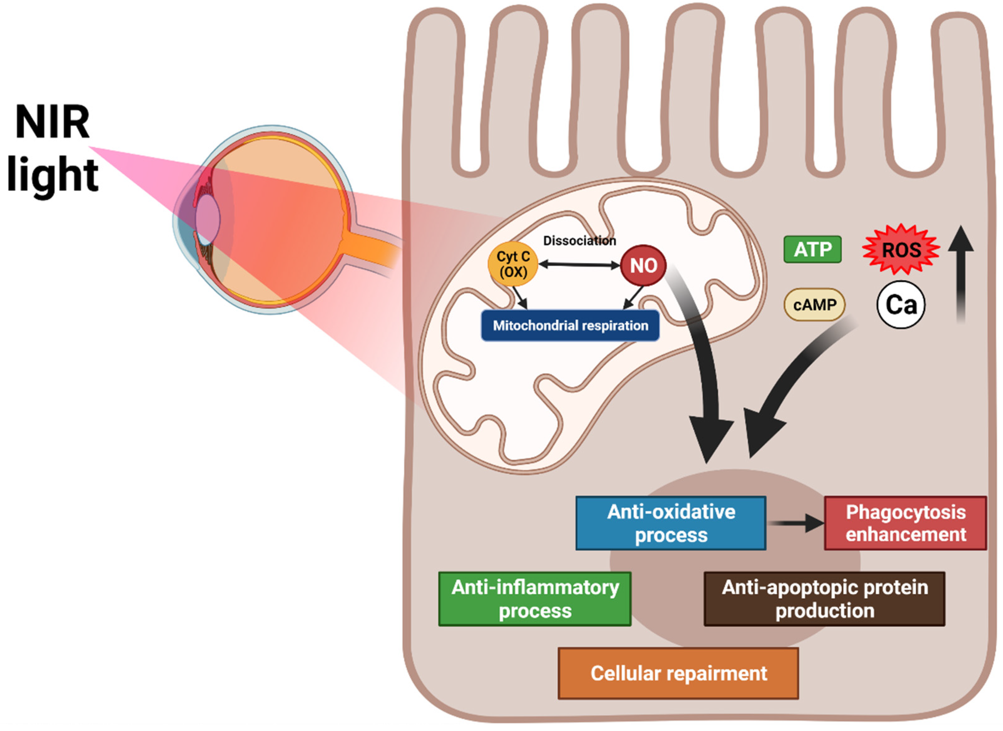
Figure 2. Overview of the mechanisms of photobiomodulation (PBM). PBM activates mitochondrial cytochrome oxidase C and increases mitochondrial respiration and nitric oxide dissociation. These processes elevate ATP, cAMP, reactive oxygen species, and intracellular calcium levels, promoting anti-inflammation, antioxidation, protein synthesis, anti-apoptosis, and cellular metabolism. The antioxidation effect of PBM increases RPE phagocytosis.

6.1.2. Application of PBM at AMD

PBM prevents AMD pathogenesis through its effect on cellular oxidative stress, apoptosis, and inflammation. Furthermore, PBM removes drusen in dry AMD by increasing RPE phagocytosis. Lavey reported that near-infrared radiation increases ATP and nitric oxide levels in RPE cells, which presumably promotes mitochondrial oxidative phosphorylation through the non-binding of nitric oxide from cytochrome oxidase C [126]. Kokkinopoulos et al. reported that after near-infrared radiation light exposure increases the mitochondrial membrane potential and decreases C3d, TNF-α, and macrophages in an animal AMD model, demonstrating an improvement in mitochondrial function and reduction in RPE inflammation [127].
In one study, 348 eyes with dry and wet type AMD were treated with radiation from a 780 nm semiconductor laser diode, and 97% of patients with cataracts and 94% of patients without cataracts showed an average visual improvement and reductions in pigmentation and cystic drusen and improvements in metamorphopsia and dyschromatopsia were observed [122].
The TORPA II study demonstrated functional changes, such as enhancement of contrast sensitivity and visual acuity, and anatomical improvements, such as reduction in drusen volume and central drusen thickness, after PBM in patients with dry AMD [128].

6.2. RPE Cell Transplantation

Unlike for early AMD and wet AMD, no appropriate treatment exists for GA. However, cell-based therapy has recently been used to treat vision loss in late AMD, especially GA. Cell transplants might be used as a rescue or replacement therapy. Rescue therapy may preserve the function of dying tissue and also restore the function of dying cells. Replacement therapy involves the replacement of dead or dying cells with normal cells for restoring the function of a tissue or organ.
Several clinical trials of RPE transplantation for late AMD have demonstrated that improvement in visual function depends on the severity of the damage before RPE cell transplantation. The subsequent sections summarize current clinical trials of RPE transplantation.

6.2.1. RPE Transplantation for Choroidal Neovascularization in AMD

Tezel et al. transplanted allogeneic RPE sheets obtained from cadavers after removing CNV lesions at the subfovea in AMD patients [129]. The patients received RPE cells from different cadavers, and no significant recovery in best-corrected visual acuity and contrast sensitivity was observed. Binder et al. transplanted RPE cells by subretinal injection of autologous RPE suspension after CNV excision at the subfovea in a prospective controlled trial [130]. One year after surgery, the best-corrected visual acuity and reading speed improved. Lu et al. also transplanted autologous RPE sheets in AMD patients after CNV excision [131] and reported recovery in best-corrected visual acuity. Kamao et al. and Nakagawa et al. transplanted autologous induced pluripotent stem cell-derived RPE sheets into the subretinal space in AMD patients after the surgical removal of subfoveal CNV [132][133][134]. Although the sheets were intact one year after surgery, no improvement was noted in the best-corrected visual acuity. Da Cruz et al. transplanted human embryonic stem cell-derived RPE in two AMD patients with subfoveal CNVs. One year after surgery, the best-corrected visual acuity and reading speed improved in only the patient with the least focal foveal atrophy [135]. Recently, the induced pluripotent stem cell (IPSc) based RPE is studied for transplantation [136][137][138]. It is expected to free from immune rejection, the most common adverse effects of transplantation, and showed well attached with AMD patients. However, their visual acuity was nearly unchanged [134].

6.2.2. RPE Transplantation for GA in AMD

Schwartz et al. transplanted embryonic stem cell-derived RPE suspensions in nine AMD patients with GA [139][140]. The improvement in best-corrected visual acuity was 14 letters in eight eyes. Kashani et al. transplanted human embryonic stem cell-derived RPE monolayer sheets (3.5 mm × 6.25 mm) into the GA lesions in five AMD patients [141]. Four of the five patients presented no improvement in vision. However, the vision of the remaining patient improved by 17 ETDRS letters.

7. Conclusions

Because AMD is a multifactorial disease with several pathways, a multifaceted intervention is required. Although the exact pathogenesis of AMD remains unknown, RPE dysfunction is a major contributor. Thus, several therapeutic strategies targeting RPE cells have been proposed. Among these, PBM is a promising noninvasive therapy to ameliorate oxidative stress, mitochondrial dysfunction, and complement dysfunction which are the main mechanisms of RPE dysfunction resulting in AMD. PBM can also remove drusen in AMD by activating RPE phagocytosis. Cell-based therapies involving transplantation of RPE sheets or cells are possible but will likely be beneficial only to patients with minimal photoreceptor atrophy in late AMD. Further research on the relationship between AMD pathogenesis and RPE dysfunction will identify other potential therapeutic targets.

References

  1. Strauss, O. The retinal pigment epithelium in visual function. Physiol. Rev. 2005, 85, 845–881.
  2. Wong, W.L.; Su, X.; Li, X.; Cheung, C.M.G.; Klein, R.; Cheng, C.-Y.; Wong, T.Y. Global prevalence of age-related macular degeneration and disease burden projection for 2020 and 2040: A systematic review and meta-analysis. Lancet Glob. Health 2014, 2, e106–e116.
  3. Klein, R.; Klein, B.E.; Linton, K.L. Prevalence of age-related maculopathy: The Beaver Dam Eye Study. Ophthalmology 1992, 99, 933–943.
  4. Mitchell, P.; Liew, G.; Gopinath, B.; Wong, T.Y. Age-related macular degeneration. Lancet 2018, 392, 1147–1159.
  5. Handa, J.T.; Rickman, C.B.; Dick, A.D.; Gorin, M.B.; Miller, J.W.; Toth, C.A.; Ueffing, M.; Zarbin, M.; Farrer, L.A. A systems biology approach towards understanding and treating non-neovascular age-related macular degeneration. Nat. Commun. 2019, 10, 1–11.
  6. Hageman, G.S.; Gehrs, K.; Johnson, L.V.; Anderson, D. Age-related macular degeneration (AMD). N. Engl. J. Med. 2008, 358, 2606–2617.
  7. Chen, M.; Luo, C.; Zhao, J.; Devarajan, G.; Xu, H. Immune regulation in the aging retina. Prog. Retin. Eye Res. 2019, 69, 159–172.
  8. Rozing, M.P.; Durhuus, J.A.; Nielsen, M.K.; Subhi, Y.; Kirkwood, T.B.; Westendorp, R.G.; Sørensen, T.L. Age-related macular degeneration: A two-level model hypothesis. Prog. Retin. Eye Res. 2020, 76, 100825.
  9. Kokotas, H.; Grigoriadou, M.; Petersen, M.B. Age-related macular degeneration: Genetic and clinical findings. Clin. Chem. Lab. Med. 2011, 49, 601–616.
  10. Bourla, D.H.; Young, T.A. Age-related macular degeneration: A practical approach to a Challenging Disease. J. Am. Geriatr. Soc. 2006, 54, 1130–1135.
  11. Age-Related Eye Disease Study Research Group. A randomized, placebo-controlled, clinical trial of high-dose supplementation with vitamins C and E, beta carotene, and zinc for age-related macular degeneration and vision loss: AREDS report no. 8. Arch. Ophthalmol. 2001, 119, 1417–1436.
  12. Age-Related Eye Disease Study Research Group. Risk factors associated with age-related macular degeneration: A case-control study in the age-related eye disease study: Age-related eye disease study report number 3. Ophthalmology 2000, 107, 2224–2232.
  13. Davis, M.D.; Gangnon, R.E.; Lee, L.Y.; Hubbard, L.D.; Klein, B.; Klein, R.; Ferris, F.L.; Bressler, S.B.; Milton, R.C. The Age-Related Eye Disease Study severity scale for age-related macular degeneration: AREDS report No. 17. Arch. Ophthalmol. 2005, 123, 1484–1498.
  14. Sunness, J.S.; Gonzalez-Baron, J.; Applegate, C.A.; Bressler, N.M.; Tian, Y.; Hawkins, B.; Barron, Y.; Bergman, A. Enlargement of atrophy and visual acuity loss in the geographic atrophy form of age-related macular degeneration. Ophthalmology 1999, 106, 1768–1779.
  15. Wong, C.W.; Liao, J.; Cheung, G.C.; Khor, C.C.; Vithana, E.N.; Wang, J.J.; Mitchell, P.; Aung, T.; Wong, T.Y.; Cheng, C.-Y. Lens status influences the association between CFH polymorphisms and age-related macular degeneration: Findings from two population-based studies in Singapore. PloS ONE 2015, 10, e0119570.
  16. Hogan, M.; Alvarado, J.; Weddell, J. Histology of the human eye. Phila. Saunders 1971.
  17. Marshall, J. The ageing retina: Physiology or pathology. Eye 1987, 1, 282–295.
  18. Streeten, B.W. Development of the human retinal pigment epithelium and the posterior segment. Arch. Ophthalmol. 1969, 81, 383–394.
  19. Boulton, M.; Dayhaw-Barker, P. The role of the retinal pigment epithelium: Topographical variation and ageing changes. Eye 2001, 15, 384–389.
  20. Baehr, W.; PALCZEWSKI, K.; WU, S.M.; BIRD, A.C. The retinoid cycle and retina disease. Vis. Res. (Oxford) 2003, 43, 2957–2958.
  21. Ach, T.; Huisingh, C.; McGwin, G.; Messinger, J.D.; Zhang, T.; Bentley, M.J.; Gutierrez, D.B.; Ablonczy, Z.; Smith, R.T.; Sloan, K.R.; et al. Quantitative autofluorescence and cell density maps of the human retinal pigment epithelium. Investig. Ophthalmol. Vis. Sci. 2014, 55, 4832–4841.
  22. Ishibashi, K.; Tian, J.; Handa, J.T. Similarity of mRNA phenotypes of morphologically normal macular and peripheral retinal pigment epithelial cells in older human eyes. Investig. Ophthalmol. Vis. Sci. 2004, 45, 3291–3301.
  23. Gao, H.; Hollyfield, J. Aging of the human retina. Differential loss of neurons and retinal pigment epithelial cells. Investig. Ophthalmol. Vis. Sci. 1992, 33, 1–17.
  24. Del Priore, L.V.; Kuo, Y.-H.; Tezel, T.H. Age-related changes in human RPE cell density and apoptosis proportion in situ. Investig. Ophthalmol. Vis. Sci. 2002, 43, 3312–3318.
  25. Feeney-Burns, L.; Hilderbrand, E.; Eldridge, S. Aging human RPE: Morphometric analysis of macular, equatorial, and peripheral cells. Investig. Ophthalmol. Vis. Sci. 1984, 25, 195–200.
  26. Caceres, P.S.; Benedicto, I.; Lehmann, G.L.; Rodriguez-Boulan, E.J. Directional fluid transport across organ–blood barriers: Physiology and cell biology. Cold Spring Harb. Perspect. Biol. 2017, 9, a027847.
  27. Bhutto, I.; Lutty, G. Understanding age-related macular degeneration (AMD): Relationships between the photoreceptor/retinal pigment epithelium/Bruch’s membrane/choriocapillaris complex. Mol. Asp. Med. 2012, 33, 295–317.
  28. Miller, S.S.; Steinberg, R.H. Active transport of ions across frog retinal pigment epithelium. Exp. Eye Res. 1977, 25, 235–248.
  29. Ved, N.; Hulse, R.P.; Bestall, S.M.; Donaldson, L.F.; Bainbridge, J.W.; Bates, D.O. Vascular endothelial growth factor-A165b ameliorates outer-retinal barrier and vascular dysfunction in the diabetic retina. Clin. Sci. 2017, 131, 1225–1243.
  30. Desjardins, D.M.; Yates, P.W.; Dahrouj, M.; Liu, Y.; Crosson, C.E.; Ablonczy, Z. Progressive early breakdown of retinal pigment epithelium function in hyperglycemic rats. Investig. Ophthalmol. Vis. Sci. 2016, 57, 2706–2713.
  31. Farjood, F.; Vargis, E. Physical disruption of cell–cell contact induces VEGF expression in RPE cells. Mol. Vis. 2017, 23, 431.
  32. Rózanowska, M.; Jarvis-Evans, J.; Korytowski, W.; Boulton, M.E.; Burke, J.M.; Sarna, T.J. Blue light-induced reactivity of retinal age pigment: IN vitro generation OF oxygen-reactive species (∗). J. Biol. Chem. 1995, 270, 18825–18830.
  33. Różanowska, M.; Wessels, J.; Boulton, M.; Burke, J.M.; Rodgers, M.A.; Truscott, T.G.; Sarna, T. Blue light-induced singlet oxygen generation by retinal lipofuscin in non-polar media. Free Radic. Biol. Med. 1998, 24, 1107–1112.
  34. Hu, D.N.; Simon, J.D.; Sarna, T. Role of ocular melanin in ophthalmic physiology and pathology. Photochem. Photobiol. 2008, 84, 639–644.
  35. Handa, J.T. How does the macula protect itself from oxidative stress? Mol. Asp. Med. 2012, 33, 418–435.
  36. Kurtz, J.; Jones, D.; Sternberg, P.; Wu, M.; Olsen, W. Antioxidant functions of glutathione in human retinal pigment epithelium in relation to age-related macular degeneration. In Retinal Pigment Epithelium and Macular Diseases; Springer: Berlin, Germany, 1998; pp. 47–57.
  37. deS Senanayake, P.; Calabro, A.; Hu, J.G.; Bonilha, V.L.; Darr, A.; Bok, D.; Hollyfield, J.G. Glucose utilization by the retinal pigment epithelium: Evidence for rapid uptake and storage in glycogen, followed by glycogen utilization. Exp. Eye Res. 2006, 83, 235–246.
  38. Shadrach, K.; Senanayake, P.; Nishiyama, K.; Lee, J.; Hu, J.; Calabro, A.; Bok, D.; Hollyfield, J. Glucose utilization by human RPE cultures. Investig. Ophthalmol. Vis. Sci. 2003, 44, 394.
  39. Khatami, M.; Stramm, L.E.; Rookey, J.H. Ascorbate transport in cultured cat retinal pigment epithelial cells. Exp. Eye Res. 1986, 43, 607–615.
  40. Bazan, N.G.; Gordon, W.C.; de Turco, E.B.R. Docosahexaenoic acid uptake and metabolism in photoreceptors: Retinal conservation by an efficient retinal pigment epithelial cell-mediated recycling process. Neurobiol. Essent. Fat. Acids 1992, 295–306.
  41. Nagelhus, E.A.; Horio, Y.; Inanobe, A.; Fujita, A.; Haug, F.m.; Nielsen, S.; Kurachi, Y.; Ottersen, O.P. Immunogold evidence suggests that coupling of K+ siphoning and water transport in rat retinal Müller cells is mediated by a coenrichment of Kir4. 1 and AQP4 in specific membrane domains. Glia 1999, 26, 47–54.
  42. Marmor, M.F. Control of subretinal fluid: Experimental and clinical studies. Eye 1990, 4, 340–344.
  43. Miller, S.S.; Edelman, J.L. Active ion transport pathways in the bovine retinal pigment epithelium. J. Physiol. 1990, 424, 283–300.
  44. Verkman, A.; Ruiz-Ederra, J.; Levin, M.H. Functions of aquaporins in the eye. Prog. Retin. Eye Res. 2008, 27, 420–433.
  45. Erickson, K.K.; Sundstrom, J.M.; Antonetti, D.A. Vascular permeability in ocular disease and the role of tight junctions. Angiogenesis 2007, 10, 103–117.
  46. Miller, S.S.; Steinberg, R.H. Passive ionic properties of frog retinal pigment epithelium. J. Membr. Biol. 1977, 36, 337–372.
  47. Finnemann, S.C. Focal adhesion kinase signaling promotes phagocytosis of integrin-bound photoreceptors. EMBO J. 2003, 22, 4143–4154.
  48. Slomiany, M.G.; Rosenzweig, S.A. Autocrine effects of IGF-I-induced VEGF and IGFBP-3 secretion in retinal pigment epithelial cell line ARPE-19. Am. J. Physiol. Cell Physiol. 2004, 287, C746–C753.
  49. Walsh, N.; Valter, K.; Stone, J. Cellular and subcellular patterns of expression of bFGF and CNTF in the normal and light stressed adult rat retina. Exp. Eye Res. 2001, 72, 495–501.
  50. Campochiaro, P.A.; Hackett, S.F.; Vinores, S.A.; Freund, J.; Csaky, C.; LaRochelle, W.; Henderer, J.; Johnson, M.; Rodriguez, I.R.; Friedman, Z. Platelet-derived growth factor is an autocrine growth stimulator in retinal pigmented epithelial cells. J. Cell Sci. 1994, 107, 2459–2469.
  51. Ahuja, P.; Caffe, A.; Holmqvist, I.; Söderpalm, A.; Singh, D.; Shinohara, T.; Van Veen, T. Lens epithelium-derived growth factor (LEDGF) delays photoreceptor degeneration in explants of rd/rd mouse retina. Neuroreport 2001, 12, 2951–2955.
  52. Adamis, A.; Shima, D.; Yeo, K.-T.; Yeo, T.; Brown, L.; Berse, B.; Damore, P.; Folkman, J. Synthesis and secretion of vascular permeability factor/vascular endothelial growth factor by human retinal pigment epithelial cells. Biochem. Biophys. Res. Commun. 1993, 193, 631–638.
  53. Tombran-Tink, J.; Chader, G.G.; Johnson, L.V. PEDF: A pigment epithelium-derived factor with potent neuronal differentiative activity. Exp. Eye Res. 1991, 53, 411–414.
  54. Tanihara, H.; Yoshida, M.; Matsumoto, M.; Yoshimura, N. Identification of transforming growth factor-beta expressed in cultured human retinal pigment epithelial cells. Investig. Ophthalmol. Vis. Sci. 1993, 34, 413–419.
  55. Witmer, A.; Vrensen, G.; Van Noorden, C.; Schlingemann, R.O. Vascular endothelial growth factors and angiogenesis in eye disease. Prog. Retin. Eye Res. 2003, 22, 1–29.
  56. Hargrave, P.A. Rhodopsin structure, function, and topography the Friedenwald lecture. Investig. Ophthalmol. Vis. Sci. 2001, 42, 3–9.
  57. Detrick, B.; Hooks, J.J. Immune regulation in the retina. Immunol. Res. 2010, 47, 153–161.
  58. Perez, V.L.; Caspi, R.R. Immune mechanisms in inflammatory and degenerative eye disease. Trends Immunol. 2015, 36, 354–363.
  59. Zavazava, N.; Halene, M.; Westphal, E.; Nölle, B.; Duncker, G.; Eckstein, E.; Harpprecht, J.; MÜLLER-RUCHHOLTZ, W. Expression of MHC class I and II molecules by cadaver retinal pigment epithelium cells: Optimization of post-mortem HLA typing. Clin. Exp. Immunol. 1991, 84, 163–166.
  60. Xu, H.; Chen, M. Targeting the complement system for the management of retinal inflammatory and degenerative diseases. Eur. J. Pharmacol. 2016, 787, 94–104.
  61. Clark, S.J.; Bishop, P.N. The eye as a complement dysregulation hotspot. Semin. Immunopathol. 2018, 40, 65–74.
  62. Maugeri, A.; Barchitta, M.; Mazzone, M.G.; Giuliano, F.; Agodi, A. Complement system and age-related macular degeneration: Implications of gene-environment interaction for preventive and personalized medicine. BioMed Res. Int. 2018, 2018, 7532507.
  63. Park, Y.-G.; Park, Y.-S.; Kim, I.-B. Complement System and Potential Therapeutics in Age-Related Macular Degeneration. Int. J. Mol. Sci. 2021, 22, 6851.
  64. Crabb, J.W.; Miyagi, M.; Gu, X.; Shadrach, K.; West, K.A.; Sakaguchi, H.; Kamei, M.; Hasan, A.; Yan, L.; Rayborn, M.E. Drusen proteome analysis: An approach to the etiology of age-related macular degeneration. Proc. Nat. Acad. Sci. USA 2002, 99, 14682–14687.
  65. Anderson, D.H.; Radeke, M.J.; Gallo, N.B.; Chapin, E.A.; Johnson, P.T.; Curletti, C.R.; Hancox, L.S.; Hu, J.; Ebright, J.N.; Malek, G.; et al. The pivotal role of the complement system in aging and age-related macular degeneration: Hypothesis re-visited. Prog. Retin. Eye Res. 2010, 29, 95–112.
  66. Lechner, J.; Chen, M.; Hogg, R.E.; Toth, L.; Silvestri, G.; Chakravarthy, U.; Xu, H. Higher plasma levels of complement C3a, C4a and C5a increase the risk of subretinal fibrosis in neovascular age-related macular degeneration. Immun. Ageing 2016, 13, 1–9.
  67. Nozaki, M.; Raisler, B.J.; Sakurai, E.; Sarma, J.V.; Barnum, S.R.; Lambris, J.D.; Chen, Y.; Zhang, K.; Ambati, B.K.; Baffi, J.Z. Drusen complement components C3a and C5a promote choroidal neovascularization. Proc. Natl. Acad. Sci. USA 2006, 103, 2328–2333.
  68. Lipo, E.; Cashman, S.M.; Kumar-Singh, R. Aurintricarboxylic acid inhibits complement activation, membrane attack complex, and choroidal neovascularization in a model of macular degeneration. Investig. Ophthalmol. Vis. Sci. 2013, 54, 7107–7114.
  69. Schramm, E.C.; Clark, S.J.; Triebwasser, M.P.; Raychaudhuri, S.; Seddon, J.M.; Atkinson, J.P. Genetic variants in the complement system predisposing to age-related macular degeneration: A review. Mol. Immunol. 2014, 61, 118–125.
  70. Fritsche, L.G.; Chen, W.; Schu, M.; Yaspan, B.L.; Yu, Y.; Thorleifsson, G.; Zack, D.J.; Arakawa, S.; Cipriani, V.; Ripke, S. Seven new loci associated with age-related macular degeneration. Nat. Genet. 2013, 45, 433–439.
  71. Lin, M.K.; Yang, J.; Hsu, C.W.; Gore, A.; Bassuk, A.G.; Brown, L.M.; Colligan, R.; Sengillo, J.D.; Mahajan, V.B.; Tsang, S.H. HTRA 1, an age-related macular degeneration protease, processes extracellular matrix proteins EFEMP 1 and TSP 1. Aging Cell 2018, 17, e12710.
  72. Fukuoka, Y.; Strainic, M.; Medof, M. Differential cytokine expression of human retinal pigment epithelial cells in response to stimulation by C5a. Clin. Exp. Immunol. 2003, 131, 248–253.
  73. Yang, P.; Baciu, P.; Kerrigan, B.C.P.; Etheridge, M.; Sung, E.; Toimil, B.A.; Berchuck, J.E.; Jaffe, G.J. Retinal pigment epithelial cell death by the alternative complement cascade: Role of membrane regulatory proteins, calcium, PKC, and oxidative stress. Investig. Ophthalmol. Vis. Sci. 2014, 55, 3012–3021.
  74. Joseph, K.; Kulik, L.; Coughlin, B.; Kunchithapautham, K.; Bandyopadhyay, M.; Thiel, S.; Thielens, N.M.; Holers, V.M.; Rohrer, B.J. Oxidative stress sensitizes retinal pigmented epithelial (RPE) cells to complement-mediated injury in a natural antibody-, lectin pathway-, and phospholipid epitope-dependent manner. J. Biol. Chem. 2013, 288, 12753–12765.
  75. Lau, L.-I.; Chiou, S.-H.; Liu, C.J.-L.; Yen, M.-Y.; Wei, Y.-H. The effect of photo-oxidative stress and inflammatory cytokine on complement factor H expression in retinal pigment epithelial cells. Investig. Ophthalmol. Vis. Sci. 2011, 52, 6832–6841.
  76. Thurman, J.M.; Renner, B.; Kunchithapautham, K.; Ferreira, V.P.; Pangburn, M.K.; Ablonczy, Z.; Tomlinson, S.; Holers, V.M.; Rohrer, B.J. Oxidative stress renders retinal pigment epithelial cells susceptible to complement-mediated injury. J. Biol. Chem. 2009, 284, 16939–16947.
  77. Terluk, M.R.; Kapphahn, R.J.; Soukup, L.M.; Gong, H.; Gallardo, C.; Montezuma, S.R.; Ferrington, D.A. Investigating mitochondria as a target for treating age-related macular degeneration. J. Neurosci. 2015, 35, 7304–7311.
  78. Brown, E.E.; DeWeerd, A.J.; Ildefonso, C.J.; Lewin, A.S.; Ash, J.D. Mitochondrial oxidative stress in the retinal pigment epithelium (RPE) led to metabolic dysfunction in both the RPE and retinal photoreceptors. Redox Biol. 2019, 24, 101201.
  79. Fisher, C.R.; Ferrington, D.A. Perspective on AMD pathobiology: A bioenergetic crisis in the RPE. Investig. Ophthalmol. Vis. Sci. 2018, 59, AMD41–AMD47.
  80. Adijanto, J.; Du, J.; Moffat, C.; Seifert, E.L.; Hurley, J.B.; Philp, N.J. The retinal pigment epithelium utilizes fatty acids for ketogenesis: Implications for metabolic coupling with the outer retina. J. Biol. Chem. 2014, 289, 20570–20582.
  81. Toms, M.; Burgoyne, T.; Tracey-White, D.; Richardson, R.; Dubis, A.M.; Webster, A.R.; Futter, C.; Moosajee, M. Phagosomal and mitochondrial alterations in RPE may contribute to KCNJ13 retinopathy. Sci. Rep. 2019, 9, 1–15.
  82. King, A.; Gottlieb, E.; Brooks, D.G.; Murphy, M.P.; Dunaief, J.L. Mitochondria-derived Reactive Oxygen Species Mediate Blue Light-induced Death of Retinal Pigment Epithelial Cells. Photochem. Photobiol. 2004, 79, 470–475.
  83. Liang, F.-Q.; Godley, B.F. Oxidative stress-induced mitochondrial DNA damage in human retinal pigment epithelial cells: A possible mechanism for RPE aging and age-related macular degeneration. Exp. Eye Res. 2003, 76, 397–403.
  84. Karunadharma, P.P.; Nordgaard, C.L.; Olsen, T.W.; Ferrington, D.A. Mitochondrial DNA damage as a potential mechanism for age-related macular degeneration. Investig. Ophthalmol. Vis. Sci. 2010, 51, 5470–5479.
  85. Kaarniranta, K.; Pawlowska, E.; Szczepanska, J.; Jablkowska, A.; Blasiak, J. Role of mitochondrial DNA damage in ROS-mediated pathogenesis of age-related macular degeneration (AMD). Int. J. Mol. Sci. 2019, 20, 2374.
  86. Starkov, A.A. The role of mitochondria in reactive oxygen species metabolism and signaling. Ann. N.Y. Acad. Sci. 2008, 1147, 37.
  87. Kasahara, E.; Lin, L.-R.; Ho, Y.-S.; Reddy, V.N. SOD2 protects against oxidation-induced apoptosis in mouse retinal pigment epithelium: Implications for age-related macular degeneration. Investig. Ophthalmol. Vis. Sci. 2005, 46, 3426–3434.
  88. Lascaratos, G.; Ji, D.; Wood, J.P.; Osborne, N.N. Visible light affects mitochondrial function and induces neuronal death in retinal cell cultures. Vis. Res. 2007, 47, 1191–1201.
  89. Jarrett, S.G.; Lewin, A.S.; Boulton, M.E. The importance of mitochondria in age-related and inherited eye disorders. Ophthalmic Res. 2010, 44, 179–190.
  90. Candas, D.; Li, J.J. MnSOD in oxidative stress response-potential regulation via mitochondrial protein influx. Antioxid. Redox Signal. 2014, 20, 1599–1617.
  91. Nita, M.; Grzybowski, A. The role of the reactive oxygen species and oxidative stress in the pathomechanism of the age-related ocular diseases and other pathologies of the anterior and posterior eye segments in adults. Oxid. Med. Cell. Longev. 2016, 2016, 3164734.
  92. Godley, B.F.; Shamsi, F.A.; Liang, F.-Q.; Jarrett, S.G.; Davies, S.; Boulton, M.J. Blue light induces mitochondrial DNA damage and free radical production in epithelial cells. J. Biol. Chem. 2005, 280, 21061–21066.
  93. Newsholme, P.; Haber, E.; Hirabara, S.; Rebelato, E.; Procopio, J.; Morgan, D.; Oliveira-Emilio, H.; Carpinelli, A.; Curi, R. Diabetes associated cell stress and dysfunction: Role of mitochondrial and non-mitochondrial ROS production and activity. J. physiol. 2007, 583, 9–24.
  94. Hanus, J.; Anderson, C.; Wang, S. RPE necroptosis in response to oxidative stress and in AMD. Ageing Res. Rev. 2015, 24, 286–298.
  95. Man, S.M.; Karki, R.; Kanneganti, T.D. Molecular mechanisms and functions of pyroptosis, inflammatory caspases and inflammasomes in infectious diseases. Immunol. Rev. 2017, 277, 61–75.
  96. Hanus, J.; Zhang, H.; Wang, Z.; Liu, Q.; Zhou, Q.; Wang, S. Induction of necrotic cell death by oxidative stress in retinal pigment epithelial cells. Cell Death Dis. 2013, 4, e965.
  97. Guang-Yu, L.; Bin, F.; Zheng, Y.-C. Calcium overload is a critical step in programmed necrosis of ARPE-19 cells induced by high-concentration H2O2. Biomed. Environ. Sci. 2010, 23, 371–377.
  98. Elmore, S. Apoptosis: A review of programmed cell death. Toxicol. Pathol. 2007, 35, 495–516.
  99. Reed, J.C. Mechanisms of apoptosis. Am. J. Pathol. 2000, 157, 1415–1430.
  100. Kaneko, H.; Dridi, S.; Tarallo, V.; Gelfand, B.D.; Fowler, B.J.; Cho, W.G.; Kleinman, M.E.; Ponicsan, S.L.; Hauswirth, W.W.; Chiodo, V.A. DICER1 deficit induces Alu RNA toxicity in age-related macular degeneration. Nature 2011, 471, 325–330.
  101. Ho, D.T.; Bardwell, A.J.; Grewal, S.; Iverson, C.; Bardwell, L. Interacting JNK-docking sites in MKK7 promote binding and activation of JNK mitogen-activated protein kinases. J. Biol. Chem. 2006, 281, 13169–13179.
  102. Gao, J.; Cui, J.Z.; To, E.; Cao, S.; Matsubara, J.A. Evidence for the activation of pyroptotic and apoptotic pathways in RPE cells associated with NLRP3 inflammasome in the rodent eye. J. Neuroinflamm. 2018, 15, 1–12.
  103. Tseng, W.A.; Thein, T.; Kinnunen, K.; Lashkari, K.; Gregory, M.S.; D'Amore, P.A.; Ksander, B.R. NLRP3 inflammasome activation in retinal pigment epithelial cells by lysosomal destabilization: Implications for age-related macular degeneration. Investig. Ophthalmol. Vis. Sci. 2013, 54, 110–120.
  104. Cai, J.; Nelson, K.C.; Wu, M.; Sternberg, P., Jr.; Jones, D.P. Oxidative damage and protection of the RPE. Prog. Retin. Eye Res. 2000, 19, 205–221.
  105. Brandstetter, C.; Patt, J.; Holz, F.G.; Krohne, T.U. Inflammasome priming increases retinal pigment epithelial cell susceptibility to lipofuscin phototoxicity by changing the cell death mechanism from apoptosis to pyroptosis. J. Photochem. Photobiol. B Biol. 2016, 161, 177–183.
  106. Kivinen, N. The Role of Autophagy in Age-Related Macular Degeneration (AMD)–Studies into the Pathogenesis of AMD. Ph.D. dissertation, University of Michigan, Ann Arbor, MI, USA, 2018.
  107. Kaarniranta, K.; Tokarz, P.; Koskela, A.; Paterno, J.; Blasiak, J. Autophagy regulates death of retinal pigment epithelium cells in age-related macular degeneration. Cell Biol. Toxicol. 2017, 33, 113–128.
  108. Mitter, S.K.; Rao, H.V.; Qi, X.; Cai, J.; Sugrue, A.; Dunn, W.A.; Grant, M.B.; Boulton, M.E. Autophagy in the retina: A potential role in age-related macular degeneration. Retin. Degener. Dis. 2012, 83–90.
  109. Krohne, T.U.; Stratmann, N.K.; Kopitz, J.; Holz, F.G. Effects of lipid peroxidation products on lipofuscinogenesis and autophagy in human retinal pigment epithelial cells. Exp. Eye Res. 2010, 90, 465–471.
  110. Wang, A.L.; Lukas, T.J.; Yuan, M.; Du, N.; Tso, M.O.; Neufeld, A.H. Autophagy and exosomes in the aged retinal pigment epithelium: Possible relevance to drusen formation and age-related macular degeneration. PloS ONE 2009, 4, e4160.
  111. Alge, C.S.; Priglinger, S.G.; Neubauer, A.S.; Kampik, A.; Zillig, M.; Bloemendal, H.; Welge-Lussen, U. Retinal pigment epithelium is protected against apoptosis by αB-crystallin. Investig. Ophthalmol. Vis. Sci. 2002, 43, 3575–3582.
  112. Gangalum, R.K.; Schibler, M.J.; Bhat, S.P. Small heat shock protein αB-crystallin is part of cell cycle-dependent Golgi reorganization. J. Biol. Chem. 2004, 279, 43374–43377.
  113. De, S.; Rabin, D.M.; Salero, E.; Lederman, P.L.; Temple, S.; Stern, J.H. Human retinal pigment epithelium cell changes and expression of αB-crystallin: A biomarker for retinal pigment epithelium cell change in age-related macular degeneration. Arch. Ophthalmol. 2007, 125, 641–645.
  114. Kannan, R.; Sreekumar, P.G.; Hinton, D.R. Alpha crystallins in the retinal pigment epithelium and implications for the pathogenesis and treatment of age-related macular degeneration. Biochim. Biophys. Acta (BBA)-Gen. Subj. 2016, 1860, 258–268.
  115. Dimberg, A.; Rylova, S.; Dieterich, L.C.; Olsson, A.-K.; Schiller, P.; Wikner, C.; Bohman, S.; Botling, J.; Lukinius, A.; Wawrousek, E.F. αB-crystallin promotes tumor angiogenesis by increasing vascular survival during tube morphogenesis. J. Am. Soc. Hematol. 2008, 111, 2015–2023.
  116. Kase, S.; He, S.; Sonoda, S.; Kitamura, M.; Spee, C.; Wawrousek, E.; Ryan, S.J.; Kannan, R.; Hinton, D.R. αB-crystallin regulation of angiogenesis by modulation of VEGF. J. Am. Soc. Hematol. 2010, 115, 3398–3406.
  117. Yaung, J.; Kannan, R.; Wawrousek, E.F.; Spee, C.; Sreekumar, P.G.; Hinton, D.R. Exacerbation of retinal degeneration in the absence of alpha crystallins in an in vivo model of chemically induced hypoxia. Exp. Eye Res. 2008, 86, 355–365.
  118. Watanabe, G.; Kato, S.; Nakata, H.; Ishida, T.; Ohuchi, N.; Ishioka, C. αB-crystallin: A novel p53-target gene required for p53-dependent apoptosis. Cancer Sci. 2009, 100, 2368–2375.
  119. Li, D.W.-C.; Liu, J.-P.; Mao, Y.-W.; Xiang, H.; Wang, J.; Ma, W.-Y.; Dong, Z.; Pike, H.M.; Brown, R.E.; Reed, J.C. Calcium-activated RAF/MEK/ERK signaling pathway mediates p53-dependent apoptosis and is abrogated by αB-crystallin through inhibition of RAS activation. Mol. Biol. Cell 2005, 16, 4437–4453.
  120. Rojas, J.C.; Gonzalez-Lima, F. Low-level light therapy of the eye and brain. Eye Brain 2011, 3, 49.
  121. Tata, D.B.; Waynant, R.W. Laser therapy: A review of its mechanism of action and potential medical applications. Laser Photonics Rev. 2011, 5, 1–12.
  122. Ivandic, B.T.; Ivandic, T. Low-level laser therapy improves vision in patients with age-related macular degeneration. Photomed. Laser Surg. 2008, 26, 241–245.
  123. Merry, G.; Dotson, R.; Devenyi, R.; Markowitz, S.; Reyes, S. Photobiomodulation as a new treatment for dry age related macular degeneration. results from the toronto and Oak ridge photobimodulation study in AMD (TORPA). Investig. Ophthalmol. Vis. Sci. 2012, 53, 2049.
  124. Tang, J.; Herda, A.A.; Kern, T.S. Photobiomodulation in the treatment of patients with non-center-involving diabetic macular oedema. Br. J. Ophthalmol. 2014, 98, 1013–1015.
  125. Fuma, S.; Murase, H.; Kuse, Y.; Tsuruma, K.; Shimazawa, M.; Hara, H. Photobiomodulation with 670 nm light increased phagocytosis in human retinal pigment epithelial cells. Mol. Vis. 2015, 21, 883.
  126. Lavey, B.J.; Estlack, L.E.; Schuster, K.J.; Rockwell, B.A.; Wigle, J.C. The response of human retinal pigmented epithelial cells in vitro to changes in nitric oxide concentration stimulated by low levels of red light. Proceedings of Mechanisms for Low-Light Therapy VIII, San Francisco, CA, USA, 2–3 February 2013; p. 85690.
  127. Kokkinopoulos, I.; Colman, A.; Hogg, C.; Heckenlively, J.; Jeffery, G. Age-related retinal inflammation is reduced by 670 nm light via increased mitochondrial membrane potential. Neurobiol. Aging 2013, 34, 602–609.
  128. Merry, G.F.; Munk, M.R.; Dotson, R.S.; Walker, M.G.; Devenyi, R.G. Photobiomodulation reduces drusen volume and improves visual acuity and contrast sensitivity in dry age-related macular degeneration. Acta Ophthalmol. 2017, 95, e270–e277.
  129. Tezel, T.H.; Del Priore, L.V.; Berger, A.S.; Kaplan, H.J. Adult retinal pigment epithelial transplantation in exudative age-related macular degeneration. Am. J. Ophthalmol. 2007, 143, 584–595.
  130. Binder, S.; Krebs, I.; Hilgers, R.-D.; Abri, A.; Stolba, U.; Assadoulina, A.; Kellner, L.; Stanzel, B.V.; Jahn, C.; Feichtinger, H.; et al. Outcome of transplantation of autologous retinal pigment epithelium in age-related macular degeneration: A prospective trial. Investig. Ophthalmol. Vis. Sci. 2004, 45, 4151–4160.
  131. Lu, Y.; Han, L.; Wang, C.; Dou, H.; Feng, X.; Hu, Y.; Feng, K.; Wang, X.; Ma, Z. A comparison of autologous transplantation of retinal pigment epithelium (RPE) monolayer sheet graft with RPE–Bruch's membrane complex graft in neovascular age-related macular degeneration. Acta Ophthalmol. 2017, 95, e443–e452.
  132. Kamao, H.; Mandai, M.; Okamoto, S.; Sakai, N.; Suga, A.; Sugita, S.; Kiryu, J.; Takahashi, M. Characterization of human induced pluripotent stem cell-derived retinal pigment epithelium cell sheets aiming for clinical application. Stem Cell Rep. 2014, 2, 205–218.
  133. Nakagawa, M.; Koyanagi, M.; Tanabe, K.; Takahashi, K.; Ichisaka, T.; Aoi, T.; Okita, K.; Mochiduki, Y.; Takizawa, N.; Yamanaka, S. Generation of induced pluripotent stem cells without Myc from mouse and human fibroblasts. Nat. Biotechnol. 2008, 26, 101–106.
  134. da Cruz, L.; Fynes, K.; Georgiadis, O.; Kerby, J.; Luo, Y.H.; Ahmado, A.; Vernon, A.; Daniels, J.T.; Nommiste, B.; Hasan, S.M. Phase 1 clinical study of an embryonic stem cell–derived retinal pigment epithelium patch in age-related macular degeneration. Nat. Biotechnol. 2018, 36, 328–337.
  135. Surendran, H.; Nandakumar, S.; Stoddard, J.; Mohan, V.; Upadhyay, P.K.; McGill, T.J.; Pal, R. Therapy. Transplantation of retinal pigment epithelium and photoreceptors generated concomitantly via small molecule-mediated differentiation rescues visual function in rodent models of retinal degeneration. Stem Cell Rep. 2021, 12, 1–17.
  136. Shrestha, R.; Wen, Y.-T.; Tsai, R.-K. Effective differentiation and biological characterization of retinal pigment epithelium derived from human induced pluripotent stem cells. Curr. Eye Res. 2020, 45, 1155–1167.
  137. Sharma, R.; Khristov, V.; Rising, A.; Jha, B.S.; Dejene, R.; Hotaling, N.; Li, Y.; Stoddard, J.; Stankewicz, C.; Wan, Q. Clinical-grade stem cell–derived retinal pigment epithelium patch rescues retinal degeneration in rodents and pigs. Sci. Trans. Med. No. 475 (eaat550). 2019, 11.
  138. Mandai, M.; Watanabe, A.; Kurimoto, Y.; Hirami, Y.; Morinaga, C.; Daimon, T.; Fujihara, M.; Akimaru, H.; Sakai, N.; Shibata, Y. Autologous induced stem-cell–derived retinal cells for macular degeneration. N. Engl. J. Med. 2017, 376, 1038–1046.
  139. Schwartz, S.D.; Regillo, C.D.; Lam, B.L.; Eliott, D.; Rosenfeld, P.J.; Gregori, N.Z.; Hubschman, J.-P.; Davis, J.L.; Heilwell, G.; Spirn, M. Human embryonic stem cell-derived retinal pigment epithelium in patients with age-related macular degeneration and Stargardt's macular dystrophy: Follow-up of two open-label phase 1/2 studies. Lancet 2015, 385, 509–516.
  140. Schwartz, S.D.; Tan, G.; Hosseini, H.; Nagiel, A. Subretinal transplantation of embryonic stem cell–derived retinal pigment epithelium for the treatment of macular degeneration: An assessment at 4 years. Investig. Ophthalmol. Vis. Sci. 2016, 57, ORSFc1–ORSFc9.
  141. Kashani, A.H.; Lebkowski, J.S.; Rahhal, F.M.; Avery, R.L.; Salehi-Had, H.; Dang, W.; Lin, C.-M.; Mitra, D.; Zhu, D.; Thomas, B.B. A bioengineered retinal pigment epithelial monolayer for advanced, dry age-related macular degeneration. Sci. Trans. Med. No. 435 (eaao4097). 2018, 10.
More
Information
Subjects: Ophthalmology
Contributor MDPI registered users' name will be linked to their SciProfiles pages. To register with us, please refer to https://encyclopedia.pub/register :
View Times: 492
Entry Collection: Neurodegeneration
Revisions: 3 times (View History)
Update Date: 24 Nov 2021
1000/1000