Submitted Successfully!
To reward your contribution, here is a gift for you: A free trial for our video production service.
Thank you for your contribution! You can also upload a video entry or images related to this topic.
Version Summary Created by Modification Content Size Created at Operation
1 + 1373 word(s) 1373 2021-10-19 09:56:55 |
2 Format correct Meta information modification 1373 2021-10-29 03:35:07 | |
3 Format correct + 12 word(s) 1385 2021-10-29 03:39:27 |

Video Upload Options

Do you have a full video?

Confirm

Are you sure to Delete?
Cite
If you have any further questions, please contact Encyclopedia Editorial Office.
Costagliola, G. Primary Immunodeficiency Disorders and Autoimmunity. Encyclopedia. Available online: https://encyclopedia.pub/entry/15518 (accessed on 19 April 2024).
Costagliola G. Primary Immunodeficiency Disorders and Autoimmunity. Encyclopedia. Available at: https://encyclopedia.pub/entry/15518. Accessed April 19, 2024.
Costagliola, Giorgio. "Primary Immunodeficiency Disorders and Autoimmunity" Encyclopedia, https://encyclopedia.pub/entry/15518 (accessed April 19, 2024).
Costagliola, G. (2021, October 28). Primary Immunodeficiency Disorders and Autoimmunity. In Encyclopedia. https://encyclopedia.pub/entry/15518
Costagliola, Giorgio. "Primary Immunodeficiency Disorders and Autoimmunity." Encyclopedia. Web. 28 October, 2021.
Primary Immunodeficiency Disorders and Autoimmunity
Edit

Primary immunodeficiency disorders (PIDs) patients can develop an immune dysregulation of variable degree, which is responsible for a clinical picture featured by infectious complications and autoimmunity. Autoimmune manifestations are observed with considerable frequency in patients with primary antibody deficiencies, including common variable immunodeficiency (CVID) and selective IgA deficiency (sIgAD), but can also be evidenced in patients with combined immunodeficiency disorders (CID).

22q11.2 deletion syndrome activated phosphoinositide 3-kinase d syndrome common variable immunodeficiency complement deficiency CTLA-4 Immune dysregulation LRBA selective IgA deficiency severe combined immunodeficiency X-linked agammaglobulinemia

1. Introduction

In recent years, the association between primary immunodeficiency disorders (PIDs) and autoimmunity has been extensively studied. Notably, autoimmunity can represent the presentation sign of PIDs in a significant number of patients [1]. The molecular mechanisms responsible for the immune dysregulation in patients with PIDs are multiple and not completely elucidated; impaired B cell differentiation and germ-center reactions, altered T cell central or peripheral tolerance, uncontrolled lymphocyte proliferation and differentiation, dysfunctional complement, and innate immune activation can participate in the complex pathogenic process leading to autoimmunity. In patients with PIDs, the association with autoimmunity leads to a significant impact on the quality of life, higher medicalization, and increased mortality [2]. Furthermore, the increasing use of new sequencing techniques allowed the identification of different monogenic causes of PID, the better understanding of genotype-phenotype correlations, and the improvement of the therapeutic strategies targeting the immune dysregulation in PIDs [3][4].

2. Clinical implications

The pathogenic and clinical associations carry different significant implications for the approach to children with autoimmunity, highlighting that it should represent a warning sign for the presence of a PID, particularly in pediatric age. Identifying a condition of PID in children presenting with autoimmunity offers the opportunity to provide an adequate treatment of the underlying disease (Ig replacement therapy, targeted treatments, HSCT (hematopoietic stem cell transplantation), gene therapy), to offer supplementary immunization if needed, and optimize the management of autoimmunity itself (Figure 1). On the other hand, as different studies have demonstrated a reduced survival in the subgroup of patients with PIDs showing autoimmunity, its finding in patients with already diagnosed PID could lead to significant changes in the follow-up strategy and therapeutic approach [5][6].
Jcm 10 04729 g002
Figure 1. Therapeutic strategies for autoimmunity in patients with PIDs. The figure shows the current therapeutic options for specific PIDs. The choice of the therapeutic strategy (immunosuppressive agents, biologic drugs, HSCT, gene therapy) depends on the clinical severity, comorbidities and also on the availability and physician’s experience. * In patients with refractory autoimmune cytopenia. APDS: Activated phosphoinositide 3-kinase d syndrome; APS-1: Autoimmune polyendocrine syndrome 1; CTLA-4: Cytotoxic lymphocyte antigen 4; CVID: Common variable immunodeficiency; HCQ: Hydroxychloroquine; HIGM: Hyper-IgM syndromes; HSCT: hematopoietic stem cell transplantation; JAK: Janus kinase; LRBA: LPS-responsive beige-like anchor protein; MMF: mycophenolate mofetil; PI3K: Phosphoinositide 3-kinase; PKCD: protein kinase C δ deficiency; SCID: severe combined immunodeficiency; sIgAD: selective IgA deficiency; STAT: Signal Transducers and Activator of Transcription; Tregs: regulatory T cells; WAS: Wiskott-Aldrich syndrome; XLA: X-linked agammaglobulinemia.

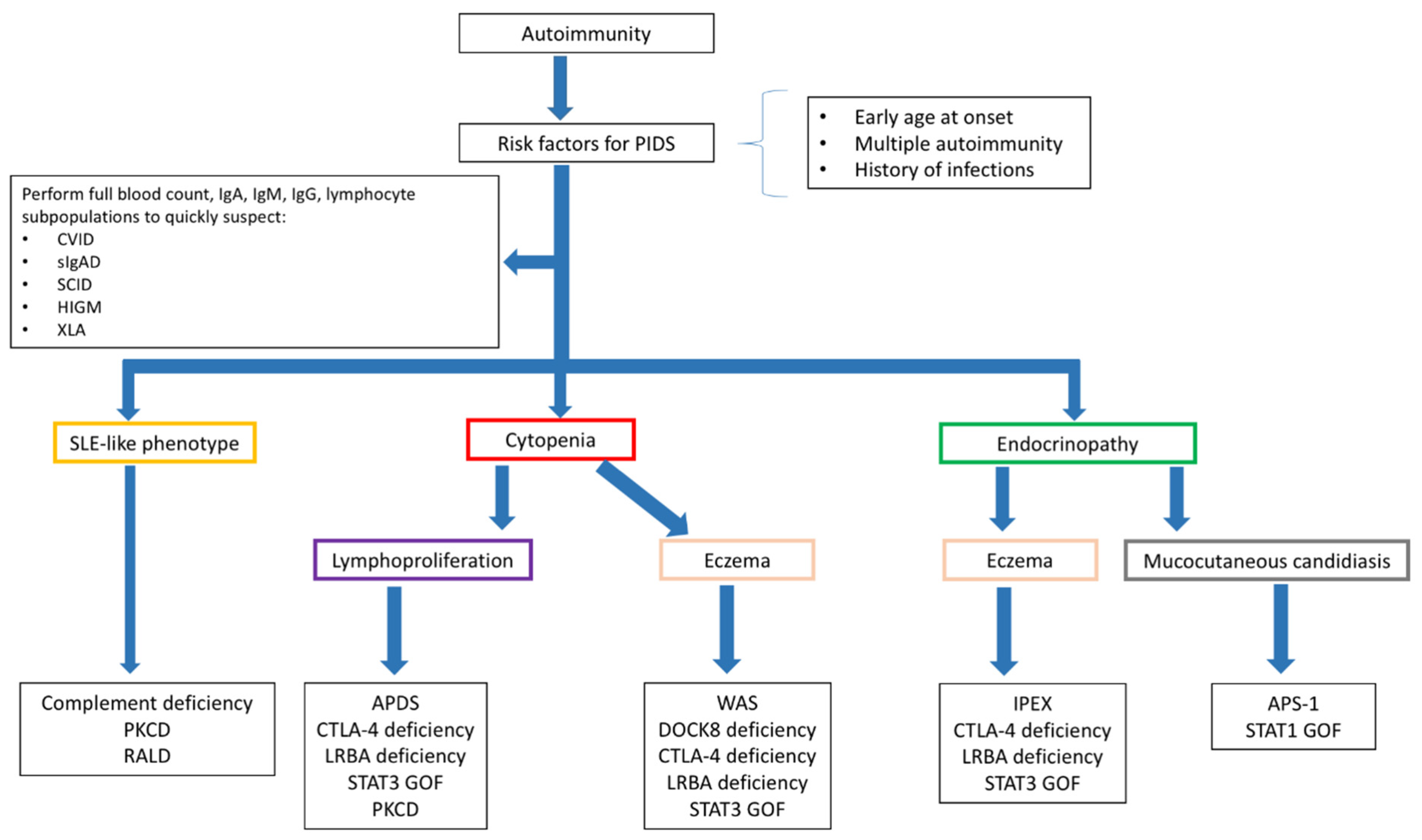
2.1. Diagnosing PIDs in Children Presenting with Autoimmunity

The diagnostic approach to a child with autoimmune manifestations should include the detailed analysis of the clinical history to evidence the occurrence of infections, quantify their impact, and point out the associated clinical features suggestive for a PID (growth delay, high frequency of infections, need of hospitalization for infections, prolonged use of antibiotics, infections by unusual pathogens, and others). Additionally, a baseline immunological assessment including the determination of serum Ig levels and lymphocyte subpopulations should be performed in all the children with autoimmune manifestations. The need for other specific investigations (Figure 2) varies depending on the specific autoimmune phenotype. The approach to children with immune cytopenia is of particular interest, since it can represent the presentation sign of a wide spectrum of PIDs, but also the first manifestation of a systemic connective tissue disease, such as SLE (systemic lupus erythematosus)[7]. As a consequence, the clinical and laboratory assessment of a child with suspected immune cytopenia should comprehend both the analysis of the immune response and the determination of the most relevant autoantibody subclasses, including antinucleous antibodies and antithyroid antibodies [8]. Interestingly, patients with PID have a 120-fold high risk of developing autoimmune cytopenia compared to the general population, with a higher increase in risk observed for autoimmune hemolytic anemia (AIHA) [9]. The case of children presenting with multiple cytopenias is of significant interest. Indeed, studies on Evans syndromes showed that almost half of the children with this condition have e positive genetic testing for PID and that in this cohort of patients the incidence of systemic autoimmune diseases is also considerable [10][11]. In patients with autoimmune endocrinopathy, the suspect of PID should be posed when the disease onset is earlier than usual, when there is an association of two or more endocrine disorders, and when other signs suggestive for PID are present. In this subset of patients, the finding of eczema, elevated serum IgE levels, and peripheral eosinophilia should induce the suspect of a Treg-mediated disorder [12], while the association with chronic mucocutaneous candidiasis is observed in patients with APS-1 and STAT1 gain of function (GOF) [13]. Finally, in the case of SLE-like manifestations, the serum levels of the complement fractions need to be determined [14]. Specific investigations to allow a definitive diagnosis, including the analysis of the immune response to vaccines (for the clinical diagnosis of CVID), extended determination of the lymphocyte subpopulations (including memory B and T cells), functional analysis, and cytogenetic and genetic testing, should be performed based on the clinical suspect.
Jcm 10 04729 g003
Figure 2. Diagnostic approach to autoimmunity in patients with suspect immunodeficiency. Figure legend: APDS: Activated phosphoinositide 3-kinase d syndrome; APS-1: Autoimmune polyendocrine syndrome 1; CTLA-4: Cytotoxic lymphocyte antigen 4; CVID: Common variable immunodeficiency; HIGM: Hyper-IgM syndrome; LRBA: LPS-responsive beige-like anchor protein; PKCD: protein kinase C δ deficiency; RALD: Ras-associated leukoproliferative disorder; SCID: severe combined immunodeficiency; sIgAD: Selective IgA deficiency; SLE: systemic lupus erythematosus; STAT: Signal Transducers and Activator of Transcription; WAS: Wiskott-Aldrich syndrome; XLA: X-linked agammaglobulinemia.

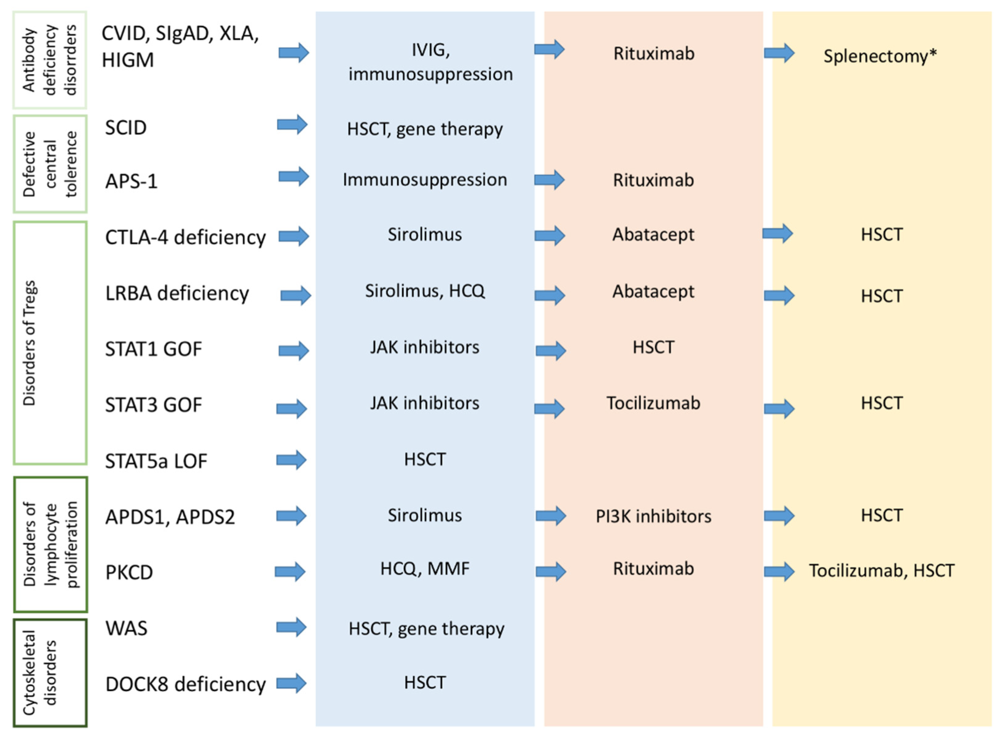
2.2. Diagnosing Autoimmunity in Children with PIDs

Periodic surveillance for autoimmune manifestations is mandatory in all patients with PIDs. This is particularly relevant for children carrying genetic mutations with a well-defined association with autoimmunity (i.e., CTLA-4, LRBA, PI3K mutations). Moreover, there is increasing interest in the identification of potential immunological predictors of autoimmunity in patients with PIDs. Although there are no specific immunological markers with high predictive value for the development of autoimmunity, literature data from patients with primary antibody deficiencies and CID have identified several potential candidates.
A large cohort study on CVID patients with immune cytopenia demonstrated higher levels of serum immunoglobulin, CD19hi B cells, and T CD4 effector T cells, accompanied by reduced naïve T cells [15]. Absence or reduced switched memory B cells have been associated with autoimmune cytopenias, systemic autoimmune diseases, splenomegaly, granulomatous diseases, and lymphadenopathy [16][17]. An expansion of CD21low B cells has been described in association with reduced Tregs in CVID patients with autoimmunity [18]. It has been found that CD21low B cells produce significantly more IgM than naïve B cells after stimulation with CD40L, IL-2, and IL-10 and that CVID patients with autoimmunity have higher levels of IgM compared with non-autoimmune phenotypes [19][20], thus suggesting that increased IgM levels may be a marker of autoimmunity and they may have a pathogenic role. Additionally, lower naïve CD4+ and CD8+ T cells and increased differentiated T cells have been described in CVID patients with autoimmunity [21].
Concerning combined immunodeficiencies, a recent study by Montin et al. in patients with 22q11.2DS highlighted that some immunological features, including a reduced number of naïve T cells, reduced RTE, and elevation of naïve B cells are associated with the development of hematologic autoimmunity and can be evidenced significantly before the onset of autoimmunity [22]. Moreover, the degree of T-cell lymphopenia has been suggested as a contributing factor for the development of autoimmunity in this disease [23].

3. Conclusions

It is well recognized that autoimmune manifestations are observed in a significant percentage of patients with PIDs, often representing the first sign of these conditions. Patients with early-onset autoimmunity, an association between two or more autoimmune manifestations, or increased susceptibility to infections should be promptly screened for PIDs. Although the intriguing mechanisms underlying the development of autoimmunity in patients with PIDs are far to be completely elucidated, the rapidly evolving knowledge in the genetic background of PIDs will hopefully help to characterize the defects linking immunodeficiency and autoimmunity, thus providing interesting diagnostic and therapeutic implications.

References

  1. Walter, J.E.; Ayala, I.A.; Milojevic, D. Autoimmunity as a continuum in primary immunodeficiency. Curr. Opin. Pediatr. 2019, 31, 851–862.
  2. Amaya-Uribe, L.; Rojas, M.; Azizi, G.; Anaya, J.M.; Gershwin, M.E. Primary immunodeficiency and autoimmunity: A comprehensive review. J. Autoimmun. 2019, 99, 52–72.
  3. Notarangelo, L.D.; Uzel, G.; Rao, V.K. Primary immunodeficiencies: Novel genes and unusual presentations. Hematol. Am. Soc. Hematol. Educ. Program 2019, 2019, 443–448.
  4. Bousfiha, A.; Jeddane, L.; Picard, C.; Ailal, F.; Bobby Gaspar, H.; Al-Herz, W. The 2017 IUIS Phenotypic Classification for Primary Immunodeficiencies. J. Clin. Immunol. 2018, 38, 129–143.
  5. Azizi, G.; Ziaee, V.; Tavakol, M.; Alinia, T.; Yazdai, R.; Mohammadi, H.; Abolhassani, H.; Aghamohammadi, A. Approach to the Management of Autoimmunity in Primary Immunodeficiency. Scand. J. Immunol. 2017, 85, 13–29.
  6. Walter, J.E.; Farmer, J.R.; Foldvari, Z.; Torgerson, T.R.; Cooper, M.A. Mechanism-Based Strategies for the Management of Autoimmunity and Immune Dysregulation in Primary Immunodeficiencies. J. Allergy Clin. Immunol. Pract. 2016, 4, 1089–1100.
  7. Abraham, R.S. How to evaluate for immunodeficiency in patients with autoimmune cytopenias: Laboratory evaluation for the diagnosis of inborn errors of immunity associated with immune dysregulation. Hematol. Am. Soc. Hematol. Educ. Program 2020, 2020, 661–672.
  8. Consolini, R.; Costagliola, G.; Spatafora, D. The Centenary of Immune Thrombocytopenia-Part 2: Revising Diagnostic and Therapeutic Approach. Front Pediatr. 2017, 5, 179.
  9. Fischer, A.; Provot, J.; Jais, J.P.; Alcais, A.; Mahlaoui, N. Autoimmune and inflammatory manifestations occur frequently in patients with primary immunodeficiencies. J. Allergy Clin. Immunol. 2017, 140, 1388–1393.e8.
  10. Besnard, C.; Levy, E.; Aladjidi, N.; Stolzenberg, M.C.; Magerus-Chatinet, A.; Alibeu, O.; Nitschke, P.; Blanche, S.; Hermine, O.; Jeziorski, E.; et al. Pediatric-onset Evans syndrome: Heterogeneous presentation and high frequency of monogenic disorders including LRBA and CTLA4 mutations. Clin. Immunol. 2018, 188, 52–57.
  11. Hadjadj, J.; Aladjidi, N.; Fernandes, H.; Leverger, G.; Magérus-Chatinet, A.; Mazerolles, F.; Hussey, A.A.; Evbuomwan, M.O.; Long Priel, D.A.; Kuhns, D.B.; et al. Pediatric Evans syndrome is associated with a high frequency of potentially damaging variants in immune genes. J. Am. Soc. Hematol. 2019, 134, 9–21.
  12. Costagliola, G.; Marco, S.D.; Comberiati, P.; D’Elios, S.; Petashvili, N.; Di Cicco, M.E.; Peroni, D. Practical Approach to Children Presenting with Eosinophila and Hypereosinophilia. Curr. Pediatr. Rev. 2020, 16, 81–88.
  13. Shamriz, O.; Tal, Y.; Talmon, A.; Nahum, A. Chronic Mucocutaneous Candidiasis in Early Life: Insights Into Immune Mechanisms and Novel Targeted Therapies. Front Immunol. 2020, 11, 593289.
  14. Leffler, J.; Bengtsson, A.A.; Blom, A.M. The complement system in systemic lupus erythematosus: An update. Ann. Rheum. Dis. 2014, 73, 1601–1606.
  15. Boileau, J.; Mouillot, G.; Gerard, L.; Carmagnat, M.; Rabian, C.; Oksenhendler, E.; Pasquali, J.-L.; Korganow, A.-S.; DEFI Study Group. Autoimmunity in common variable immunodeficiency: Correlation with lymphocyte phenotype in the French DEFI study. J. Autoimmun. 2011, 36, 25–32.
  16. Sánchez-Ramón, S.; Radigan, L.; Yu, J.E.; Bard, S.; Cunningham-Rundles, C. Memory B cells in common variable immunodeficiency: Clinical associations and sex differences. Clin. Immunol. 2008, 128, 314–321.
  17. Ahn, S.; Cunningham-Rundles, C. Role of B cells in common variable immune deficiency. Expert Rev. Clin. Immunol. 2009, 5, 557–564.
  18. Arumugakani, G.; Wood, P.M.; Carter, C.R. Frequency of Treg cells is reduced in CVID patients with autoimmunity and splenomegaly and is associated with expanded CD21lo B lymphocytes. J. Clin. Immunol. 2010, 30, 292–300.
  19. Abolhassani, H.; Amirkashani, D.; Parvaneh, N.; Mohammadinejad, P.; Gharib, B.; Shahinpour, S. Autoimmune phenotype in patients with common variable immunodeficiency. J. Investig. Allergol. Clin. Immunol. 2013, 23, 323–329.
  20. Picchianti Diamanti, A.; Rosado, M.M.; Scarsella, M.; Ceccarelli, S.; Laganà, B.; D’Amelio, R.; Carsetti, R. Increased serum IgM, immunodeficiency, and autoimmunity: A clinical series. Int. J. Immunopathol. Pharmacol. 2015, 28, 547–556.
  21. Warnatz, K.; Voll, R.E. Pathogenesis of autoimmunity in common variable immunodeficiency. Front Immunol. 2012, 3, 210.
  22. Montin, D.; Marolda, A.; Licciardi, F.; Robasto, F.; Di Cesare, S.; Ricotti, E.; Ferro, F.; Scaioli, G.; Giancotta, C.; Amodio, D.; et al. Immunophenotype Anomalies Predict the Development of Autoimmune Cytopenia in 22q11.2 Deletion Syndrome. J. Allergy Clin. Immunol. Pract. 2019, 7, 2369–2376.
  23. McLean-Tooke, A.; Spickett, G.P.; Gennery, A.R. Immunodeficiency and autoimmunity in 22q11.2 deletion syndrome. Scand. J. Immunol. 2007, 66, 1–7.
More
Information
Subjects: Allergy
Contributor MDPI registered users' name will be linked to their SciProfiles pages. To register with us, please refer to https://encyclopedia.pub/register :
View Times: 409
Revisions: 3 times (View History)
Update Date: 29 Oct 2021
1000/1000