Submitted Successfully!
To reward your contribution, here is a gift for you: A free trial for our video production service.
Thank you for your contribution! You can also upload a video entry or images related to this topic.
Version Summary Created by Modification Content Size Created at Operation
1 + 2851 word(s) 2851 2021-09-14 05:20:50 |
2 format correct -13 word(s) 2838 2021-09-15 03:17:59 |

Video Upload Options

Do you have a full video?

Confirm

Are you sure to Delete?
Cite
If you have any further questions, please contact Encyclopedia Editorial Office.
Alshawsh, M. Smoothened-Dependent/-Independent Hedgehog Signaling Pathway. Encyclopedia. Available online: https://encyclopedia.pub/entry/14196 (accessed on 18 April 2024).
Alshawsh M. Smoothened-Dependent/-Independent Hedgehog Signaling Pathway. Encyclopedia. Available at: https://encyclopedia.pub/entry/14196. Accessed April 18, 2024.
Alshawsh, Mohammed. "Smoothened-Dependent/-Independent Hedgehog Signaling Pathway" Encyclopedia, https://encyclopedia.pub/entry/14196 (accessed April 18, 2024).
Alshawsh, M. (2021, September 15). Smoothened-Dependent/-Independent Hedgehog Signaling Pathway. In Encyclopedia. https://encyclopedia.pub/entry/14196
Alshawsh, Mohammed. "Smoothened-Dependent/-Independent Hedgehog Signaling Pathway." Encyclopedia. Web. 15 September, 2021.
Smoothened-Dependent/-Independent Hedgehog Signaling Pathway
Edit

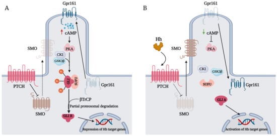
The Hedgehog (Hh)-glioma-associated oncogene homolog (GLI) signaling pathway is highly conserved among mammals, with crucial roles in regulating embryonic development as well as in cancer initiation and progression. The GLI transcription factors (GLI1, GLI2, and GLI3) are effectors of the Hh pathway and are regulated via Smoothened (SMO)-dependent and SMO-independent mechanisms. The SMO-dependent route involves the common Hh-PTCH-SMO axis, and mutations or transcriptional and epigenetic dysregulation at these levels lead to the constitutive activation of GLI transcription factors. Conversely, the SMO-independent route involves the SMO bypass regulation of GLI transcription factors by external signaling pathways and their interacting proteins or by epigenetic and transcriptional regulation of GLI transcription factors expression. Both routes of GLI activation, when dysregulated, have been heavily implicated in tumorigenesis of many known cancers, making them important targets for cancer treatment. 

GLI1 protein hedgehog pathway mutations epigenetic regulation glioma-associated oncogene noncanonical cancer clinical trial hedgehog inhibitors

1. Introduction

The hedgehog (Hh) signaling pathway was first discovered in Drosophila melanogaster’s embryonic cell [1]. Its evolution across various species is relatively conserved; however, duplication of the Hh gene in mammals revealed the involvement of three other members of the Hh family, namely the Sonic (Shh), Desert (Dhh), and Indian hedgehog (Ihh) [2]. Each of these genes has a diverse function in developing various tissues and organs; nevertheless, they utilize a similar pathway to be activated. In mammals, the Hh signaling is a rather complex relay mechanism that occurs in the primary cilium. Without the binding of Hh ligands to the Patched (PTCH), a 12-pass transmembrane protein receptor, the pathway remains suppressed due to the inhibitory effect of PTCH on the seven-pass transmembrane protein Smoothened (SMO) [3]. The binding of Hh ligands to PTCH relieves the inhibition of SMO protein, allowing its translocation into the primary cilium, where it rapidly accumulates [4]. Subsequently, activated SMO interferes with the proteolytic processing of glioma-associated oncogene homolog (GLI) proteins and promotes their dissociation from Suppressor of Fused (SUFU), allowing their translocation into the nucleus [5]. Through their DNA-binding domains, GLI activators (GLIAs) then bind to the GLI-binding consensus sequence 5′-GACCACCCA-3′ residing within promoters of target genes to initiate their gene transcription, such as cyclins (CCND1, CCND2), antiapoptotic factors (BCL2, BCLX), migratory genes (SNAI1, ZEB1), and its own pathway genes (PTCH1, GLI1) [6][7].
However, the diverse response of GLI in tissues is very dependent on the delicate balance between GLIAs and GLI repressors (GLIRs) combined. The negative regulation of GLI protein (Figure 1A) is regulated by its interaction with SUFU by virtue of its SUFU-binding domain. In the absence of the Hh ligand, SMO remains inactivated, which allows the tight association of SUFU with GLI [8]. GLI bound to SUFU is susceptible to phosphorylation events that promote its processing into repressors. G-protein coupled receptor 61 (Gpr161) localizes to the primary cilia to maintain high cyclic adenosine monophosphate (CAMP) levels and protein kinase A (PKA) activity [9], which phosphorylate P1-6 clusters located on GLI2/3 [10]. Their phosphorylation by PKA primes their subsequent phosphorylation by glycogen synthase kinase 3 beta (GSK3β) and casein kinase I (CKI) [11]. Phosphorylated GLI2/3 are recognized by the Cul1/β-TrCP complex, promoting their ubiquitination and subsequent proteasomal-dependent processing into GLIRs [12][13]. GLIRs then bind to the promoters of target genes to repress their transcription. In the presence of an Hh ligand, however, activation of SMO leads to the dephosphorylation of GLI2/3 P1-6 clusters and their dissociation from SUFU [10], favoring the translocation of GLIAs into the nucleus to initiate the transcription of target genes (Figure 1B). The expression of GLI1, a primary Hh target gene, serves to amplify Hh transduction at the transcriptional level further [14].
Figure 1. (A) The repression of Smoothened (SMO) by the Patched (PTCH) receptor in the absence of hedgehog (Hh) ligands promotes the interaction of Suppressor of Fused (SUFU) and glioma-associated oncogene homolog (GLI). G-protein coupled receptor 61 (GPR161) translocates to the primary cilium, which triggers high levels of cyclic adenosine monophosphate (CAMP). Elevated ciliary levels of CAMP maintain high levels of protein kinase A (PKA) activity, which phosphorylate GLI at P1-6 clusters. Consequently, phosphorylation of GLI by PKA prime its phosphorylation by casein kinase I (CKI) and glycogen synthase kinase 3 beta (GSK3β) further. Phosphorylated GLI is recognized by the β-TrCP, promoting its ubiquitination and partial proteasomal processing into a repressor. GLI repressor (GLIR) then translocates into the nucleus to repress target gene transcription. (B) The binding of the Hh ligand to the PTCH receptor alleviates its repression of SMO, allowing SMO translocation to the primary cilium. Activated SMO inhibits SUFU, allowing the dissociation of GLI from SUFU. Additionally, Gpr161 is removed from the primary cilium, causing low CAMP levels and PKA activity. The release of GLI from SUFU and low PKA activity results in the dephosphorylation of GLI, preventing its proteasomal processing into a repressor. Full-length GLI or GLI activator (GLIA) then translocates into the nucleus to transcribe target genes. Red upward triangle-headed arrow: upregulation; green downward triangle-headed arrow: downregulation; dotted black triangle-headed arrow: inactivation; bar-headed arrow: inhibition; dotted bar-headed arrow: loss of inhibition.
Typically, Hh signaling activation is classified into two general models: ligand-independent (Type I) and ligand-dependent (Type II and III) Hh signaling. This model centers around the various Hh pathway components leading to GLI activation, which can occur either through mutations in PTCH or SMO (ligand independent) or Hh ligand stimulation (ligand dependent); however, the transcriptional or epigenetic dysregulation of Hh pathway-related genes (e.g., aberrant methylation or excessive transcription factor activation) leading to GLI activation and the regulation of GLI beyond SMO transduction are often overlooked in this model. In this review, we describe a Hh signaling model that focuses on two different GLI regulation levels relevant to carcinogenesis: SMO-dependent and SMO-independent GLI activation. Arguably, these models provide a holistic view of the paradigms of hedgehog signaling networks involving GLI regulation at the SMO level or beyond and may be more relevant to current therapeutic strategies involving the development of SMO and GLI inhibitors for treating Hh-dependent cancers. Additionally, we present the latest clinical trial findings for the recent development of Hh inhibitors in cancer treatment and provide a comprehensive review concerning the relevance, limitations, and future perspective of SMO/GLI inhibitors as targeted cancer therapy. Importantly, GLI inhibitors have shown superior anticancer activity compared to inhibitors targeting upstream (Hh and SMO) of GLI in preclinical studies [15][16]. Furthermore, GLI inhibitors effectively suppress cancer growth in many GLI-dependent cancers that utilize an SMO-independent route of GLI regulation, of which treatment with upstream inhibitors has proven ineffective [17]. Thus, understanding GLI regulation paradigms is fundamental to developing novel GLI inhibitors worthy of moving forward to clinical settings, which may help set a new stage for Hh therapy in the future.

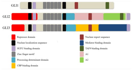
2. GLI Proteins and Their Domains

GLI is a part of the GLI-Kruppel family, characterized by the presence of C2H2-Kruppel-type zinc-finger (ZF) motifs [18]. Three homologs exist in vertebrates, namely GLI1, GLI2, and GLI3 (Figure 2). These proteins consist of overlapping domains, including a repressor and transactivation domain, and possess distinct but partially redundant functions. Since GLI1 lacks the repressor domain, it acts as a sole transcriptional enhancer. By contrast, GLI2 protein possesses both a repressor and two transactivation domains (TADs), A1 and A2, and acts as both a repressor and an activator. However, GLI2 mostly behaves as a transcriptional activator due to the inefficient processing of GLI2 into GLI2 repressor (GLI2R) [19]. Likewise, GLI3 protein possesses both repressor and activation domains but serves primarily as a transcriptional repressor due to an active processing determinant domain (PDD) that allows efficient processing of GLI3 into GLI3 repressor (GLI3R) [19][20]. Of note, GLI2 also contains a PDD but is inefficiently processed due to differences in amino residues critical for preventing complete degradation by the proteasome [20].
Figure 2. Schematic representation of the domains and motifs in glioma-associated oncogene homolog (GLI) proteins. All GLI proteins contain a well-conserved Supressor of Fused (SUFU)-binding domain, zinc finger motifs, nuclear localization sequences, and a nuclear export sequence. GLI2 and GLI3 contain both an N-terminal repressor and several C-terminal transactivation domains, unlike GLI1, which contains only a single transactivation domain reported so far. Additionally, GLI2 and GLI3 contain a second SUFU-binding domain at the C-terminal end critical for regulating nuclear GLI function. Both GLI2 and GLI3 contain a processing determinant domain that contributes to the proteolytic processing of these proteins into their repressor form with a more active role in GLI3 than GLI2. GLI2 contains two major transactivation domains, termed A1 and A2, while the GLI3 transactivation domain includes a CREB-binding protein (CBP)-binding domain and mediator-binding domain. Both GLI1 and GLI2 contain an α-helical herpes simplex viral protein 16-like activation domain that binds to TATA-box binding protein associated factor 9 (TAF9) due to the presence of a highly conserved FXXΦΦ (F = phenylalanine; X = any residue; Φ = any hydrophobic residue) motif in the domain. The FXXΦΦ motif is also conserved in GLI3 but does not bind to TAF9.
All three GLI homologs contain five Krüppel-like ZF motifs that recognize and bind to a nine base-pair DNA consensus motif 5′-GACCACCCA-3′. ZF4 and ZN5 mediate the binding of GLI proteins to the conserved DNA motif, while ZNF1-3 stabilizes the DNA domain through interaction with the phosphate backbone [21][22]. All three GLI homologs also contained a highly conserved SUFU-binding domain and two putative nuclear localization sequences (NLSs), including NLS1 and NLS2. NSL2 is a canonical bipartite NLS containing two basic clusters mapped to the fifth ZF motif in Ci and C-terminal side of GLI proteins, while NLS1 has features of both a canonical monopartite NLS and a noncanonical proline-tyrosine (PY)-NLS located just upstream of the SUFU-binding domain [23]. Both NLSs play a cooperative functional role in regulating the nuclear localization of GLI proteins, likely through importin (Imp)-α/β1 mediated nuclear import classic of canonical NLSs [24].
Mutations in either the NLS1 or NLS2 have been shown to partially impair the nuclear transportation of both GLI1 and GLI2, while the loss of both NLSs results in a drastic increase in cytoplasmic localization of both GLI proteins [25][26]. The function of NLS2 is also heavily regulated by a Thr374 residue adjacent to the first basic cluster of the bipartite motif. Phosphorylation of Thr374 residue by PKA enhanced the local negative charge nearby the NLS2, thus inhibiting NLS2 function and consequently inhibiting GLI1 nuclear accumulation [27]. The noncanonical PY-NLS feature of NLS1 has also been shown to mediate GLI2 and GLI3 nuclear transport by cooperating with karyopherin-β2 (Kapβ2) known to recognize PY-NLSs [28]. Besides regulating nuclear transportation, the interaction of PY-NLS and Kapβ2 also plays a major role in regulating the ciliary localization of all three GLI proteins independently of the Imp-α/β1 nuclear import system [25][29].
A leucine-rich nuclear export sequence (NES) is also found in all three mammalian GLI proteins, and their functional coordination with NLSs plays a major role in regulating the nuclear-cytoplastic shuttling of GLI proteins. The nuclear export of GLI1 and GLI2 is chromosomal region maintenance 1 (CRM1)-dependent, and inactivation of CRM1 with Leptomycin B (LMB) or the substitution of conserved leucine residues within the GLI1/GLI2 NES was shown to enhance the nuclear accumulation and transcriptional activities of both GLI proteins [26][30]. Interestingly, the shuttling of the SUFU-GLI1 complex between the cytoplasmic and nuclear compartments was found to depend on functional NES and CRM1, whereby the loss of either function led to an impaired cytoplasmic sequestration of GLI1 by SUFU and enhanced colocalization of SUFU and GLI1 in the nucleus [26].
SUFU binds to SUFU-binding domains located at N- and C-terminals of GLI proteins to regulate their activities through various mechanisms. Firstly, SUFU sequesters GLI proteins in the cytoplasm by binding to the SYGH core motif within the N-terminal domain of GLI proteins [31]. This interaction facilitates the phosphorylation of GLI proteins by PKA, GSK3β, and CK1, leading to their proteasomal degradations or processing into repressors [8][12]. Secondly, SUFU prevents the nuclear translocation of GLI proteins by masking their NLSs. The binding of SUFU to the SYGH core motif within the N-terminal domain of GLI proteins blocks the binding of Impβ1 and Kapβ2 to NLSs, which impedes the nuclear import of GLI proteins [24][28]. Thirdly, SUFU has been shown to regulate Ci/GLI transcriptional activity inside the nucleus. The binding of SUFU to a second conserved C-terminal SUFU-binding domain of Ci masked the Drosophila CREB-binding protein (CBP)-binding domain (dCBD) located near the SUFU-binding domain, impeding the recruitment of the transcriptional coactivator CBP. In turn, the loss of CBP recruitment inhibited the transcriptional activity of Ci; the C-terminal SUFU-binding site was also conserved in GLI2 and GLI3, and deletion of this site partially blocked the SUFU-mediated inhibition of GLI2 transcriptional activity [31], recapitulating the effect of SUFU-binding at the C-terminal SUFU-binding domain in the impediment of CBP recruitment and suppression of Ci transcriptional activity.
SUFU also negatively regulates the transcriptional activity of GLI1 in the nucleus despite the loss of CRM1-dependent nuclear export (inhibited by LMB), suggesting that the repressive activity of SUFU can still occur in the nucleus independent of cytoplasmic sequestration [26]. In further support of this finding, SUFU was also shown to interact directly with GLI1 bound to DNA, raising the possibility that this interaction may hinder the binding of other transcription activators with GLI1 [30]. By contrast, Zhang et al. argued that SUFU does not serve merely as a negative regulator but rather as a chaperone protein with a unifying role in regulating the function of GLI proteins [32]. Evidently, SUFU accompanied the translocation of GLI1 into and GLIR out of the nucleus. Furthermore, SUFU also accompanied GLI1 but not GLIR to the ciliary tip, a necessary step that precedes the translocation of GLI1 into the nucleus. Importantly, SUFU facilitates the binding of GLI1 to GLI-binding sites on the chromatin, while concomitantly reducing GLI3 binding, and intact SUFU expression is required for maximal Shh signaling output needed for the specification of the most ventral neurons [32].
The Drosophila CBP (dCBP) has been shown to bind to the dCBD of Ci as a coactivator, while the loss of dCBP abolished Hh signaling [33]. Sequence alignment revealed a motif fairly well conserved between the dCBP-binding domain of Ci and the A1 domain of GLI2 [34], but the role of CBP in GLI2 activity has yet to be elucidated. Like Ci, GLI3 also possesses a CBP-binding domain (CBD) and utilizes CBP as a coactivator for its transcriptional activity [35]. By contrast, Zhou et al. reported that CBD showed weak transactivation in vivo, but CBP could bind efficiently to the Mediator-binding domain (MBD) located upstream of CBD to promote GLI3 transactivation, suggesting a concerted functional interaction between CBP and RNA polymerase II transcriptional mediator complex [36]. Besides binding CBP, MBD also physically targeted and inhibited the MED12 interface in the mediator complex, which in turn reversed the mediator-dependent suppression of GLI3 transactivation activity [36]. By contrast, CBP does not bind to GLI1 [35], suggesting the lack of a CBD or MBD in GLI1.
The C-terminal end of the GLI1 contains an α-helical herpes simplex viral protein 16-like activation domain, including a highly conserved FXXΦΦ (F = phenylalanine; X = any residue; Φ = any hydrophobic residue) motif recognizing TAFII31/TATA-box binding protein associated factor 9 (TAF9) subunit of general transcription factor II D [37]. This motif is fairly conserved in the A2 domain of GLI2 and the C-terminal end of GLI3 [37][38]. However, TAF9 binds only to GLI1 and GLI2 but not GLI3 to promote their transcriptional activities, suggesting a redundancy of the FXXΦΦ motif in GLI3. Conversely, binding interference between GLI proteins and TAF9 by mutating the FXXΦΦ motif resulted in the loss of transcriptional activities of GLI proteins [37][38][39].
Both GLI2 and GLI3 contain an N-terminal repressor domain (RD) that exerts repressive transcriptional activity upon proteolytic removal of their C-terminal TADs. In contrast to the TAD of GLI proteins, their RDs are less well characterized in terms of their motifs and binding partners. The RD is most well defined for its interaction with the histone deacetylase (HDAC) complex. Ski was shown to interact directly with the N-terminal domain of both GLI3R and full-length GLI3 and to form a complex with HDAC1 to promote GLI3-mediated transcriptional repression. Additionally, a Ski-binding site was also mapped to the N-terminal RD of GLI2. Conversely, Ski-deficient mouse embryonic fibroblast (MEF) efficiently abrogated GLI3 and GLI2 transcriptional repressive activities. Ski forms complexes with corepressors such as N-CoR/SMRT, mSin3, and Sno to recruit HDACs necessary to mediate transcriptional repression activities of other repressors [40]. Mouse SUFU has been shown to interact with SAP18, a member of the mSin3-HDAC corepressor complex, to enhance GLI3-mediated transcriptional repression and impaired GLI1 transcriptional activity. Functionally, mouse SUFU interacted with GLI1, possibly via the SYGF motif in the N-terminal SUFU-binding domain and recruited the mSin3-HDAC complex through interaction with SAP18 to impede GLI1 transcriptional activity. It is conceivable that the same process may also occur in GLI3 to potentiate GLI3 transcriptional repressive activity, as both GLI1 and GLI3 interact with SUFU via the same SYGH motif at the N-terminal end. Furthermore, the Ski binding site overlaps the SUFU-binding domain at the N-terminal region of GLI3, suggesting a possible functional cooperative role between SUFU and Ski in recruiting the HDAC corepressor complex to promote GLI3-mediated transcriptional repression activity [41].

References

  1. Nüsslein-volhard, C.; Wieschaus, E. Mutations Affecting Segment Number and Polarity in Drosophila. Nature 1980, 287, 795–801.
  2. Carballo, G.B.; Honorato, J.R.; De Lopes, G.P.F.; de Sampaio, E.; Spohr, T.C.L. A Highlight on Sonic Hedgehog Pathway. Cell Commun. Signal. 2018, 16, 11.
  3. Mastronardi, F.G.; Dimitroulakos, J.; Kamel-Reid, S.; Manoukian, A.S. Co-Localization of Patched and Activated Sonic Hedgehog to Lysosomes in Neurons. Neuroreport 2000, 11, 581–585.
  4. Rohatgi, R.; Milenkovic, L.; Corcoran, R.B.; Scott, M.P. Hedgehog Signal Transduction by Smoothened: Pharmacologic Evidence for a 2-Step Activation Process. Proc. Natl. Acad. Sci. USA 2009, 106, 3196–3201.
  5. Hsu, S.H.C.; Zhang, X.; Yu, C.; Li, Z.J.; Wunder, J.S.; Hui, C.C.; Alman, B.A. Kif7 Promotes Hedgehog Signaling in Growth Plate Chondrocytes by Restricting the Inhibitory Function of Sufu. Development 2011, 138, 3791–3801.
  6. Skoda, A.M.; Simovic, D.; Karin, V.; Kardum, V.; Vranic, S.; Serman, L. The Role of the Hedgehog Signaling Pathway in Cancer: A Comprehensive Review. Bosn. J. Basic Med. Sci. 2018, 18, 8–20.
  7. Gonnissen, A.; Isebaert, S.; Haustermans, K. Targeting the Hedgehog Signaling Pathway in Cancer: Beyond Smoothened. Oncotarget 2015, 6, 13899–13913.
  8. Jia, J.; Zhang, L.; Zhang, Q.; Tong, C.; Wang, B.; Hou, F.; Amanai, K.; Jiang, J. Phosphorylation by Double-Time/CKIε and CKIα Targets Cubitus Interruptus for Slimb/β-TRCP-Mediated Proteolytic Processing. Dev. Cell 2005, 9, 819–830.
  9. Tschaikner, P.; Enzler, F.; Torres-Quesada, O.; Aanstad, P.; Stefan, E. Hedgehog and Gpr161: Regulating CAMP Signaling in the Primary Cilium. Cells 2020, 9, 118.
  10. Niewiadomski, P.; Kong, J.H.; Ahrends, R.; Ma, Y.; Humke, E.W.; Khan, S.; Teruel, M.N.; Novitch, B.G.; Rohatgi, R. Gli Protein Activity Is Controlled by Multisite Phosphorylation in Vertebrate Hedgehog Signaling. Cell Rep. 2014, 6, 168–181.
  11. Price, M.A.; Kalderon, D. Proteolysis of the Hedgehog Signaling Effector Cubitus Interruptus Requires Phosphorylation by Glycogen Synthase Kinase 3 and Casein Kinase 1. Cell 2002, 108, 823–835.
  12. Shafique, S.; Rashid, S. Structural Basis of ΒTrCP1-Associated GLI3 Processing. Sci. Rep. 2019, 9, 6865.
  13. Pan, Y.; Wang, C.; Wang, B. Phosphorylation of Gli2 by Protein Kinase A Is Required for Gli2 Processing and Degradation and the Sonic Hedgehog-Regulated Mouse Development. Dev. Biol. 2009, 326, 177–189.
  14. Sabol, M.; Trnski, D.; Musani, V.; Ozretić, P.; Levanat, S. Role of GLI Transcription Factors in Pathogenesis and Their Potential as New Therapeutic Targets. Int. J. Mol. Sci. 2018, 19, 2562.
  15. Zubčić, V.; Rinčić, N.; Kurtović, M.; Trnski, D.; Musani, V.; Ozretić, P.; Levanat, S.; Leović, D.; Sabol, M. GANT61 and Lithium Chloride Inhibit the Growth of Head and Neck Cancer Cell Lines Through the Regulation of GLI3 Processing by GSK3β. Int. J. Mol. Sci. 2020, 21, 6410.
  16. Miyazaki, Y.; Matsubara, S.; Ding, Q.; Tsukasa, K.; Yoshimitsu, M.; Kosai, K.i.; Takao, S. Efficient Elimination of Pancreatic Cancer Stem Cells by Hedgehog/GLI Inhibitor GANT61 in Combination with MTOR Inhibition. Mol. Cancer 2016, 15, 49.
  17. Pietrobono, S.; Gagliardi, S.; Stecca, B. Non-Canonical Hedgehog Signaling Pathway in Cancer: Activation of GLI Transcription Factors beyond Smoothened. Front. Genet. 2019, 10, 556.
  18. Zhu, H.; Lo, H.-W. The Human Glioma-Associated Oncogene Homolog 1 (GLI1) Family of Transcription Factors in Gene Regulation and Diseases. Curr. Genomics 2010, 11, 238–245.
  19. Niewiadomski, P.; Niedziółka, S.M.; Markiewicz, Ł.; Uśpieński, T.; Baran, B.; Chojnowska, K. Gli Proteins: Regulation in Development and Cancer. Cells 2019, 8, 147.
  20. Pan, Y.; Wang, B. A Novel Protein-Processing Domain in Gli2 and Gli3 Differentially Blocks Complete Protein Degradation by the Proteasome. J. Biol. Chem. 2007, 282, 10846–10852.
  21. Kinzler, K.W.; Vogelstein, B. The GLI Gene Encodes a Nuclear Protein Which Binds Specific Sequences in the Human Genome. Mol. Cell. Biol. 1990, 10, 634.
  22. Pavletich, N.; Pabo, C. Crystal Structure of a Five-Finger GLI-DNA Complex: New Perspectives on Zinc Fingers. Science 1993, 261, 1701–1707.
  23. Hatayama, M.; Aruga, J. Gli Protein Nuclear Localization Signal. Vitam. Horm. 2012, 88, 73–89.
  24. Szczepny, A.; Wagstaff, K.M.; Dias, M.; Gajewska, K.; Wang, C.; Davies, R.G.; Kaur, G.; Ly-Huynh, J.; Loveland, K.L.; Jans, D.A. Overlapping Binding Sites for Importin Β1 and Suppressor of Fused (SuFu) on Glioma-Associated Oncogene Homologue 1 (Gli1) Regulate Its Nuclear Localization. Biochem. J. 2014, 461, 469–476.
  25. Torrado, B.; Graña, M.; Badano, J.L.; Irigoín, F. Ciliary Entry of the Hedgehog Transcriptional Activator Gli2 Is Mediated by the Nuclear Import Machinery but Differs from Nuclear Transport in Being Imp-α/Β1-Independent. PLoS ONE 2016, 11, e0162033.
  26. Barnfield, P.C.; Zhang, X.; Thanabalasingham, V.; Yoshida, M.; Hui, C. Negative Regulation of Gli1 and Gli2 Activator Function by Suppressor of Fused through Multiple Mechanisms. Differentiation 2005, 73, 397–405.
  27. Sheng, T.; Chi, S.; Zhang, X.; Xie, J. Regulation of Gli1 Localization by the CAMP/Protein Kinase A Signaling Axis through a Site Near the Nuclear Localization Signal. J. Biol. Chem. 2006, 281, 9–12.
  28. Shi, Q.; Han, Y.; Jiang, J. Suppressor of Fused Impedes Ci/Gli Nuclear Import by Opposing Trn/Kapb2 in Hedgehog Signaling. J. Cell Sci. 2014, 127, 1092–1103.
  29. Han, Y.; Xiong, Y.; Shi, X.; Wu, J.; Zhao, Y.; Jiang, J. Regulation of Gli Ciliary Localization and Hedgehog Signaling by the PY-NLS/Karyopherin-Β2 Nuclear Import System. PLOS Biol. 2017, 15, e2002063.
  30. Kogerman, P.; Grimm, T.; Kogerman, L.; Krause, D.; Undén, A.B.; Sandstedt, B.; Toftgård, R.; Zaphiropoulos, P.G. Mammalian Suppressor-of-Fused Modulates Nuclear-Cytoplasmic Shuttling of GLI-1. Nat. Cell Biol. 1999, 1, 312–319.
  31. Han, Y.; Shi, Q.; Jiang, J. Multisite Interaction with Sufu Regulates Ci/Gli Activity through Distinct Mechanisms in Hh Signal Transduction. Proc. Natl. Acad. Sci. USA 2015, 112, 6383–6388.
  32. Zhang, Z.; Shen, L.; Law, K.; Zhang, Z.; Liu, X.; Hua, H.; Li, S.; Huang, H.; Yue, S.; Hui, C.; et al. Suppressor of Fused Chaperones Gli Proteins To Generate Transcriptional Responses to Sonic Hedgehog Signaling. Mol. Cell. Biol. 2017, 37, e00421-16.
  33. Akimaru, H.; Chen, Y.; Dai, P.; Hou, D.-X.; Nonaka, M.; Smolik, S.M.; Armstrong, S.; Goodman, R.H.; Ishii, S. Drosophila CBP Is a Co-Activator of Cubitus Interruptus in Hedgehog Signalling. Nature 1997, 386, 735–738.
  34. Hughes, D.C.; Allen, J.; Morley, G.; Sutherland, K.; Ahmed, W.; Prosser, J.; Lettice, L.; Allan, G.; Mattei, M.G.; Farrall, M.; et al. Cloning and Sequencing of the Mouse Gli2 Gene: Localization to the Dominant Hemimelia Critical Region. Genomics 1997, 39, 205–215.
  35. Dai, P.; Akimaru, H.; Tanaka, Y.; Maekawa, T.; Nakafuku, M.; Ishii, S. Sonic Hedgehog-Induced Activation of the Gli1Promoter Is Mediated by GLI3. J. Biol. Chem. 1999, 274, 8143–8152.
  36. Zhou, H.; Kim, S.; Ishii, S.; Boyer, T.G. Mediator Modulates Gli3-Dependent Sonic Hedgehog Signaling. Mol. Cell. Biol. 2006, 26, 8667.
  37. Yoon, J.W.; Liu, C.Z.; Yang, J.T.; Swart, R.; Iannaccone, P.; Walterhouse, D. GLI Activates Transcription through a Herpes Simplex Viral Protein 16-Like Activation Domain. J. Biol. Chem. 1998, 273, 3496–3501.
  38. Yoon, J.W.; Lamm, M.; Iannaccone, S.; Higashiyama, N.; Leong, K.F.; Iannaccone, P.; Walterhouse, D. P53 Modulates The Activity Of The GLI1 Oncogene Through Interactions With The Shared Coactivator TAF9. DNA Repair 2015, 34, 9.
  39. Bosco-Clément, G.; Zhang, F.; Chen, Z.; Zhou, H.M.; Li, H.; Mikami, I.; Hirata, T.; Yagui-Beltran, A.; Lui, N.; Do, H.T.; et al. Targeting Gli Transcription Activation by Small Molecule Suppresses Tumor Growth. Oncogene 2014, 33, 2087–2097.
  40. Dai, P.; Shinagawa, T.; Nomura, T.; Harada, J.; Kaul, S.C.; Wadhwa, R.; Khan, M.M.; Akimaru, H.; Sasaki, H.; Colmenares, C.; et al. Ski Is Involved in Transcriptional Regulation by the Repressor and Full-Length Forms of Gli3. Genes Dev. 2002, 16, 2843.
  41. Cheng, S.Y.; Bishop, J.M. Suppressor of Fused Represses Gli-Mediated Transcription by Recruiting the SAP18-MSin3 Corepressor Complex. Proc. Natl. Acad. Sci. USA 2002, 99, 5442–5447.
More
Information
Subjects: Oncology
Contributor MDPI registered users' name will be linked to their SciProfiles pages. To register with us, please refer to https://encyclopedia.pub/register :
View Times: 481
Revisions: 2 times (View History)
Update Date: 15 Sep 2021
1000/1000