Submitted Successfully!
To reward your contribution, here is a gift for you: A free trial for our video production service.
Thank you for your contribution! You can also upload a video entry or images related to this topic.
Version Summary Created by Modification Content Size Created at Operation
1 + 1750 word(s) 1750 2021-07-31 16:15:54

Video Upload Options

Do you have a full video?

Confirm

Are you sure to Delete?
Cite
If you have any further questions, please contact Encyclopedia Editorial Office.
Arrigo, A. Retinal Diseases. Encyclopedia. Available online: https://encyclopedia.pub/entry/13209 (accessed on 20 April 2024).
Arrigo A. Retinal Diseases. Encyclopedia. Available at: https://encyclopedia.pub/entry/13209. Accessed April 20, 2024.
Arrigo, Alessandro. "Retinal Diseases" Encyclopedia, https://encyclopedia.pub/entry/13209 (accessed April 20, 2024).
Arrigo, A. (2021, August 16). Retinal Diseases. In Encyclopedia. https://encyclopedia.pub/entry/13209
Arrigo, Alessandro. "Retinal Diseases." Encyclopedia. Web. 16 August, 2021.
Retinal Diseases
Edit

The human retina may be affected by two macro groups of diseases, namely maculopathies and retinopathies. Whereas maculopathies are confined to the central part of the retina, bounded by the vascular arcades, retinopathies may extend up to the extreme retinal periphery. These two categories can be further subdivided according to the main features characterizing the disease, thus taking into consideration exudative or atrophic phenomena.

retinal diseases anti-VEGF corticosteroids intravitreal injections complement inhibitors chemokine receptor inhibitors integrins inhibitors tyrosine kinase inhibitors nutraceutics

1. Introduction

The management of exudative retinal diseases underwent a revolution due to the introduction of intravitreal treatments. There are two main classes of intravitreal drugs, namely anti-vascular endothelial growth factors (anti-VEGF) and corticosteroids molecules. The clinical course and the outcome of retinal diseases radically changed thanks to the efficacy of these molecules in determining the regression of the exudation and the restoration of the macular profile.

Exudation is an active process, and its nature depends on each specific retinal disease, causing fluid to accumulate within the retina or in the subretinal space. It mainly involves variable amounts of fluid, the major pathogenic features of which are the breakdown of the blood-retinal barrier and increased inflammation [1][2][3]. Retinal diseases can also be characterized by other types of debris, including lipofuscin and lipidic and proteinaceous materials [3][4]. Retinal diseases can also be characterized by the progressive degeneration of inner and outer retinal layers. These atrophic changes may occur independently or in the context of an initial exudative disease [3][5].

Current retinal therapeutic approaches are based on these premises and designed to prompt the exudation to regress, stimulate debris reabsorption or prevent the atrophy from expanding.

2. Retinal Drugs for Exudative Diseases

The prognosis of retinal exudative diseases changed radically after the introduction of intravitreal therapies. While the old laser-based treatments were effective in blocking exudation, they were associated with an extremely poor visual outcome [6][7][8]; nowadays, patients can expect to preserve their quality of life and a good visual function. The current intravitreal therapeutic bullets consist of anti-vascular endothelial growth factor (anti-VEGF) and corticosteroids. The pros of anti-VEGF drugs are their easier management and the low instance of side effects; the cons comprise their limited duration, meaning a large number of injections are required, and their contraindication in patients displaying a high risk of cardiovascular dysfunction. In contrast, the pros of corticosteroids include their longer duration, thus reducing the number of injections administered and their greater anti-inflammatory action. Conversely, corticosteroids are closely associated with an increase in intraocular pressure and a faster progression of cataracts.

3. The Role of Inflammation in the Human Retina

All retinal diseases are characterized by pro-inflammatory alterations, differing according to the specific pathogenic features. Inflammation may derive from progressive accumulation of pathologic debris, such as in the first stages of age-related macular degeneration; in these cases, pro-inflammatory mediators are stimulated by progressive and chronic increases of oxidative stress and cytotoxicity produced by these accumulations [9]. Furthermore, the progressive degeneration of retinal cells may stimulate complement activation and lead to the accumulation of microglia and inflammatory cells, and the onset and progression of pro-inflammatory phenomena [10]. At the same time, some retinal diseases reveal major pro-inflammatory sources. Inflammation is a key component of diabetes mellitus and diabetic retinopathy; progressive metabolic dysfunctions leading to increasing levels of advanced glycation end products and free radicals causes a chronic pro-inflammatory status, with progressive increases of pro-inflammatory cytokines, chemokines, and other inflammatory mediators, the accumulation of inflammatory cells and increases in the vascular permeability [11][12]. High pro-inflammatory mediator release is also a major phenomenon found in another frequent retinal disorder, namely retinal vein occlusion, where increasing levels of pro-inflammatory cytokines, interleukins and other factors have been encountered [13]. Inflammation is the key mechanism in uveitis [14] and is also involved in the pathogenesis of retinal dystrophies [15].

4. Emerging Therapies for Exudative Retinal Diseases
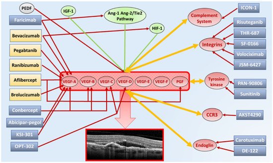
A comprehensive scenario regarding the present and future intravitreal drugs for exudative retinal diseases, with particular regards to exudative AMD, are reported in Figure 3.
Figure 3. Summary diagram showing present and experimental molecular targets and corresponding intravitreal drugs for exudative age-related macular degeneration (AMD). The upregulation of vascular endothelial grow factor (VEGF) family expression, made by VEGF-A, VEGF-B, VEGF-C, VEGF-D, VEGF-E, VEGF-F and PGF, is responsible for the onset and progression of macular neovascularization and fluid production in exudative AMD. All VEGF molecule subtypes bind to different types of VEGF tyrosine kinase trans-membrane receptors. VEGF family production and release is further stimulated by increased levels of angiotensin 1-2 (Ang-1-Ang-2)/Tie2 pathway, insulin-like growth factor 1 (IGF-1) and hypoxia inducible factor-1 (HIF-1) (green arrows). Furthermore, a natural VEGF inhibitor is represented by PEDF (red arrow), resulting dysregulated in exudative AMD. Currently, approved anti-VEGF molecules include bevacizumab, pegaptanib, ranibizumab, aflibercept and brolucizumab. Although most of current anti-VEGF drugs mainly bind VEGF-A isoforms (red arrows), bevacizumab shows mild affinity also for HIF-1, while aflibercept also binds PGF. Anti-VEGF molecules under investigation include faricimab (VEGF-A isoforms + Ang-1-Ang-2/Tie2 pathway), conbercept (VEGF-A + VEGF-B + VEGF-C isoforms), abicipar-pegol (VEGF-A isoforms), KSI-301 (VEGF-A isoforms) and OPT-302 (VEGF-C + VEGF-D isoforms). Exudative AMD is also characterized by upregulation of complement system, integrins, tyrosine kinase, eotaxin C-C chemokine receptor type 3 (CCR3) and endoglin, which contribute to the increased production and release of VEGF family and often are themselves stimulated by the increased level of VEGF isoforms (orange arrows). Several molecules are currently under investigation through multicenter clinical trials, targeting these cofactors of the neovascular process, including complement system inhibitors (ICON-1), integrins inhibitors (risuteganib, THR-687, SF-0166, volociximab, JSM-6427), tyrosine kinase inhibitors (PAN-0806, sunitinib), CCR3 inhibitors (AKST4290) and endoglin inhibitors (carotuximab, DE-122).

4.1. AKST4290

AKST4290 (Alkahest, San Carlos, CA, USA) is the inhibitor of the natural receptor for eotaxin C-C chemokine receptor type 3 (CCR3), a molecule proving to be highly expressed in choroidal neovascularization [16]. This molecule is under investigation as an oral formulation (400 mg), combined with intravitreal anti-VEGF injections, in AKST4290–201 (NCT03558061) and AKST4290–202 (NCT03558074) phase 2a clinical trials.

4.2. Carotuximab

Carotuximab (DE-122) (SANTEN, Osaka, Japan; TRACON Pharmaceuticals, San Diego, CA, USA) is an antibody directed against endoglin, and was developed on the basis of evidence showing endoglin to be actively involved in angiogenesis [17]. It has been found that a hypoxic status may stimulate the production of endoglin, which is also enhanced in actively proliferating endothelial cells. A mouse model of neovascularization has provided promising results regarding the use of anti-endoglin in combination with anti-VEGF injections [18]. DE-122 is under investigation in a phase 2a randomized controlled trial (NCT03211234).

4.3. Complement Inhibitors

The complement system is made up of an extremely complex network of molecules that play a fundamental role in innate immunity and in the activation of the inflammatory cascade. The role of the complement system has been widely demonstrated in both exudative and dry retinal diseases; complement activation is a major factor in enhancing pro-inflammatory mechanisms and in the onset and progression of cell death [19][20][21]. For these reasons, complement inhibitors are currently under investigation for both forms of retinal diseases, through the development of different molecules acting at multiple levels in the complement system’s activation cascade. The evidence regarding the efficacy of complement inhibitor drugs is not conclusive as yet, and there are many ongoing clinical trials designed to demonstrate the rationale for employing these molecules in wet and dry retinal diseases [22][23][24].

4.4. Integrins Inhibitors

Integrins are a major class of cell adhesion receptors for extracellular matrix molecules. They are heavily involved in retinal development, as well as being major regulatory factors in cell adhesion, migration, proliferation, invasion and apoptosis [25][26]. These features make the molecules promising targets for several retinal diseases. Many integrin inhibitor molecules are currently under investigation, including Risuteganib (Luminate, Allegro Ophthalmics, CA, USA), THR-687 (Oxurion, Leuven, Belgium), SF-0166 (SciFluor Life Sciences, Boston, MA, USA), Volociximab (Ophthotech Corporation, New York, NY, USA, Now Iveric Bio, New York, NY, USA) and JSM-6427 (Takeda Pharmaceutical Company, Tokyo, Japan) [26].

4.5. KSI-301

KSI-301 (KODIAK sciences, Palo Alto, CA, USA), is a new generation antibody biopolymer conjugate resulting from the combination of humanized anti-VEGF monoclonal antibody and a phosphorylcholine-based polymer, which has been developed to increase the duration of anti-VEGF action. This molecule is under investigation in a phase 1b trial (NCT03790852), showing promising results in neovascular age-related macular degeneration, diabetic macular edema and retinal vein occlusion, and in a DAZZLE Phase 2 trial (NCT04049266) involving only neovascular age-related macular degeneration patients.

5. Retinal Drugs for Non-Exudative Diseases

Unlike exudative retinal diseases, non-exudative retinal diseases lack any approved treatment, to date. These disorders, caused by multifactorial etiopathogenesis, including environmental and genetic factors, mainly involve the progressive degeneration of retinal cells, with onset of retinal atrophy. Most non-exudative retinal diseases start with the degeneration of the retinal pigment epithelium-photoreceptor complex; outer retinal impairment and atrophy is followed by damage to the inner retinal cells, with irremediable loss of retinal function [27][28][29].
Treatment for non-exudative retinal diseases is based on nutraceutics, which employs a combination of different natural substances known to slow down degenerative processes. Although several molecules are under investigation, most of the evidence concerns formulations developed for dry age-related macular degeneration by Age-Related Eye Disease Studies 1 and 2 (AREDS1 and AREDS2) [30]. The AREDS1 formulation contained vitamin C (500 mg), vitamin E (273 mg/473 IU), beta-carotene (15 mg), zinc (80 mg), and copper (2 mg) [31], whereas the AREDS2 formulation differed in the removal of beta-carotene and addition of lutein/zeaxanthin and omega-3 long-chain polyunsaturated fatty acid [32]. Vitamins and ions are important antioxidants and promote enzymatic functions. Lutein/zeaxanthin produce many effects, including removing reactive oxygen species and protecting against photooxidative stress. Omega-3 long-chain polyunsaturated fatty acids are key cell membrane components and are involved in many metabolic pathways. These nutraceutical supplements have been associated with a slowing in the progression of geographic atrophy and a reduced probability of experiencing advances in the stages of age-related macular degeneration. Different formulations of vitamins, polyunsaturated fatty acids, minerals and other compounds have been proposed for inherited retinal dystrophies, supported by modest evidence regarding their clinical efficacy [33].

5.1. Complement Inhibitors

Complement inhibitors [34] have already been discussed as a potential new treatment for exudative retinal diseases. In the context of dry retinal diseases, their role should cover the inhibition of the molecular mechanisms leading to cell degeneration and apoptosis. Current complement inhibitor drugs under investigation include Lampalizumab (NCT01229215), Zimura (ARC-1905) (NCT02686658), APL-4 (POT-4/AL-78898A) (NCT03525613-NCT03525600), CLG561 (NCT02515942) and LFG316 (NCT01527500).

5.2. Brimonidine

Brimonidine is a selective α2 adrenergic (α2A) receptor agonist used as intraocular pressure-lowering medication in glaucoma. Previous evidence suggested this molecule plays a neuroprotective role through as yet poorly understood mechanisms [35][36]. This molecule is under investigation in a Brimonidine Intravitreal Implant in Geographic Atrophy Secondary to Age-related Macular Degeneration (BEACON) phase 2 study (NCT02087085).

References

  1. Díaz-Coránguez, M.; Ramos, C.; Antonetti, D.A. The inner blood-retinal barrier: Cellular basis and development. Vision Res. 2017, 139, 123–137.
  2. Antonetti, D.A.; Klein, R.; Gardner, T.W. Diabetic retinopathy. N. Engl. J. Med. 2012, 366, 1227–1239.
  3. De Jong, P.T. Age-related macular degeneration. N. Engl. J. Med. 2006, 355, 1474–1485.
  4. Davoudi, S.; Papavasileiou, E.; Roohipoor, R.; Cho, H.; Kudrimoti, S.; Hancock, H.; Hoadley, S.; Andreoli, C.; Husain, D.; James, M.; et al. Optical coherence tomography characteristics of macular edema and hard exudates and their association with lipid serum levels in type 2 diabetes. Retina 2016, 36, 1622–1629.
  5. Jager, R.D.; Mieler, W.F.; Miller, J.W. Age-related macular degeneration. N. Engl. J. Med. 2008, 358, 2606–2617.
  6. Jost, B.F.; Alexander, M.F.; Maguire, M.G.; Fine, S.L.; Chamberlin, J.A.; Murphy, R.P. Laser treatment for choroidal neovascularization outside randomized clinical trials. Arch. Ophthalmol. 1988, 106, 357–361.
  7. Shah, C.P.; Chen, C. Review of therapeutic advances in diabetic retinopathy. Ther. Adv. Endocrinol. Metab. 2011, 2, 39–53.
  8. Stenner, A.M.; Frederiksen, K.H.; Grauslund, J. Is there still a role of macular laser treatment in branch retinal vein occlusion in the era of intravitreal injections? Acta Ophthalmol. 2020, 98, 9–21.
  9. Hageman, G.S.; Luthert, P.J.; Victor Chong, N.H.; Johnson, L.V.; Anderson, D.H.; Mullins, R.F. An integrated hypothesis that considers drusen as biomarkers of immune-mediated processes at the RPE-Bruch’s membrane interface in aging and age-related macular degeneration. Prog. Retin. Eye Res. 2001, 20, 705–732.
  10. Xu, H.; Chen, M.; Forrester, J.V. Para-inflammation in the aging retina. Prog. Retin. Eye Res. 2009, 28, 348–368.
  11. Tang, J.; Kern, T.S. Inflammation in diabetic retinopathy. Prog. Retin. Eye Res. 2011, 30, 343–358.
  12. Joussen, A.M.; Murata, T.; Tsujikawa, A.; Kirchhof, B.; Bursell, S.E.; Adamis, A.P. Leukocyte-mediated endothelial cell injury and death in the diabetic retina. Am. J. Pathol. 2001, 158, 147–152.
  13. Koss, M.J.; Pfister, M.; Rothweiler, F.; Michaelis, M.; Cinatl, J.; Schubert, R.; Koch, F.H. Comparison of cytokine levels from undiluted vitreous of untreated patients with retinal vein occlusion. Acta Ophthalmol. 2012, 90, e98–e103.
  14. De Smet, M.D.; Taylor, S.R.; Bodaghi, B.; Miserocchi, E.; Murray, P.I.; Pleyer, U.; Zierhut, M.; Barisani-Asenbauer, T.; LeHoang, P.; Lightman, S. Understanding uveitis: The impact of research on visual outcomes. Prog. Retin. Eye Res. 2011, 30, 452–470.
  15. Olivares-González, L.; Velasco, S.; Campillo, I.; Rodrigo, R. Retinal Inflammation, Cell Death and Inherited Retinal Dystrophies. Int. J. Mol. Sci. 2021, 22, 2096.
  16. Sharma, N.K.; Prabhakar, S.; Gupta, A.; Singh, R.; Gupta, P.K.; Gupta, P.K.; Anand, A. New biomarker for neovascular age-related macular degeneration: Eotaxin-2. DNA Cell Biol. 2012, 31, 1618–1627.
  17. Grisanti, S.; Canbek, S.; Kaiserling, E.; Adam, A.; Lafaut, B.; Gelisken, F.; Szurman, P.; Henke-Fahle, S.; Oficjalska-Mlynczak, J.; Bartz-Schmidt, K.U. Expression of endoglin in choroidal neovascularization. Exp. Eye Res. 2004, 78, 207–213.
  18. Shen, W.; Lee, S.R.; Yam, M.; Zhu, L.; Zhang, T.; Pye, V.; Mathai, A.E.; Shibagaki, K.; Zhang, J.Z.; Matsugi, T.; et al. A Combination Therapy Targeting Endoglin and VEGF-A Prevents Subretinal Fibro-Neovascularization Caused by Induced Müller Cell Disruption. Investig. Ophthalmol. Vis. Sci. 2018, 59, 6075–6088.
  19. Frederick, P.A.; Kleinman, M.E. The Immune System and AMD. Curr. Ophthalmol. Rep. 2014, 2, 14–19.
  20. Pan, W.W.; Lin, F.; Fort, P.E. The innate immune system in diabetic retinopathy. Prog. Retin. Eye Res. 2021, 100940.
  21. Sodi, A.; Passerini, I.; Bacherini, D.; Boni, L.; Palchetti, S.; Murro, V.; Caporossi, O.; Mucciolo, D.P.; Franco, F.; Vannozzi, L.; et al. CFH Y402H polymorphism in Italian patients with age-related macular degeneration, retinitis pigmentosa, and Stargardt disease. Ophthalm. Genet. 2018, 39, 699–705.
  22. Williams, M.A.; McKay, G.J.; Chakravarthy, U. Complement inhibitors for age-related macular degeneration. Cochrane Database Syst. Rev. 2014, 2014, CD009300.
  23. Park, D.H.; Connor, K.M.; Lambris, J.D. The Challenges and Promise of Complement Therapeutics for Ocular Diseases. Front. Immunol. 2019, 10, 1007.
  24. Wu, J.; Sun, X. Complement system and age-related macular degeneration: Drugs and challenges. Drug Des. Devel. Ther. 2019, 13, 2413–2425.
  25. Li, M.; Sakaguchi, D.S. Inhibition of integrin-mediated adhesion and signaling disrupts retinal development. Dev. Biol. 2004, 275, 202–214.
  26. Bhatwadekar, A.D.; Kansara, V.; Luo, Q.; Ciulla, T. Anti-integrin therapy for retinovascular diseases. Exp. Opin. Investig. Drugs 2020, 29, 935–945.
  27. Boyer, D.S.; Schmidt-Erfurth, U.; van Lookeren Campagne, M.; Henry, E.C.; Brittain, C. The pathophysiology of geographic atrophy secondary to age-related macular degeneration and the complement pathway as a therapeutic target. Retina 2017, 37, 819–835.
  28. Boon, C.J.; den Hollander, A.I.; Hoyng, C.B.; Cremers, F.P.; Klevering, B.J.; Keunen, J.E. The spectrum of retinal dystrophies caused by mutations in the peripherin/RDS gene. Prog. Retin. Eye Res. 2008, 27, 213–235.
  29. Bird, A.C. Retinal photoreceptor dystrophies. Am. J. Ophthalmol. 1995, 119, 543–562.
  30. Parodi, M.B.; Zucchiatti, I.; Cicinelli, M.V.; Cascavilla, M.L.; Bandello, F. Nutritional supplementation in age-related macular degeneration. Retina 2016, 36, 1119–1125.
  31. Age-Related Eye Disease Study Research Group. A randomized, placebo-controlled, clinical trial of high-dose supplementation with vitamins C and E, beta carotene, and zinc for age-related macular degeneration and vision loss: AREDS report no. 8. Arch. Ophthalmol. 2001, 119, 1417–1436.
  32. Group, A.R.; Chew, E.Y.; Clemons, T.; SanGiovanni, J.P.; Danis, R.; Domalpally, A.; McBee, W.; Sperduto, R.; Ferris, F.L. The Age-Related Eye Disease Study 2 (AREDS2): Study design and baseline characteristics (AREDS2 report number 1). Ophthalmology 2012, 119, 2282–2289.
  33. Brito-García, N.; Del Pino-Sedeño, T.; Trujillo-Martín, M.M.; Coco, R.M.; Rodríguez de la Rúa, E.; Del Cura-González, I.; Serrano-Aguilar, P. Effectiveness and safety of nutritional supplements in the treatment of hereditary retinal dystrophies: A systematic review. Eye 2017, 31, 273–285.
  34. Liao, D.S.; Grossi, F.V.; El Mehdi, D.; Gerber, M.R.; Brown, D.M.; Heier, J.S.; Wykoff, C.C.; Singerman, L.J.; Abraham, P.; Grassmann, F.; et al. Complement C3 Inhibitor Pegcetacoplan for Geographic Atrophy Secondary to Age-Related Macular Degeneration: A Randomized Phase 2 Trial. Ophthalmology 2020, 127, 186–195.
  35. Saylor, M.; McLoon, L.K.; Harrison, A.R.; Lee, M.S. Experimental and clinical evidence for brimonidine as an optic nerve and retinal neuroprotective agent: An evidence-based review. Arch. Ophthalmol. 2009, 127, 402–406.
  36. Nizari, S.; Guo, L.; Davis, B.M.; Normando, E.M.; Galvao, J.; Turner, L.A.; Bizrah, M.; Dehabadi, M.; Tian, K.; Cordeiro, M.F. Non-amyloidogenic effects of α2 adrenergic agonists: Implications for brimonidine-mediated neuroprotection. Cell Death Dis. 2016, 7, e2514.
More
Information
Subjects: Pathology
Contributor MDPI registered users' name will be linked to their SciProfiles pages. To register with us, please refer to https://encyclopedia.pub/register :
View Times: 435
Revision: 1 time (View History)
Update Date: 16 Aug 2021
1000/1000