Submitted Successfully!
To reward your contribution, here is a gift for you: A free trial for our video production service.
Thank you for your contribution! You can also upload a video entry or images related to this topic.
Version Summary Created by Modification Content Size Created at Operation
1 + 2914 word(s) 2914 2021-08-11 07:49:41 |
2 format correct Meta information modification 2914 2021-08-12 03:54:00 |

Video Upload Options

Do you have a full video?

Confirm

Are you sure to Delete?
Cite
If you have any further questions, please contact Encyclopedia Editorial Office.
Hasan, T. Photoimmunotherapy of Ovarian Cancer. Encyclopedia. Available online: https://encyclopedia.pub/entry/13078 (accessed on 23 April 2024).
Hasan T. Photoimmunotherapy of Ovarian Cancer. Encyclopedia. Available at: https://encyclopedia.pub/entry/13078. Accessed April 23, 2024.
Hasan, Tayyaba. "Photoimmunotherapy of Ovarian Cancer" Encyclopedia, https://encyclopedia.pub/entry/13078 (accessed April 23, 2024).
Hasan, T. (2021, August 12). Photoimmunotherapy of Ovarian Cancer. In Encyclopedia. https://encyclopedia.pub/entry/13078
Hasan, Tayyaba. "Photoimmunotherapy of Ovarian Cancer." Encyclopedia. Web. 12 August, 2021.
Photoimmunotherapy of Ovarian Cancer
Edit

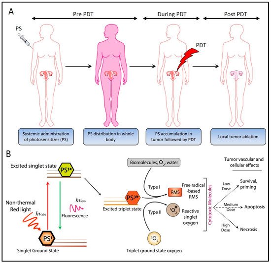
Ovarian cancer (OvCa) is the leading cause of gynecological cancer-related deaths in the United States, with five-year survival rates of 15–20% for stage III cancers and 5% for stage IV cancers. The standard of care for advanced OvCa involves surgical debulking of disseminated disease in the peritoneum followed by chemotherapy. Despite advances in treatment efficacy, the prognosis for advanced stage OvCa patients remains poor and the emergence of chemoresistant disease localized to the peritoneum is the primary cause of death. Therefore, a complementary modality that is agnostic to typical chemo- and radio-resistance mechanisms is urgently needed. Photodynamic therapy (PDT), a photochemistry-based process, is an ideal complement to standard treatments for residual disease. The confinement of the disease in the peritoneal cavity makes it amenable for regionally localized treatment with PDT. PDT involves photochemical generation of cytotoxic reactive molecular species (RMS) by non-toxic photosensitizers (PSs) following exposure to non-harmful visible light, leading to localized cell death. However, due to the complex topology of sensitive organs in the peritoneum, diffuse intra-abdominal PDT induces dose-limiting toxicities due to non-selective accumulation of PSs in both healthy and diseased tissue. In an effort to achieve selective damage to tumorous nodules, targeted PS formulations have shown promise to make PDT a feasible treatment modality in this setting. This targeted strategy involves chemical conjugation of PSs to antibodies, referred to as photoimmunoconjugates (PICs), to target OvCa specific molecular markers leading to enhanced therapeutic outcomes while reducing off-target toxicity. 

Ovarian cancer targeted therapy photodynamic therapy photoimmunoconjugates photoimmunotherapy EGFR

1. Introduction

Ovarian carcinoma (OvCa) is the leading cause of death from gynecological cancers and is the fifth most frequent cause of cancer-related deaths among women in the United States. It is estimated that in 2019, there will be 22,530 new cases of OvCa and 13,980 deaths in the US (www.cancer.org). The overall five-year survival of ~45% has not improved significantly in the past few decades due to the presence of advanced disease at the time of diagnosis and acquired resistance to the spectrum of currently available chemotherapeutic agents [1][2]. OvCa is especially dangerous as the disease can invade neighboring organs in the abdominal cavity and can enter the bloodstream and lymphatic system to form distant metastases. Making matters worse, early-stage disease is often asymptomatic and misdiagnosed as less deadly digestive issues, accounting for late-diagnosis in its advanced stages. About 80% of the patients are diagnosed at an advanced stage after the cancer has spread throughout the peritoneal cavity. These patients are typically treated with aggressive surgical resection followed by chemotherapy [3]. A number of chemotherapeutic regimens exist for OvCa, and the reader is referred to excellent reviews on the current state of chemotherapy regimens [4][5][6][7]. However, even among patients with negative follow up exams, 50% of patients later present with incurable radio- and chemo-resistant disease.
Photodynamic therapy (PDT) is a therapeutic and diagnostic modality utilizing the photochemical properties of small molecule photosensitizers (PS). PDT is particularly beneficial for cancer treatment as it imparts two degrees of selectivity—(i) certain formulations of PS preferentially accumulate in cancerous lesions, and (ii) the phototoxicity is limited to regions of tissue irradiated during treatment (Figure 1A). PDT is an FDA-approved anti-cancer treatment modality that has been investigated in preclinical and clinical settings for the management of ovarian [8][9][10][11][12][13][14][15][16][17][18][19][20] and other cancers [21][22]. It is clinically approved in the US for the treatment of numerous cancerous (esophageal and lung cancer) and non-cancerous indications.
Figure 1. Photobiological and photochemical overview of photodynamic therapy (PDT). (A) Process of clinical PDT; the photosensitizer (PS) is administered systemically, followed by an appropriate PS-light interval, where the administered PS preferentially accumulates at the tumor site. Near-infrared (NIR) irradiation of the target tissue leads to localized tumor destruction. (B) Schematic representation of the Jablonski diagram showing the ground state of the PS (PS1) and the subsequent shift to a high energy excited state (PS1*) upon NIR irradiation. The PS in the excited state (PS1*) can either emit energy in the form of fluorescence radiation and relax to the ground state or undergo intersystem crossing to generate a long-lived excited triplet state (PS3*). Energy and electron transfer from the excited triplet state (PS3*) to biomolecules, water, triplet ground state oxygen, etc. leads to the formation of cytotoxic reactive molecular species (RMS). Depending on the dose of the RMS, the target tissues may either survive or undergo apoptosis/necrosis. Adapted from Nath et al. (2019) [23].
PDT has shown particular promise in treating OvCa in preclinical studies [15][20] and clinical trials. Although phase I and II trials in OvCa and other intraperitoneal cancers demonstrated promising results, significant dose-limiting toxicities were also observed [8][9][11]. Major complications included cutaneous phototoxicity and bowel perforation due to non-specific localization of the PS and inadequate light dosimetry. These trials highlighted the clinical potential of PDT in prolonging disease-free survival, with tissue selectivity being a major challenge. In this context, encouraging reports were published by Schmidt et al. [24][25], wherein photoimmunoconjugates (PICs, discussed in Section 3), prepared by the conjugation of phthalocyanine to monoclonal antibodies (MABs) recognizing CA125, were shown to be comparatively more effective in pre-clinical and clinical studies. Since these early studies, considerable advances have been made in PIC-based PDT, or photo-immunotherapy (PIT, discussed in Section 3), suggesting the potential of PIT in the targeted therapy of OvCa. The high degree of spatial and temporal control of cytotoxicity afforded by PIT and its unique mechanism of cell killing make it an ideal candidate for treating regionally localized and resistant disease often seen in OvCa patients. The high recurrence rate even among patients with negative second-look laparotomies is partly attributed to micrometastatic tumor nodules in the peritoneum that are invisible to the eye [26]. Strategies to address these deadly pockets of disease utilizing mechanistically distinct combination therapies that exploit non-overlapping molecular targets represent a promising direction of research in PIT-based treatment of OvCa. PIT has demonstrated promise in overcoming the challenges associated with this deadly disease, and this review will give an overview of preclinical and clinical developments of this therapy and perspective of PIT’s potential role in treating OvCa.

2. Photoimmunoconjugates and Photoimmunotherapy

A limitation of intraperitoneal PDT in OvCa is the lack of compounds that are both good PSs and good tumor localizers. Although initial preclinical and clinical studies with free PSs demonstrated promising results, their use was limited due to the dose-limiting damage to internal organs [9][11]. PIT has been explored to circumvent this problem by combining the tumor-localization properties of monoclonal antibodies (MAB) with phototoxic properties of PSs. PIT involves the covalent linkage of a PS to an antibody, creating a PIC. An incubation period following PIC administration allows the antibody to potentially block the targeted receptor activation, followed by uptake and processing of the PIC by the target tissue. By combining the individual modalities, the efficacy of each is improved. Erbitux, for example, recognizes and blocks the epidermal growth factor receptor (EGFR) and is FDA approved for the treatment of metastatic colorectal cancer. EGFR is a rational target because it is overexpressed in 70–90% of advanced OvCa and is associated with aggressive and resistant tumors. Interestingly, some of the early work explored the inhibition of EGFR signaling in combination with benzoporphyrin derivative (BPD)-based PDT. A synergistic response between immunotherapy and PDT was observed with increased survival in vivo [27]. This was followed by conjugation of PSs to the EGFR antibody to achieve synchronized pharmacokinetics and enhance synergistic treatment outcomes. Growth factor receptor–targeted immunotherapy is suggested to be particularly promising because it exploits blocking the dependence of tumor cells on specific molecular pathways critical for survival and growth. This is made possible by synthesizing PICs in a way that the MAB remains functional in its ability to block receptor signaling. The persistent phototoxicity of PICs due to the “always-on” nature of the PS molecules can be minimized by using tumor-targeted activatable PIT (taPIT) where PS loading on the antibody can be optimized to attain a self-quenched “off state”, which can be activated to an “on state” following target site binding, internalization and lysosomal degradation of the PICs (Figure 4A–C). This approach of lysosome activated probes has been demonstrated in various studies to enhance specificity and selectivity in therapeutic and diagnostic applications (Figure 4D,E) [28][29][30]. While no therapy can be entirely selective, use of EGFR-targeted PIC in OvCa has some significant advantages—(i) EGFR is often overexpressed in OvCa leading to highly selective PS delivery in cancer cells compared to normal tissues, (ii) greater dependence of cancer cells on EGFR signaling, and (iii) confinement of light to the peritoneal cavity [31].
Figure 4. Tumor-targeted activatable photoimmunotherapy. (A) Pictorial representation of PICs. (B) Mechanism of tumor-targeted PIC activation. (C) The PS in PICs stay quenched under normal conditions. Once internalized in the target cells, they are dequenched through lysosomal degradation for tumor-targeted activatable photoimmunotherapy (taPIT). (D) Mouse model of micrometastatic OvCa and the scheme for endoscopic fluorescence imaging. (E) Comparison of fluorescence (red) from free benzoporphyrin derivative (BPD) (upper panel) and PIC (lower panel) administered to mice in no tumor controls (left panel) and with disseminated tumors (right panel). Adapted from Spring et al. 2014 [30].
In addition to providing an additional level of selectivity, the use of MAB-PS conjugates increases the versatility of PDT by allowing the use of compounds that are good PSs but have poor biodistribution properties on their own. MABs have been used for improved diagnosis and therapy of cancer for some time. Radiolabeled and fluorescent MABs [32][33] have been extensively used for diagnosis and image-guided surgeries [34][35]. For therapeutic applications, conjugates with radioisotopes, cytotoxic drugs, protein toxins [36], cytokines [37], and boron compounds for neutron capture [38][39] have been investigated. Besides increased selectivity, other potential advantages of PIT over conventional therapy with MAB alone or with MAB coupled to radioisotopes, drugs, or toxins are (i) since it is primarily used as a carrier, the MABs do not require any in vivo effector activity (e.g., complement fixation); (ii) in contrast to most drugs and toxins, the PS can act at the cell membrane as well as intracellularly [40]; (iii) PIT may stimulate the host immune response which may help eliminate the tumor as demonstrated by Steele et al. [41] using MAB-HP conjugates. Since the initial report by Mew et al. [42], we [14][20][30][43][44][45][46] and others [47][48][49][50][51][52][53] have demonstrated the feasibility of PICs for the preferential killing of selected cell populations in various systems [16][19][30][54][55][56]. In the 1990s, Schmidt et al. [24][25] prepared PICs of MABs recognizing CA125 on human OvCa cells and performed pilot studies of PIT in OvCa patients. While the results were encouraging, technical challenges such as uniform light delivery and toxicity remained. 

3. The Rationale to Target EGFR for OvCa Photoimmunotherapy

Growth factor receptors have been recognized as important targets in cancer treatment [57][58][59][60]. Amongst these, the EGFR family of receptors has attracted significant attention [61]. The ERBB family of proteins comprises four closely related receptor tyrosine kinases with structural similarity to the epidermal growth factor receptor (EGFR). The four members are human epidermal growth factor receptor 1 (HER1; ERBB1 also called EGFR), HER2 (ERBB2), HER3 (ERBB3), and HER4 (ERBB4) [62]. Upon ligand binding, EGFR elicits cellular responses through multiple divergent pathways. These pathways control a number of cellular processes, such as growth, motility, and production of growth factors [63]. The ectodomain of the receptor contains ligand-binding sites, while the protein-kinase catalytic sites are in the intracellular domains. Receptor signaling follows five major pathways viz. 1) The Ras/Raf/MEK/Erk pathway, 2) STAT pathway, 3) PI3K/AKT pathway, 4) Src kinase pathway, and 5) PLCγ/PKC pathway [64] (Figure 6). While the Ras/Raf/MEK/Erk and STAT pathways are involved in cellular differentiation and proliferation, the PI3K/AKT and PLCγ/PKC pathways are important for cell survival and motility, respectively.
Figure 6. Multi-functional epidermal growth factor receptor (EGFR)-targeted PIT. (A) In cancer cells, overexpressed EGFRs bind to the corresponding ligands and promote cell growth, proliferation, metastasis, angiogenesis, etc. (B) The administration of PICs targeting EGFR leads to selective accumulation of the PS in the malignant tissue and inhibition of EGFR signaling pathway and induces localized cell death upon irradiation (right panel).
Under normal conditions, the expression of EGFR in the epithelial lining of ovarian tissue is generally low. However, its overexpression has been reported in 30–98% of OvCa cases [65] and is thus considered a strong prognostic indicator for OvCa. It binds to various ligands, including EGF and TGF alpha, and contributes to the active malignancy of OvCa by promoting cell growth, cell migration, angiogenesis, and conferring resistance to apoptosis.
Overexpression of EGFR is an attractive and reasonable target for OvCa management; therefore, many antibodies targeting EGFR have been developed and are in clinical use. These include cetuximab, panitumumab, and necitumumab which act by competing with the ligand-binding sites on the extracellular domains of EGFR and inhibiting downstream signaling pathways. Previous studies from our group and others have demonstrated improved therapeutic outcomes with the combination of EGFR inhibition and PDT as compared to monotherapy [27]. The study by Del Carmen et al. showed a synergistic enhancement of tumor control (Figure 7A), increased survival, and a 33% cure (Figure 7B) in mice with disseminated disease treated with both modalities. Conjugation of PS to therapeutic EGFR antibody (Cetuximab), as in PICs, therefore provides a combination therapy with a single therapeutic agent along with a targeting specificity which may overcome the limitations in previous clinical studies [9][11][66]. Although PDT has been shown to degrade EGFR [67][68], it also sensitizes cells to EGFR-based inhibitors, thus highlighting the potential of this combinatorial approach. Given the selectivity afforded by the EGFR-based targeting and the simultaneous inhibition of the EGFR-based survival signaling pathways, PIT with EGFR targeting holds great clinical potential where phototoxicity due to non-specific PS distribution has been a limiting factor. Most studies related to PIT of OvCa have been performed with EGFR antibodies, discussed in detail in Section 3 and Section 4. Other molecular targets that have been explored in this disease context are discussed in the following section.
Figure 7. (A) Mean tumor burden for mice treated with either C225 or PDT monotherapy, compared with a combination therapy of C225 and PDT. (B) Kaplan–Meier survival curves for mice treated with photodynamic therapy only, C225 only, and mice treated with a combination therapy of PDT and C225. Combination treatment with PDT and C225 resulted in a significant enhancement in survival as compared to the individual monotherapies. BPD = benzoporphyrin derivative. PDT = photodynamic therapy. C225 (Cetuximab, Anti-EGFR antibody). Adapted from del Carmen et al. 2005 [27].

4. PIC-Based Combination Therapies

While it has shown considerable promise as a standalone therapy in OvCa, preclinical evidence suggests that PIT is most likely to be effective in combination with other cytotoxic therapies. PIT itself is inherently a combination therapy, as it is composed of a receptor blocking antibody and a phototoxic sensitizer. The therapeutic interaction of these two core components of PIT was examined by del Carmen and colleagues (del Carmen-Hasan 2005) in which anti-EGFR antibody was combined with untargeted PDT in an orthotopic xenograft mouse model. The combination significantly reduced tumor burden and increased animal survival compared to either therapy alone, providing a strong therapeutic rationale for combining PDT and immunotherapy in addition to the inherent selectivity of PIT. PIT has been shown to act synergistically with chemotherapy as well. Both untargeted and targeted forms of the cytotoxic drug SOS thiophene and the PS mesochlorin-e6 were demonstrated to be highly synergistic at low doses by Hongrapipat et al. [69], suggesting that PIT-based combinations may enable dose reductions of toxic chemotherapies while maintaining efficacy in this highly resistant disease. This effect was confirmed in vivo by Rizvi and colleagues [70], whereby cisplatin followed by PIT demonstrated comparable or better antitumor effects than 2 cycles of chemotherapy. Additionally, numerous studies have shown that PDT synergizes with various chemo- and radio-therapies in a range of cancer types [71][72], indicating that this is a fairly generalizable phenomenon.
The resistance to radio- and chemo-therapy agents frequently observed in recurrent OvCa leaves this patient population with few or no treatment options. In this context, PIT makes a compelling case to improve survival outlooks for this disease. Studies by Goff et al. [20] and Duska et al. [15] investigated the effects of combined PIT and cisplatin treatment on ex vivo samples from OvCa patients. In both studies, patient tissues resistant to standard chemo- and radio-therapy were demonstrated to be responsive to PIT. In the study by Duska et al., platinum treatment following PIT was demonstrated to have an additive therapeutic effect in cisplatin sensitive cancer cells, while, remarkably, a strong synergistic effect was observed in platinum-resistant cells, suggesting that PIT can resensitize these tumors to chemotherapy (Figure 3). These data suggest that the use of PIT fills in a crucial niche in the treatment of OvCa, where resistance to chemo- or radio-therapy is a major problem. These types of PIT-based resensitization strategies may hold promise for increasing treatment options in these patients.
The synergistic and re-sensitization effects observed in PIT-based combination therapies such as these may represent a new strategy for managing patients with advanced disease. Typical treatment regimens involve administering the maximum tolerated dose of toxic chemo- or radio-therapies with the hope of eliminating any remaining cancer cells. However, this approach comes with dangerous and often dose-limiting side effects, negatively impacting patient quality of life and limiting these treatments only to those with favorable health status. The non-overlapping toxicity profiles of PIT and chemotherapy, and the ability to reduce the dose of these cytotoxic drugs following PIT-based “priming” of the disease may open up new treatment options for patients who may otherwise not be eligible or responsive to classical therapeutic regimens. Furthermore, optimized PIT regimens that enable dose de-escalation of toxic chemotherapies while maintaining antitumor efficacy hold promise as a means for improving patient quality of life and more humanely treating this devastating disease.

References

  1. Christie, E.; Bowtell, D. Acquired chemotherapy resistance in ovarian cancer. Ann. Oncol. 2017, 28, viii13–viii15.
  2. Cooke, S.L.; Brenton, J.D. Evolution of platinum resistance in high-grade serous ovarian cancer. Lancet Oncol. 2011, 12, 1169–1174.
  3. Jelovac, D.; Armstrong, D.K. Recent progress in the diagnosis and treatment of ovarian cancer. CA Cancer J. Clin. 2011, 61, 183–203.
  4. Cortez, A.J.; Tudrej, P.; Kujawa, K.A.; Lisowska, K.M. Advances in ovarian cancer therapy. Cancer Chemother. Pharmacol. 2018, 81, 17–38.
  5. Brackmann, M.; Stasenko, M.; Uppal, S.; Erba, J.; Reynolds, R.K.; McLean, K. Comparison of first-line chemotherapy regimens for ovarian carcinosarcoma: A single institution case series and review of the literature. BMC Cancer 2018, 18, 172.
  6. Kelland, L. The resurgence of platinum-based cancer chemotherapy. Nat. Rev. Cancer 2007, 7, 573.
  7. Yap, T.A.; Carden, C.P.; Kaye, S.B. Beyond chemotherapy: Targeted therapies in ovarian cancer. Nat. Rev. Cancer 2009, 9, 167.
  8. Hendren, S.K.; Hahn, S.M.; Spitz, F.R.; Bauer, T.W.; Rubin, S.C.; Zhu, T.; Glatstein, E.; Fraker, D.L. Phase II trial of debulking surgery and photodynamic therapy for disseminated intraperitoneal tumors. Ann. Surg. Oncol. 2001, 8, 65–71.
  9. Sindelar, W.F.; DeLaney, T.F.; Tochner, Z.; Thomas, G.F.; Dachoswki, L.J.; Smith, P.D.; Friauf, W.S.; Cole, J.W.; Glatstein, E. Technique of photodynamic therapy for disseminated intraperitoneal malignant neoplasms: Phase I study. Arch. Surg. 1991, 126, 318–324.
  10. Dougherty, T.J.; Gomer, C.J.; Henderson, B.W.; Jori, G.; Kessel, D.; Korbelik, M.; Moan, J.; Peng, Q. Photodynamic therapy. JNCI J. Natl. Cancer Inst. 1998, 90, 889–905.
  11. Delaney, T.F.; Sindelar, W.F.; Tochner, Z.; Smith, P.D.; Friauf, W.S.; Thomas, G.; Dachowski, L.; Cole, J.W.; Steinberg, S.M.; Glatstein, E. Phase I study of debulking surgery and photodynamic therapy for disseminated intraperitoneal tumors. Int. J. Radiat. Oncol. Biol. Phys. 1993, 25, 445–457.
  12. Wierrani, F.; Fiedler, D.; Grin, W.; Henry, M.; Dienes, E.; Gharehbaghi, K.; Krammer, B.; Grünberger, W. Clinical effect of meso-tetrahydroxyphenylchlorine based photodynamic therapy in recurrent carcinoma of the ovary: Preliminary results. BJOG Int. J. Obstet. Gynaecol. 1997, 104, 376–378.
  13. Tochner, Z.; Mitchell, J.; Hoekstra, H.; Smith, P.; DeLuca, A.; Barnes, M.; Harrington, F.; Manyak, M.; Russo, D.; Russo, A. Photodynamic therapy of the canine peritoneum: Normal tissue response to intraperitoneal and intravenous photofrin followed by 630 nm light. Lasers Surg. Med. 1991, 11, 158–164.
  14. Goff, B.; Hermanto, U.; Rumbaugh, J.; Blake, J.; Bamberg, M.; Hasan, T. Photoimmunotherapy and biodistribution with an OC125-chlorin immunoconjugate in an in vivo murine ovarian cancer model. Br. J. Cancer 1994, 70, 474.
  15. Duska, L.R.; Hamblin, M.R.; Miller, J.L.; Hasan, T. Combination photoimmunotherapy and cisplatin: Effects on human ovarian cancer ex vivo. J. Natl. Cancer Inst. 1999, 91, 1557–1563.
  16. Molpus, K.L.; Hamblin, M.R.; Rizvi, I.; Hasan, T. Intraperitoneal Photoimmunotherapy of Ovarian Carcinoma Xenografts in Nude Mice Using Charged Photoimmunoconjugates. Gynecol. Oncol. 2000, 76, 397–404.
  17. Hamblin, M.R.; Miller, J.L.; Hasan, T. Effect of charge on the interaction of site-specific photoimmunoconjugates with human ovarian cancer cells. Cancer Res. 1996, 56, 5205–5210.
  18. Tochner, Z.A.; Hahn, S.; Glatstein, E. Photoimmunotherapy and ovarian cancer: An improbable fiction or a palpable hit? J. Natl. Cancer Inst. 1999, 91, 1526–1527.
  19. Duska, L.; Hamblin, M.; Bamberg, M.; Hasan, T. Biodistribution of charged F (ab’) 2 photoimmunoconjugates in a xenograft model of ovarian cancer. Br. J. Cancer 1997, 75, 837.
  20. Goff, B.A.; Bamberg, M.; Hasan, T. Photoimmunotherapy of human ovarian carcinoma cells ex vivo. Cancer Res. 1991, 51, 4762–4767.
  21. DeWitt, J.M.; Sandrasegaran, K.; O’Neil, B.; House, M.G.; Zyromski, N.J.; Sehdev, A.; Perkins, S.M.; Flynn, J.; McCranor, L.; Shahda, S. Phase 1 study of EUS-guided photodynamic therapy for locally advanced pancreatic cancer. Gastrointest. Endosc. 2019, 89, 390–398.
  22. Huggett, M.T.; Jermyn, M.; Gillams, A.; Illing, R.; Mosse, S.; Novelli, M.; Kent, E.; Bown, S.; Hasan, T.; Pogue, B. Phase I/II study of verteporfin photodynamic therapy in locally advanced pancreatic cancer. Br. J. Cancer 2014, 110, 1698.
  23. Nath, S.; Obaid, G.; Hasan, T. The Course of Immune Stimulation by Photodynamic Therapy: Bridging fundamentals of photochemically-induced Immunogenic Cell Death to the Enrichment of T Cell Repertoire. Photochem. Photobiol. 2019.
  24. Schmidt, S.; Wagner, U.; Oehr, P.; Krebs, D. Clinical use of photodynamic therapy in gynecologic tumor patients—Antibody-targeted photodynamic laser therapy as a new oncologic treatment procedure. Zent. Fur Gynakol. 1992, 114, 307–311.
  25. Schmidt, S.; Wagner, U.; Schultes, B.; Oehr, P.; Decleer, W.; Ertmer, W.; Lubaschowski, H.; Biersack, H.J.; Krebs, D. [Photodynamic laser therapy with antibody-bound dyes. A new procedure in therapy of gynecologic malignancies]. Fortschr. Der Med. 1992, 110, 298–301.
  26. Löning, M.; Diddens, H.; Küpker, W.; Diedrich, K.; Hüttmann, G. Laparoscopic fluorescence detection of ovarian carcinoma metastases using 5-aminolevulinic acid-induced protoporphyrin IX. Cancer Interdiscip. Int. J. Am. Cancer Soc. 2004, 100, 1650–1656.
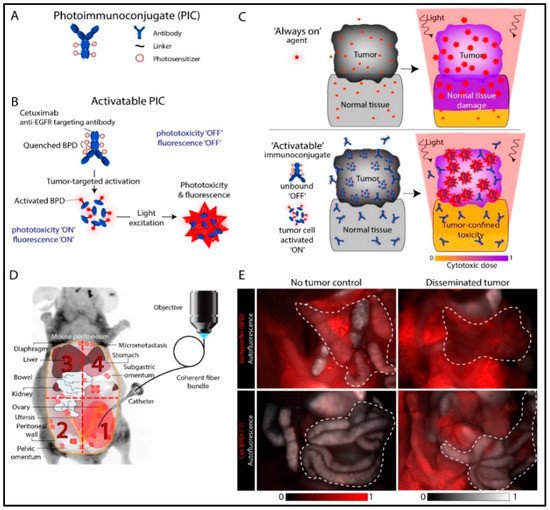
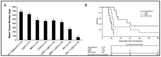
  27. del Carmen, M.G.; Rizvi, I.; Chang, Y.; Moor, A.C.; Oliva, E.; Sherwood, M.; Pogue, B.; Hasan, T. Synergism of epidermal growth factor receptor-targeted immunotherapy with photodynamic treatment of ovarian cancer in vivo. J Natl Cancer Inst 2005, 97, 1516–1524.
  28. Obaid, G.; Spring, B.Q.; Bano, S.; Hasan, T. Activatable clinical fluorophore-quencher antibody pairs as dual molecular probes for the enhanced specificity of image-guided surgery. J. Biomed. Opt. 2017, 22, 121607.
  29. Urano, Y.; Asanuma, D.; Hama, Y.; Koyama, Y.; Barrett, T.; Kamiya, M.; Nagano, T.; Watanabe, T.; Hasegawa, A.; Choyke, P.L. Selective molecular imaging of viable cancer cells with pH-activatable fluorescence probes. Nat. Med. 2009, 15, 104.
  30. Spring, B.Q.; Abu-Yousif, A.O.; Palanisami, A.; Rizvi, I.; Zheng, X.; Mai, Z.; Anbil, S.; Sears, R.B.; Mensah, L.B.; Goldschmidt, R.; et al. Selective treatment and monitoring of disseminated cancer micrometastases in vivo using dual-function, activatable immunoconjugates. Proc. Natl. Acad. Sci. USA 2014, 111, E933–E942.
  31. Patterson, M.S.; Wilson, B.C.; Graff, R. In vivo tests of the concept of photodynamic threshold dose in normal rat liver photosensitized by aluminum chlorosulphonated phthalocyanine. Photochem. Photobiol. 1990, 51, 343–349.
  32. McGuire, W.P.; Hoskins, W.J.; Brady, M.F.; Kucera, P.R.; Partridge, E.E.; Look, K.Y.; Clarke-Pearson, D.L.; Davidson, M. Cyclophosphamide and cisplatin compared with paclitaxel and cisplatin in patients with stage III and stage IV ovarian cancer. New Engl. J. Med. 1996, 334, 1–6.
  33. Folli, S.; Westermann, P.; Braichotte, D.; Pèlegrin, A.; Wagnières, G.; van den Bergh, H.; Mach, J.-P. Antibody-indocyanin conjugates for immunophotodetection of human squamous cell carcinoma in nude mice. Cancer Res. 1994, 54, 2643–2649.
  34. Rosenthal, E.L.; Warram, J.M.; De Boer, E.; Chung, T.K.; Korb, M.L.; Brandwein-Gensler, M.; Strong, T.V.; Schmalbach, C.E.; Morlandt, A.B.; Agarwal, G. Safety and tumor specificity of cetuximab-IRDye800 for surgical navigation in head and neck cancer. Clin. Cancer Res. 2015, 21, 3658–3666.
  35. Nagaya, T.; Nakamura, Y.A.; Choyke, P.L.; Kobayashi, H. Fluorescence-guided surgery. Front. Oncol. 2017, 7, 314.
  36. COGLIATI, T.; BRUSA, P.; CANEVARI, S.; CALDERA, M. Preparation and Biological Characterization of Conjugates Consisting of Ricin and a Tumor-Specific Non-Internalizing. Anticancer Res. 1991, 11, 417–422.
  37. Ozzello, L.; De Rosa, C.; Blank, E.; Cantell, K.; Ceriani, R.; Habif, D. The use of natural interferon alpha conjugated to a monoclonal antibody anti mammary epithelial mucin (Mc5) for the treatment of human breast cancer xenografts. Breast Cancer Res. Treat. 1993, 25, 265–276.
  38. Alam, F.; Soloway, A.H.; Barth, R.F.; Mafune, N.; Adams, D.M.; Knoth, W.H. Boron neutron capture therapy: Linkage of a boronated macromolecule to monoclonal antibodies directed against tumor-associated antigens. J. Med. Chem. 1989, 32, 2326–2330.
  39. Barth, R.F.; Adams, D.M.; Soloway, A.H.; Alam, F.; Darby, M.V. Boronated starburst dendrimer-monoclonal antibody immunoconjugates: Evaluation as a potential delivery system for neutron capture therapy. Bioconj. Chem. 1994, 5, 58–66.
  40. Oseroff, A.R.; Ohuoha, D.; Hasan, T.; Bommer, J.C.; Yarmush, M.L. Antibody-targeted photolysis: Selective photodestruction of human T-cell leukemia cells using monoclonal antibody-chlorin e6 conjugates. Proc. Natl. Acad. Sci. 1986, 83, 8744–8748.
  41. Steele, J.K.; Liu, D.; Stammers, A.T.; Whitney, S.; Levy, J.G. Suppressor deletion therapy: Selective elimination of T suppressor cells in vivo using a hematoporphyrin conjugated monoclonal antibody permits animals to reject syngeneic tumor cells. Cancer Immunol. Immunother. 1988, 26, 125–131.
  42. Mew, D.; Lum, V.; Wat, C.; Towers, G.; Sun, C.C.; Walter, R.; Wright, W.; Berns, M.; Levy, J. Ability of specific monoclonal antibodies and conventional antisera conjugated to hematoporphyrin to label and kill selected cell lines subsequent to light activation. Cancer Res. 1985, 45, 4380–4386.
  43. Goff, B.A.; Bachor, R.; Kollias, N.; Hasan, T. Effects of photodynamic therapy with topical application of 5-aminolevulinic acid on normal skin of hairless guinea pigs. J. Photochem. Photobiol. B Biol. 1992, 15, 239–251.
  44. Hasan, T.; Lin, C.; Lin, A. Laser-induced selective cytotoxicity using monoclonal antibody-chromophore conjugates. Prog. Clin. Biol. Res. 1989, 288, 471.
  45. Hasan, T.; Lin, A.; Yarmush, D.; Oseroff, A.; Yarmush, M. Monoclonal antibody-chromophore conjugates as selective phototoxins. J. Control. Release 1989, 10, 107–117.
  46. Hasan, T.; Sherwood, M.; Anderson, T.; Bamberg, M.; Flotte, T.J.; Zurawski, V.R. Cellular response of ovarian carcinoma cells to antibody-photosensitizer-mediated injury. In Proceedings of the Photodynamic Therapy: Mechanisms II; pp. 136–144. Available online: https://spie.org/Publications/Proceedings/Volume/1203?SSO=1 (accessed on 26 November 2019).
  47. Vrouenraets, M.B.; Visser, G.W.; Stewart, F.A.; Stigter, M.; Oppelaar, H.; Postmus, P.E.; Snow, G.B.; van Dongen, G.A. Development of meta-tetrahydroxyphenylchlorin-monoclonal antibody conjugates for photoimmunotherapy. Cancer Res. 1999, 59, 1505–1513.
  48. Jiang, F.N.; Liu, D.J.; Neyndorff, H.; Chester, M.; Jiang, S.-y.; Levy, J.G. Photodynamic killing of human squamous cell carcinoma cells using a monoclonal antibody-photosensitizer conjugate. JNCI J. Natl. Cancer Inst. 1991, 83, 1218–1225.
  49. Krinick, N.; Sun, Y.; Joyner, D.; Spikes, J.; Straight, R.; Kopeček, J. A polymeric drug delivery system for the simultaneous delivery of drugs activatable by enzymes and/or light. J. Biomater. Sci. Polym. Ed. 1994, 5, 303–324.
  50. Lu, X.-M.; Fischman, A.; Stevens, E.; Lee, T.; Strong, L.; Tompkins, R.; Yarmush, M. Sn-chlorin e6 antibacterial immunoconjugates. An in vitro and in vivo analysis. J. Immunol. Methods 1992, 156, 85–99.
  51. Morgan, J.; Gray, A.; Huehns, E. Specific targeting and toxicity of sulphonated aluminium phthalocyanine photosensitised liposomes directed to cells by monoclonal antibody in vitro. Br. J. Cancer 1989, 59, 366.
  52. Sandland, J.; Boyle, R.W. Photosensitizer Antibody–Drug Conjugates: Past, Present, and Future. Bioconj. Chem. 2019, 30, 975–993.
  53. Wang, S.; Hüttmann, G.M.; Rudnitzki, F.; Diddens-Tschoeke, H.; Zhang, Z.; Rahmanzadeh, R. Indocyanine green as effective antibody conjugate for intracellular molecular targeted photodynamic therapy. J. Biomed. Opt. 2016, 21, 078001.
  54. Goff, B.A.; Blake, J.; Bamberg, M.P.; Hasan, T. Treatment of ovarian cancer with photodynamic therapy and immunoconjugates in a murine ovarian cancer model. Br. J. Cancer 1996, 74, 1194.
  55. Pogrebniak, H.; Matthews, W.; Black, C.; Russo, A.; Mitchell, J.; Smith, P.; Roth, J.; Pass, H. Targetted phototherapy with sensitizer-monoclonal antibody conjugate and light. Surg. Oncol. 1993, 2, 31–42.
  56. Del Governatore, M.; Hamblin, M.R.; Shea, C.R.; Rizvi, I.; Molpus, K.G.; Tanabe, K.K.; Hasan, T. Experimental photoimmunotherapy of hepatic metastases of colorectal cancer with a 17.1 A chlorine6 immunoconjugate. Cancer Res. 2000, 60, 4200–4205.
  57. Fan, Z.; Mendelsohn, J. Therapeutic application of anti-growth factor receptor antibodies. Curr. Opin. Oncol. 1998, 10, 67–73.
  58. Mendelsohn, J. Use of an antibody to target geldanamycin; Oxford University Press: Oxford, UK, 2000.
  59. Mendelsohn, J. Jeremiah Metzger Lecture. Targeted cancer therapy. Trans. Am. Clin. Climatol. Assoc. 2000, 111, 95.
  60. Mendelsohn, J. Blockade of receptors for growth factors: An anticancer therapy—the fourth annual Joseph, H. Burchenal American Association for Cancer Research Clinical Research Award Lecture. Clin. Cancer Res. 2000, 6, 747–753.
  61. Ciardiello, F.; Tortora, G. A novel approach in the treatment of cancer: Targeting the epidermal growth factor receptor. Clin. Cancer Res. 2001, 7, 2958–2970.
  62. Baselga, J.; Swain, S.M. Novel anticancer targets: Revisiting ERBB2 and discovering ERBB3. Nat. Rev. Cancer 2009, 9, 463.
  63. Wells, A. Tumor invasion: Role of growth factor-induced cell motility. Adv. Cancer Res. 1999, 78, 31–101.
  64. Teplinsky, E.; Muggia, F. EGFR and HER2: Is there a role in ovarian cancer? Transl. Cancer Res 2015, 4.
  65. Gui, T.; Shen, K. The epidermal growth factor receptor as a therapeutic target in epithelial ovarian cancer. Cancer Epidemiol. 2012, 36, 490–496.
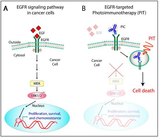
  66. Abu-Yousif, A.O.; Moor, A.C.; Zheng, X.; Savellano, M.D.; Yu, W.; Selbo, P.K.; Hasan, T. Epidermal growth factor receptor-targeted photosensitizer selectively inhibits EGFR signaling and induces targeted phototoxicity in ovarian cancer cells. Cancer Lett. 2012, 321, 120–127.
  67. Bhuvaneswari, R.; Gan, Y.Y.; Soo, K.C.; Olivo, M. Targeting EGFR with photodynamic therapy in combination with Erbitux enhances in vivo bladder tumor response. Mol. Cancer 2009, 8, 94.
  68. Ahmad, N.; Kalka, K.; Mukhtar, H. In vitro and in vivo inhibition of epidermal growth factor receptor-tyrosine kinase pathway by photodynamic therapy. Oncogene 2001, 20, 2314.
  69. Hongrapipat, J.; Kopecková, P.; Liu, J.; Prakongpan, S.; Kopecek, J. Combination chemotherapy and photodynamic therapy with Fab′ fragment targeted HPMA copolymer conjugates in human ovarian carcinoma cells. Mol. Pharm. 2008, 5, 696–709.
  70. Rizvi, I.; Dinh, T.A.; Yu, W.; Chang, Y.; Sherwood, M.E.; Hasan, T. Photoimmunotherapy and irradiance modulation reduce chemotherapy cycles and toxicity in a murine model for ovarian carcinomatosis: Perspective and results. Israel J. Chem. 2012, 52, 776–787.
  71. Celli, J.P.; Solban, N.; Liang, A.; Pereira, S.P.; Hasan, T. Verteporfin-based photodynamic therapy overcomes gemcitabine insensitivity in a panel of pancreatic cancer cell lines. Lasers Surg. Med. 2011, 43, 565–574.
  72. Huang, H.-C.; Mallidi, S.; Liu, J.; Chiang, C.-T.; Mai, Z.; Goldschmidt, R.; Ebrahim-Zadeh, N.; Rizvi, I.; Hasan, T. Photodynamic therapy synergizes with irinotecan to overcome compensatory mechanisms and improve treatment outcomes in pancreatic cancer. Cancer Res. 2016, 76, 1066–1077.
More
Information
Contributor MDPI registered users' name will be linked to their SciProfiles pages. To register with us, please refer to https://encyclopedia.pub/register :
View Times: 480
Revisions: 2 times (View History)
Update Date: 12 Aug 2021
1000/1000