Submitted Successfully!
To reward your contribution, here is a gift for you: A free trial for our video production service.
Thank you for your contribution! You can also upload a video entry or images related to this topic.
Version Summary Created by Modification Content Size Created at Operation
1 + 2405 word(s) 2405 2021-07-23 05:12:56 |
2 format correct Meta information modification 2405 2021-08-05 04:36:39 | |
3 move out from EC Meta information modification 2405 2021-09-22 04:37:55 |

Video Upload Options

Do you have a full video?

Confirm

Are you sure to Delete?
Cite
If you have any further questions, please contact Encyclopedia Editorial Office.
Strashilov, S. Pathogenesis of Cutaneous Melanoma. Encyclopedia. Available online: https://encyclopedia.pub/entry/12780 (accessed on 19 April 2024).
Strashilov S. Pathogenesis of Cutaneous Melanoma. Encyclopedia. Available at: https://encyclopedia.pub/entry/12780. Accessed April 19, 2024.
Strashilov, Strahil. "Pathogenesis of Cutaneous Melanoma" Encyclopedia, https://encyclopedia.pub/entry/12780 (accessed April 19, 2024).
Strashilov, S. (2021, August 04). Pathogenesis of Cutaneous Melanoma. In Encyclopedia. https://encyclopedia.pub/entry/12780
Strashilov, Strahil. "Pathogenesis of Cutaneous Melanoma." Encyclopedia. Web. 04 August, 2021.
Pathogenesis of Cutaneous Melanoma
Edit

Melanoma develops from malignant transformations of the pigment-producing melanocytes. If located in the basal layer of the skin epidermis, melanoma is referred to as cutaneous, which is more frequent. However, as melanocytes are be found in the eyes, ears, gastrointestinal tract, genitalia, urinary system, and meninges, cases of mucosal melanoma or other types (e.g., ocular) may occur. The incidence and morbidity of cutaneous melanoma (cM) are constantly increasing worldwide.

aetiology pathogenesis melanoma skin melanoma

1. Introduction

The term melanoma was first used in 1812 by René Laennec to describe a case of metastatic dissemination of the disease [1]. Cutaneous melanoma (cM) develops from malignant transformations of pigment-producing melanocytes in the basal layer of the skin epidermis. Non-cutaneous melanoma arises from malignantly transformed melanocytes in the uvea [2][3], gastrointestinal tract [4][5], genitalia [6][7], urinary system [8][9], meninges [10][11], etc. The incidence and morbidity of cM are constantly increasing worldwide. Australia and New Zealand are world leaders in this regard with a morbidity rate of 54/100,000 and a mortality rate of 5.6/100,000 for 2015 [12].

2. Pathogenesis of Cutaneous Melanoma
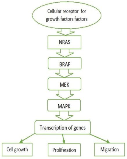
2.1. MAPK Pathway

The mitogen-activated protein kinase (MAPK) cascade regulates cell proliferation, growth, and migration and is activated in almost all types of melanomas. This pathway is active under normal conditions, but in the cases of melanoma it is associated with excessive activation [13]. It is activated after binding of growth factors to tyrosine kinase receptors. Stimulation of these receptors activates Ras family proteins—monomeric G proteins (NRAS)—causing the cascade activation of serine/threonine kinases (BRAF) and results in the activation of ERK kinase (also known as MAPK). Serine/threonine kinase activates transcription factors, thereby intensifying the transcription of genes involved in cell growth, proliferation, and migration (Figure 1).
Figure 1. MAPK pathway. NRAS—Neuroblastoma RAS viral oncogene homolog. BRAF—v-Raf murine sarcoma viral oncogene homolog B. MEK—Mitogen-activated protein kinase kinase. MAPK—Mitogen-activated protein kinase.

2.2. BRAF

BRAF is a serine/threonine kinase that is activated directly by RAS and is strongly expressed in melanocytes, neural tissue, testes, and hematopoietic cells. BRAF has been found in phosphorylate and activates MEK (a kinase component of the MAPK pathway), which in turn activates ERK (MAPK) by phosphorylation and thus stimulates growth and transformation. This is crucial for the pathogenesis of melanomas [14][15][16]. The conversion of thymidine to adenine (T → A) is the most common mutation in the BRAF gene (~70%). It results in the substitution of valine with glutamate (V600E) in the protein molecule, resulting in the activation of its kinase domain. These mutations indirectly results in BRAF activation by disrupting the normal intramolecular interactions that hold BRAF in inactive configurations [14][17]. Mutations in the BRAF gene (V600E) are more common in melanoma that develops in parts of the body that are exposed to solar radiation. Although BRAF mutations may be associated with sun exposure, the transversion (T → A), as mentioned earlier, is not classically related to UV exposure [14][16]. Other amino acid substitutions in the protein are possible, such as those observed in V600K mutations (representing ~20% of BRAF mutations in melanoma). They are mainly found in melanoma patients exposed to chronic sun exposure. BRAF V600 mutations represent an early event in the development of melanoma. They are less common in its initial stages—in 10% of the cases with radial growth—and in 6% of in situ melanomas. However, they are very common in metastatic melanoma. These mutations occur in approximately 80% of the benign and the dysplastic nevi, but they alone are not sufficient for the development of cutaneous melanoma [17].

2.3. RAS

Mutations, leading to increased RAS activity in melanomas also increase cell proliferation, but this occurs significantly less frequently than in other solid tumours [16]. The Q16R mutation is the most common NRAS mutation in melanoma. Somatic NRAS gene mutations may cause increased activity of the NRAS protein, which cannot “shut down”. This results in a serial activation of serine/threonine kinases, stimulating cell cycle progression, cell transformation, and cell survival. The cascade of events can also be caused by overexpression and/or hyperactivation of various growth factor receptors such as c-Met, epidermal growth factor receptor (EGFR), and c-KIT as well as the functional loss of neurofibromatosis type 1 (NF1) tumour suppressor gene, which suppresses NRAS signalling [18][19]. Activating RAS mutations were observed in only 10–20% of melanomas (mostly in amelanotic nodular subtypes), with NRAS mutations being the most common. BRAF mutations only activate the MAPK signalling pathway, while activating NRAS mutations simultaneously activates the MAPK and PI3K pathways [16][19]. NRAS and BRAF mutations have been found to rarely co-occur, indicating that a mutation in one of the two genes is sufficient to activate the MAPK pathway. Activating BRAF mutations are more common in nevus cells (70–80% of dysplastic nevi). NRAS mutations are rare in nevi and are most common in congenital ones. NRAS mutations are often associated with Spitz nevi [16].
The uncontrolled activation of the MAPK signalling pathway in melanomas may be caused by overexpression or hyperactivation of growth factor receptors such as c-Met, c-KIT, and epidermal growth factor receptor (EGFR) [19].

2.4. c-KIT

c-KIT (tyrosine kinase receptor) and its ligand (stem cell factor) play an essential role in melanocyte development [15][20]. c-KIT mutations cause insufficient pigmentation. The results of numerous immunohistochemical studies indicate that the transition from a benign condition to primary or metastatic melanoma is associated with a loss of c-KIT expression. The activating mutations and amplification of KIT genes have been observed in cutaneous melanomas in areas exposed to chronic sun exposure as well as in acral melanomas (of the hands, feet, and nail bed). KIT mutations can activate multiple signalling pathways and the PI3K-AKT pathway in particular. The presence of point mutations in the KIT gene has also been observed in gastrointestinal stromal tumours (GIST). The functional characteristics of these mutations are clinically significant as KIT inhibitors are effective in melanomas, but with a much lower degree of clinical response (10–30%) compared with GIST (>70%) [17][20].

2.5. c-MET and HGF

Overexpression of another tyrosine kinase receptor c-MET and its ligand HGF (hepatocyte growth factor) correlates with melanoma progression. c-MET is known to control a variety of biological functions such as propagation, survival, motility, and invasion. Tumours can grow and metastasize due to dysregulation caused by aberrant c-MET activation. It should be noted that c-MET (tyrosine kinase receptor) may be overactivated in the event of the excessive secretion of its HGF ligand produced by tumour cells or the tumour microenvironment of melanoma. This paracrine effect activates the PI3K-AKT pathway in tumour cells and leads to resistance to MAPK inhibitors [17][21].

2.6. Other Factors

Neurofibromatosis type 1 (NF1) tumour suppressor gene and the negative RAS regulator are other driving factors in the process. NF1 mutations were identified in 5 of 21 tumours without BRAF and NRAS mutations. In the context of BRAF (V600E) mutations, NF1 has been observed to disrupt normal MAPK and PI3K pathways by inhibiting the development and the metastatic spread of melanoma. Inactivating mutations of neurofibromatosis tumour suppressor gene 2 (NF2) have been observed in some melanomas. Initial mutations in NF1 and NF2 genes are associated with hereditary neurofibromatosis. Other somatic mutations of the melanoma cells in MAPK downstream effectors have been identified, such as MAP3K5, MAP3K9, MEK1, and MEK2 [16][22].

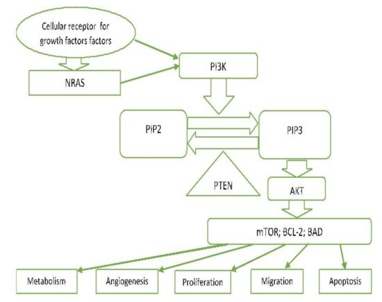
2.7. PI3K/PTEN/AKT Pathway (Phosphatidylinositol 3-kinase Pathway)

PI3K-AKT is another critical signalling pathway in the cell. It is involved in the regulation of cell survival, growth, and apoptosis [13][23] (Figure 2).

Figure 2. PI3K/PTEN/AKT pathway. NRAS—Neuroblastoma RAS viral oncogene homolog. PI3—Phosphoinositol-3-kinase. PIP2—Phosphatidylinositol 4,5-bisphosphate. PIP3—Phosphatidylinositol (3,4,5)-trisphosphate. PTEN—Phosphatase and tensin homolog deleted on chromosome 10. AKT—Protein kinase B. mTOR—The mechanistic target of rapamycin. BCL-2—B-cell lymphoma-2. BAD—proapoptotic protein.
In carcinomas, this pathway can be genetically driven by activating mutations (e.g., PIK3CA and AKT1) or by functional loss of some of the components of this pathway (e.g., PTEN). PI3 kinases (phosphatidylinositol 3-kinase-lipid kinases) are triggered directly by tyrosine kinase receptor activation or indirectly by RAS. They cause phosphorylation of PIP2 to PIP3 and subsequent phosphorylation of AKT [17][23]. PTEN lipid phosphatase plays an essential role as an agent antagonizing this pathway by converting PIP3 back to PIP2 [24]. Phosphorylation of AKT (serine/threonine protein kinase) results in the phosphorylation of multi-factor proteins that regulate cellular processes (proliferation, survival, motility, angiogenesis, and metabolism). m-TOR is one of these proteins found to be activated in 73% of human melanoma cell lines and very rarely in nevus cells. Increased activation of PI3K signalling is observed in a large number of melanomas, usually triggered by mutations, deletions, and promoter methylation of the coding genes of the PTEN inhibitor [23].

2.8. MITF Signalling

Microphthalmia-associated transcription factor (MITF) is a key regulator that is required for melanocyte differentiation, which may affect malignancy in some melanomas. The most common MITF genetic alteration is amplification, which occurs in 15–20% of melanomas (more common in metastatic melanomas) [25][26]. These changes are thought to occur later in disease progression and are associated with lower 5 year survival [27]. MITF increases the gene expression involved in the cell cycle progression, the cell proliferation, and the cell survival. It is a transcription factor for cellular cyclin-dependent kinase (CDK2), CDK inhibitors p16INK4a, and p21 as well as for the antiapoptotic mitochondrial membrane protein BCL-2 (B-cell lymphoma 2) [28][29]. MITF expression is triggered by the activation of the melanocortin 1-receptor (MC1R), which in turn is activated by the binding of melanocortins (ACTH and α-MSH) [28]. This induces adenylate cyclase activation and the production of c-AMP, which activates protein kinase A (PKA). In turn, PKA activates CREB (cAMP protein-binding response element), which acts as a transcription factor and enhances MITF expression. MITF signalling is also closely related and regulated by MAPK signalling. It has been observed that the mutated oncogene BRAF can regulate MITF expression, thus ensuring protein levels compatible with the proliferation and survival of melanoma cells. Importantly, MITF may also act as an anti-proliferative transcription factor that induces cell cycle arrest. In melanocytes, MITF expression occurs after the binding of the melanocyte-stimulating hormone (MSH) to the melanocortin 1 receptor (MC1R). In this process, MITF targets the genes involved in the regulation of differentiation and pigmentation, distribution, and survival [25][28].

2.9. p53

p53 is the primary tumour suppressor gene, associated with apoptosis. The p53 protein encoded is a major transcription factor and bears crucial responsibility for various stresses such as DNA damage, genome instability, hypoxia, and oncogenic aberrations [30]. Disorders in the control of the cell cycle, genomic instability, and abnormal proliferation are observed during the transformation of melanocytes into melanoma cells. Mutations or deletions of p53 are observed in more than 50% of carcinomas. In melanomas, they occur in only 1–5% of primary melanomas and in 11–25% of metastatic melanomas [31]. The expression of the p53 protein in melanomas is variable [32][33], while it is often absent in nevi. Melanoma often results in the overexpression of this protein than when compared to other tumours. Despite the high expression of p53, melanoma cells are highly resistant to apoptosis. This can be explained by the impaired functionality of the p53 apoptotic pathway [34].

2.10. Hypoxia-Induced Factor (HIF)

An essential factor for tumour development and progression is the environment itself. Human skin is generally hypoxic, which in turn favours melanogenesis [35]. The hypoxic response is primarily mediated by HIF. It activates many genes, associated with angiogenesis, invasion, and metastasis. Hypoxia in the microenvironment of the skin favours melanocyte transformation and tumour growth induced by the PI3K pathway. Inhibition of the HIF gene reduces this process. Tumour hypoxia occurs with melanoma growth due to poor vascularization, which increases HIF and stimulates melanogenesis and melanoma progression, thus being of negative prognostic value [36]. Melanomas located in areas of hypoxia possess the worse prognosis despite treatment [37][38].

2.11. Notch Signalling Pathway

Notch signalling pathway has a substantial potential for the development and maintenance of tissue homeostasis. It includes a family of transmembrane receptors and their ligands, negative and positive modifiers, and transcription factors. In mammals, four receptors and five ligands are known [39]. Notch can function as a tumour promoter or suppressor depending on the type of cells. Its activation through the overexpression of Notch receptor genes 1, 2, and 4 plays a role in melanoma progression and metastasis, which is a function mediated by β-catenin [40][41][42]. Notch 1 signalling pathway is enhanced in melanoma cells. Along with the AKT pathway and the pathway induced by hypoxia, Notch is involved in the transformation of a normal melanocyte into a melanoma cell [43].

3.12. Other Factors Important in the Pathogenesis of Melanoma

Telomerase reverse transcriptase (TERT) is a gene located on chromosome 5p15.33 and encodes the catalytic subunit of telomerase. Telomerase is an enzyme that adds nucleotides to telomeres. In normal cells its activity is low, which leads to cell aging and death. The promoter’s mutations of the TERT gene are observed in 77% of the pecursor lesions of the melanoma and in a relatively early stage of their genesis, as well as in a large part of the cells of the melanoma itself. This leads to high telomerases activity, which prevents cell aging and apoptosis. The melanomas that cells have mutations in the TERT gene are accompanied with a poor prognosis [44].
Numerous cellular interactions, mediated by their cell adhesion molecules (cadherins and adherents) also play an important part in the pathogenesis of melanoma. The immune system also plays a significant role in the carcinogenesis of melanoma. Essential factors for anti-melanoma immunity are two types of immune response: humoral and cell-mediated. In order to survive, cancer cells can modify the immune response by employing several mechanisms such as reduction or deactivation of antigen presentation, immunological barriers in the tumour microenvironment, negative regulatory pathways in T cells, and T cell dysfunction [45][46][47]. Cytotoxic T lymphocyte-associated antigen 4 (CTLA-4) and programmed cell death protein 1 (PD1) are the two immune checkpoints that regulate immune homeostasis by inhibiting T cell activation. The development of monoclonal antibodies directed against CTLA-4 (ipilimumab) and PD1 (nivolumab, pembrolizumab), which eliminates the inhibition of T cell activity and restores the recognition ability of T cells, is a game-changer in the treatment of this disease; the development provides better treatment response and improved long-term survival [45]. These developments have become the gold standard for treatment in most patients with advanced and metastatic melanoma and are currently used in many other solid cancers and hematological malignancies [5][48][49].
A distinctive feature of the malignant neoplasms is their ability to disseminate and metastasize. Typically, melanocytes bind to basal keratinocytes through cell–cell adhesion molecules, such as the transmembrane glycoprotein E-cadherin. Loss of E-cadherin and upregulation of N-cadherin leads to the separation of melanoma cells from the epidermis and promotes melanoma invasion. Phosphatase and tensin homologue (PTEN) are suggested as potential regulators in the cell adhesion process [24][50].

References

  1. Chin, L.; Merlino, G.; Depinho, R.A. Malignant melanoma: Modern black plague and genetic black box. Genes Dev. 1998, 12, 3467–3481.
  2. Sayan, M.; Mamidanna, S.; Oncel, D.; Jan, I.; Vergalasova, I.; Weiner, J.; Ohri, N.; Acikalin, B.; Chundury, A. Clinical management of uveal melanoma: A comprehensive review with a treatment algorithm. Radiat. Oncol. J. 2020, 38, 162–169.
  3. Jager, M.J.; Shields, C.L.; Cebulla, C.M.; Abdel-Rahman, M.H.; Grossniklaus, H.E.; Stern, M.H.; Carvajal, R.D.; Belfort, R.N.; Jia, R.; Shields, J.A.; et al. Uveal melanoma. Nat. Rev. Dis. Primers 2020, 6, 1–25.
  4. Zheng, Y.; Cong, C.; Su, C.; Sun, Y.; Xing, L. Epidemiology and survival outcomes of primary gastrointestinal melanoma: A SEER-based population study. Int. J. Clin. Oncol. 2020, 25, 1951–1959.
  5. Kahl, A.R.; Gao, X.; Chioreso, C.; Goffredo, P.; Hassan, I.; Charlton, M.E.; Lin, C. Presentation, Management, and Prognosis of Primary Gastrointestinal Melanoma: A Population-Based Study. J. Surg. Res. 2021, 260, 46–55.
  6. Wohlmuth, C.; Wohlmuth-Wieser, I.; May, T.; Vicus, D.; Gien, L.T.; LaFramboise, S. Malignant Melanoma of the Vulva and Vagina: A US Population-Based Study of 1863 Patients. Am. J. Clin. Dermatol. 2019, 21, 285–295.
  7. Sun, X.; Gu, Y.; Xie, J.; Wang, L.; Zhou, Q. Melanoma of female genital tract: A clinicopathological analysis of 5 cases. Zhonghua Bing Li Xue Za Zhi 2020, 49, 834–836.
  8. Acikalin, A.; Bagir, E.; Karim, S.; Bisgin, A.; Izol, V.; Erdogan, S. Primary melanoma of the urinary tract; Clinicopathologic and molecular review of a case series. Pathol. Res. Pract. 2020, 216, 153095.
  9. Kaboré, F.A.; Ouédraogo, B.; Ido, F.A.H.A.; Hafing, T.; Karama, H.; Traoré, O. Primary malignant melanoma of the urethra in women: About a case. Urol. Case Rep. 2021, 35, 101542.
  10. Machado, A.K.L.P.; Nunes, D.B.C.; Carneiro, F.R.O.; Mendes, A.M.D. Primary melanoma of leptomeninge in a patient with giant congenital melanocytic nevus. An. Bras. Dermatol. 2020, 95, 404–406.
  11. Cao, Y.; Wang, Y.-B.; Tan, X.-Y.; Cui, Y.-H.; Zhao, G. Multifocal primary amelanotic meningeal melanomas mimicking lymphoma: A case report and literature review. Br. J. Neurosurg. 2020, 15, 1–5.
  12. Karimkhani, C.; Green, A.; Nijsten, T.; Weinstock, M.; Dellavalle, R.; Naghavi, M.; Fitzmaurice, C. The global burden of melanoma: Results from the Global Burden of Disease Study 2015. Br. J. Dermatol. 2017, 177, 134–140.
  13. Garbe, C.; Bauer, J. Melanoma. In Dermatology, 3rd ed.; Bolognia, J., Jorizzo, J., Schaffer, J., Eds.; Elsevier: Amsterdam, The Netherlands; Saunders: Philadelphia, PA, USA, 2012; Volume 2, pp. 1885–1914.
  14. Hima, P.; Yacoub, N.; Mishra, R.; White, A.; Long, Y.; Alanazi, S.; Garrett, J.T. Current Advances in the Treatment of BRAF-Mutant Melanoma. Cancers 2020, 12, 482.
  15. Leonardi, G.C.; Falzone, L.; Salemi, R.; Zanghì, A.; Spandidos, D.A.; Mccubrey, J.A.; Candido, S.; Libra, M. Cutaneous melanoma: From pathogenesis to therapy. Int. J. Oncol. 2018, 52, 1071–1080.
  16. Lo, J.; Fisher, D. Melanoma pathogenesis. In BRAF Targets in Melanoma; Sullivan, R., Ed.; Springer: New York, NY, USA, 2015; pp. 25–45.
  17. Davies, M.; Garraway, L. Molecular biology of cutaneous melanoma. In Principles and Practice of Oncology; Devita, V., Hellman, T., Rosenberg, S., Eds.; Wolters Kluwer: Philadelphia, PA, USA, 2015; pp. 1337–1345.
  18. Sullivan, R.; Fisher, D. The Molecular Biology of Melanoma 2017. Available online: https://www.uptodate.com/contents/ (accessed on 12 June 2021).
  19. Kiuru, M.; Busam, K.J. The NF1 gene in tumor syndromes and melanoma. Lab. Investig. 2017, 97, 146–157.
  20. Meng, D.; Carvajal, R.D. KIT as an Oncogenic Driver in Melanoma: An Update on Clinical Development. Am. J. Clin. Dermatol. 2019, 20, 315–323.
  21. Czyz, M. HGF/c-MET Signaling in Melanocytes and Melanoma. Int. J. Mol. Sci. 2018, 19, 3844.
  22. Williams, E.A.; Montesion, M.; Shah, N.; Sharaf, R.; Pavlick, D.C.; Sokol, E.S.; Alexander, B.; Venstrom, J.; Elvin, J.A.; Ross, J.S.; et al. Melanoma with in-frame deletion of MAP2K1: A distinct molecular subtype of cutaneous melanoma mutually exclusive from BRAF, NRAS, and NF1 mutations. Mod. Pathol. 2020, 33, 2397–2406.
  23. Schadendorf, D.; Fisher, D.E.; Garbe, C.; Gershenwald, J.E.; Grob, J.J.; Halpern, A.; Herlyn, M.; Marchetti, M.A.; McArthur, G.; Ribas, A.; et al. Melanoma. Nat. Rev. Dis. Primers 2015, 1, 15003.
  24. Roesch, A.; Volkenandt, M. Melanoma. In Dermatology, 3rd ed.; Braun-Falco, O., Plewig, G., Wolf, H., Burgdorf, W.H.C., Eds.; Springer: Berlin, Germany, 2009; pp. 1416–1432.
  25. Ballesteros-Álvarez, J.; Dilshat, R.; Fock, V.; Möller, K.; Karl, L.; Larue, L.; Ögmundsdóttir, M.H.; Steingrímsson, E. MITF and TFEB cross-regulation in melanoma cells. PLoS ONE 2020, 15, e0238546.
  26. Garraway, L.A.; Widlund, H.; Rubin, M.A.; Getz, G.; Berger, A.J.; Ramaswamy, S.; Beroukhim, R.; Milner, J.D.A.; Granter, S.R.; Du, J.; et al. Integrative genomic analyses identify MITF as a lineage survival oncogene amplified in malignant melanoma. Nat. Cell Biol. 2005, 436, 117–122.
  27. Ugurel, S.; Houben, R.; Schrama, D.; Voigt, H.; Zapatka, M.; Schadendorf, D.; Bröcker, E.B.; Becker, J.C. Microphthalmia-Associated Transcription Factor Gene Amplification in Metastatic Melanoma Is a Prognostic Marker for Patient Survival, But Not a Predictive Marker for Chemosensitivity and Chemotherapy Response. Clin. Cancer Res. 2007, 13, 6344–6350.
  28. Du, J.; Widlund, H.; Horstmann, M.A.; Ramaswamy, S.; Ross, K.; Huber, W.E.; Nishimura, E.K.; Golub, T.R.; Fisher, D.E. Critical role of CDK2 for melanoma growth linked to its melanocyte-specific transcriptional regulation by MITF. Cancer Cell 2004, 6, 565–576.
  29. Levy, C.; Khaled, M.; Fisher, D.E. MITF: Master regulator of melanocyte development and melanoma oncogene. Trends Mol. Med. 2006, 12, 406–414.
  30. Sherr, C.J. Principles of Tumor Suppression. Cell 2004, 116, 235–246.
  31. Avery-Kiejda, K.A.; Bowden, N.A.; Croft, A.J.; Scurr, L.L.; Kairupan, C.F.; Ashton, K.A.; Talseth-Palmer, B.A.; Rizos, H.; Zhang, X.D.; Scott, R.J.; et al. P53 in human melanoma fails to regulate target genes associated with apoptosis and the cell cycle and may contribute to proliferation. BMC Cancer 2011, 11, 1–17.
  32. Sirigu, P.; Piras, F.; Minerba, L.; Murtas, D.; Maxia, C.; Colombari, R.; Corbu, A.; Perra, M.T.; Ugalde, J. Prognostic prediction of the immunohistochemical expression of p16 and p53 in cutaneous melanoma: A comparison of two populations from different geographical regions. Eur. J. Histochem. 2006, 50, 191–198.
  33. Ragnarsson-Olding, B.; Platz, A.; Olding, L.; Ringborg, U. p53 protein expression and TP53 mutations in malignant melanomas of sun-sheltered mucosal membranes versus chronically sun-exposed skin. Melanoma Res. 2004, 14, 395–401.
  34. Smalley, K.S.; Contractor, R.; Haass, N.K.; Kulp, A.N.; Atilla-Gokcumen, G.E.; Williams, U.S.; Bregman, H.; Flaherty, K.T.; Soengas, M.S.; Meggers, E.; et al. An Organometallic Protein Kinase Inhibitor Pharmacologically Activates p53 and Induces Apoptosis in Human Melanoma Cells. Cancer Res. 2007, 67, 209–217.
  35. Onder, T.; Gupta, P.B.; Mani, S.; Yang, J.; Lander, E.S.; Weinberg, R.A. Loss of E-Cadherin Promotes Metastasis via Multiple Downstream Transcriptional Pathways. Cancer Res. 2008, 68, 3645–3654.
  36. Wouters, B.G.; Koritzinsky, M. Hypoxia signalling through mTOR and the unfolded protein response in cancer. Nat. Rev. Cancer 2008, 8, 851–864.
  37. Bachmann, I.; Ladstein, R.; Straume, O.; Naumov, G.; Akslen, L. Tumor necrosis is associated with increased alphavbeta3 integrin expression and poor prognosis in nodular cutaneous melanomas. BMC Cancer 2008, 8, 1–10.
  38. Chang, S.-H.; Worley, L.A.; Onken, M.; Harbour, J.W. Prognostic biomarkers in uveal melanoma: Evidence for a stem cell-like phenotype associated with metastasis. Melanoma Res. 2008, 18, 191–200.
  39. Hurlbut, G.D.; Kankel, M.W.; Lake, R.J.; Artavanis-Tsakonas, S. Crossing paths with Notch in the hyper-network. Curr. Opin. Cell Biol. 2007, 19, 166–175.
  40. Balint, K.; Xiao, M.; Pinnix, C.C.; Soma, A.; Veres, I.; Juhasz, I.; Brown, E.J.; Capobianco, A.J.; Herlyn, M.; Liu, Z.-J. Activation of Notch1 signaling is required for beta-catenin-mediated human primary melanoma progression. J. Clin. Investig. 2005, 115, 3166–3176.
  41. Hoek, K.; Rimm, D.L.; Williams, K.R.; Zhao, H.; Ariyan, S.; Lin, A.; Kluger, H.M.; Berger, A.J.; Cheng, E.; Trombetta, E.S.; et al. Expression Profiling Reveals Novel Pathways in the Transformation of Melanocytes to Melanomas. Cancer Res. 2004, 64, 5270–5282.
  42. Pinnix, C.C.; Lee, J.T.; Liu, Z.-J.; McDaid, R.; Balint, K.; Beverly, L.J.; Brafford, P.A.; Xiao, M.; Himes, B.; Zabierowski, S.E.; et al. Active Notch1 Confers a Transformed Phenotype to Primary Human Melanocytes. Cancer Res. 2009, 69, 5312–5320.
  43. Bedogni, B.; Warneke, J.A.; Nickoloff, B.J.; Giaccia, A.J.; Powell, M.B. Notch1 is an effector of Akt and hypoxia in melanoma development. J. Clin. Investig. 2008, 118, 3660–3670.
  44. Shain, A.H.; Yeh, I.; Kovalyshyn, I.; Sriharan, A.; Talevich, E.; Gagnon, A.; Dummer, R.; North, J.P.; Pincus, L.B.; Ruben, B.S.; et al. The Genetic Evolution of Melanoma from Precursor Lesions. N. Engl. J. Med. 2015, 373, 1926–1936.
  45. Konsoulova, A. Principles of cancer immunobiology and immunotherapy of solid tumors. In Immunopathology and Immunomodulation; Metodiev, K., Ed.; 2015; pp. 77–100.
  46. Schadendorf, D.; Kochs, C.; Livingstone, E. Introduction to cutaneous melanoma. In Handbook of Cutaneous Melanoma—A Guide to Diagnosis and Treatment; Schadendorf, D., Kochs, C., Livingstone, E., Eds.; Springer Healthcare: New York, NY, USA, 2013; pp. 1–12.
  47. Lugović, L.; Situm, M.; Kos, L. Malignant melanoma--future prospects. Acta Dermatovenerol. Croat. ADC 2005, 13, 36–43.
  48. Sullivan, R.J.; Atkins, M.B.; Kirkwood, J.M.; Agarwala, S.S.; Clark, J.I.; Ernstoff, M.S.; Fecher, L.; Gajewski, T.F.; Gastman, B.; Lawson, D.H.; et al. An update on the Society for Immunotherapy of Cancer consensus statement on tumor immunotherapy for the treatment of cutaneous melanoma: Version 2.0. J. Immunother. Cancer 2018, 6, 1–23.
  49. Weiss, S.A.; Wolchok, J.D.; Sznol, M. Immunotherapy of Melanoma: Facts and Hopes. Clin. Cancer Res. 2019, 25, 5191–5201.
  50. Lade-Keller, J.; Riber-Hansen, R.; Guldberg, P.; Schmidt, H.; Hamilton-Dutoit, S.; Steiniche, T. E- to N-cadherin switch in melanoma is associated with decreased expression of phosphatase and tensin homolog and cancer progression. Br. J. Dermatol. 2013, 169, 618–628.
More
Information
Subjects: Dermatology
Contributor MDPI registered users' name will be linked to their SciProfiles pages. To register with us, please refer to https://encyclopedia.pub/register :
View Times: 611
Revisions: 3 times (View History)
Update Date: 22 Sep 2021
1000/1000