Submitted Successfully!
To reward your contribution, here is a gift for you: A free trial for our video production service.
Thank you for your contribution! You can also upload a video entry or images related to this topic.
Version Summary Created by Modification Content Size Created at Operation
1 + 2919 word(s) 2919 2021-06-29 10:39:12 |
2 format correction Meta information modification 2919 2021-07-09 05:06:49 |

Video Upload Options

Do you have a full video?

Confirm

Are you sure to Delete?
Cite
If you have any further questions, please contact Encyclopedia Editorial Office.
Ismadji, S. Nanocelluloses. Encyclopedia. Available online: https://encyclopedia.pub/entry/11853 (accessed on 24 April 2024).
Ismadji S. Nanocelluloses. Encyclopedia. Available at: https://encyclopedia.pub/entry/11853. Accessed April 24, 2024.
Ismadji, Suryadi. "Nanocelluloses" Encyclopedia, https://encyclopedia.pub/entry/11853 (accessed April 24, 2024).
Ismadji, S. (2021, July 09). Nanocelluloses. In Encyclopedia. https://encyclopedia.pub/entry/11853
Ismadji, Suryadi. "Nanocelluloses." Encyclopedia. Web. 09 July, 2021.
Nanocelluloses
Edit

Cellulose nanofiber (CNF), nanocrystal cellulose (NCC), and bacterial nanocellulose (BC) are the most common nanocellulose used as nanocarriers in drug delivery systems. Modification and functionalization using various processes and chemicals have been carried out to increase the adsorption and drug delivery performance of nanocellulose.

drug delivery drug release functionalization nanocellulose

1. Introduction

Drug delivery technology (DDT) is a cutting-edge applied science for delivering drugs to specific targets. This technology regulates the absorption and release of therapeutic drugs via various drug carriers to the desired organs, including subcellular organs, tissues, and cells, to improve human health [1]. DDT has advanced rapidly in the past few decades, enabled by various discoveries in various fields, including pharmaceutical, materials, and biomedical sciences. DDT development aims to improve therapeutic drugs’ pharmacological activity and overcome various disadvantages of conventional therapeutic drugs such as drug agglomeration, biodistribution deficiency, low bioavailability, limited solubility, and insufficient selectivity to prevent the concurrent effects of therapeutic drugs.
The majority of research studies on drug delivery technology revolve around developing materials suitable for drug delivery with desirable characteristics such as high drug adsorption capacity, targeted drug administration, controlled release, biocompatibility, and non-immunogenic and non-toxic effects that optimize therapeutic efficacy and eliminates side effects [2]. Many engineered nanomaterials have been studied for drug delivery applications [3]. Some nanomaterials have recently been undergoing development and clinical investigation; however, each nanomaterial has its various characteristics and limitations, challenging the researcher in creating a suitable drug delivery system.
Natural-based polymers have drawn considerable attention as suitable biomaterials for numerous applications in drug delivery systems. Various nature-based polymers such as polysaccharides (cellulose, chitosan, hyaluronic acid, pectins, alginate, cellulose ethers), proteins (silk fibroin and collagen), and peptides have been identified as promising biomaterials for drug delivery systems given their biocompatibility, processability, and characteristics (e.g., nanoparticles, hydrogels, aerogels, tablets, and so on) that can be regulated by modifying various polymer functional groups such as amino groups, carboxyl groups, and hydroxyl groups [4]. The current development of these mentioned various polysaccharides, proteins, and peptides for drug delivery systems have been well-reviewed elsewhere [4][5][6][7].
Several natural polymers have been shown to have a higher affinity for cell receptors and modulate cellular processes such as adhesion, migration, and proliferation. These advantages make these natural polymers attractive for effective and high-efficiency drug delivery systems [8]. They can also be degraded in the presence of in vivo enzymes, which ensures their ability to create responsive local delivery systems. However, only polysaccharides and proteins have been extensively studied in drug delivery systems (DDS). These natural polymers have unique characteristics in each tissue and have identical characteristics in the extracellular skeleton. These characteristics support these natural polymers’ utilization as drug carriers with insignificant invasive features [9][10][11].
Cellulose is the most abundant and commonly found natural polymer [12]. Its annual production is estimated at more than 7.5·1010 tons [13]. As a promising fuel and chemical precursor, cellulose has been widely utilized in various industries such as textile, pulp, paper, composite, and pharmaceutical excipients [2]. However, the development of cellulose-based materials as a direct molecule controller for drug adsorption and release had not been evaluated until the discovery of nanocellulose, which became a turning point for using carbohydrate-based nanomaterials in the field of drug delivery [14][15].

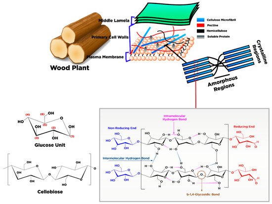
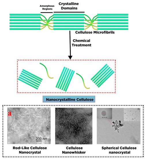
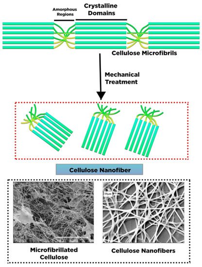
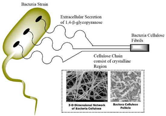
2. Conversion of Cellulose into Nanocellulose and Its Characteristic

Cellulose is the most abundant natural polymer globally and is a renewable source and essential raw material for various industries. Cellulose is a crucial constituent compound for plants, marine animals, algae, fungi, bacteria, and amoebae [12]. In 1838, French chemist Anselme Payen discovered and isolated cellulose from plant fibers using nitric acid and determined its chemical structure. The primary sources of cellulose are plant fibers with a high cellulose content, such as cotton (containing more than 90% cellulose content) [16] and wood (up to 50% cellulose). Other compounds such as hemicellulose, lignin, pectin, and wax are also present; they can be recovered during the separation process.
Recently, various agricultural wastes with high cellulose content were explored as a source of cellulose, such as oil palm empty fruit bunches (OPEFB) [17], palm and banana fronds, passionfruit peel waste [18], bagasse, wheat straw, rice straw, bamboo stalks, hemp bark, potato tubers, mulberry bark, hemp avicel, and sugar beets [19]. Cellulose derived from these non-plant precursors can have a molecular structure similar to that of plant cellulose. However, the main difference is that much less hemicellulose or lignin is present in these non-plant-based precursors; higher cellulose content with much lower impurities can be obtained from these precursors.
In terms of chemical structure, cellulose is composed of a linear homopolysaccharide consist of β-d-glucopyranose units entirely condensed and bonded through β-1,4-glycoside linkages (Figure 1). The structure foundation of the cellulose network is arranged by a chain glucose dimer comprising two anhydrous glucoses (AG) defined as cellobiose [20] (Figure 1). The raw material or the pretreatment (chemical or mechanical) of cellulose may affect the cellulose chain length and thus lead to molecular weight variation. The number of AG units in each chain is known as the polymerization degree (PD). The value of PD for cellulose powder varies from 100 to 300 units and around 26,500 for cellulose pulp [21]. The PD value for cellulose from cotton is 15,000, and wood is approximately 10,000 [22].
Figure 1. Schematic of cellulose production from wood plant and structural chemistry of exhibiting arrangement betwixt individual fibers.
Each cellulose monomer contains three reactive hydroxyl groups in the repeating chemical structure of the β-d-glucopyranose unit. In the same chain, these hydroxyl groups can make hydrogen bonds with the adjacent β-d-glucopyranose units. At different chain locations, the bonds present are intramolecular and intermolecular hydrogen bonds responsible for the crystal arrangement, determining the cellulose’s physical characteristics. Based on molecular orientation and hydrogen network between molecules and intramolecular, cellulose is classified into different types, i.e., I, II, III, IIII, IIIII, IVI, and IVII. For details about the classification of cellulose, the reader can refer to the work of Moon et al. [23]. Some of the cellulose characteristics are mainly represented by hydrogen linkage coordination [24][25].
Structurally, the cellulose is a linear chain polymer with a rod-like configuration, aided by the glucose residues’ equatorial conformation that is intensely aggregated together with the lateral size 3–5 nm [25]. Primary chains of cellulose, especially polysaccharide chains, are found on the secondary walls of plants arranged in a parallel configuration. The cellulose’s basic fibers have a cross-sectional diameter between 10–450 nm with a length of several micrometers that depend on the diversity of material sources [26]. Moreover, the elementary fibrils were arranged into large pack units called microfibrils, further foregathered into fibrils [13]. There are regions within the cellulose fibrils where the cellulose chains are organized into a highly crystalline structure with a length of 50–150 nm and disordered amorphous regions with 25–50 nm [23]. The cellulose chains construct the crystalline regions through Van der Waals forces, strong intra- and intermolecular hydrogen linkage, and β-1,4-glycosidic bonds. In contrast, amorphous regions are built up through the deficiency of hydrogen bonds in the crystalline region. The crystalline and amorphous regions in cellulose may vary depending on various sources.
The crystalline constituent within cellulose fibers can be refined through various chemical treatments by destructing and removing the disordered amorphous or para-crystalline regions. The purified crystal fragments with particle sizes on the nanometer scale are called nanocrystalline cellulose (NCC) (Figure 2). Different shapes of NCC are present such as needle and elongated rod-like shape or spindle-like shape with high stiffness of crystalline fragments [27], which are reported as cellulose whisker [28], nanowhisker [29], nanorod [30], and spherical nanocrystal [31].
Figure 2. Schematic representation nanocrystalline cellulose fabrication by chemical treatment ((a) transmission electron microscopy (TEM) images of rod-like cellulose nanocrystals [27], reprinted with permission; transmission electron microscopy (TEM) images of cellulose nano whisker reprinted with permission from [32]. Copyright © 2019 Elsevier B.V.; (b) transmission electron microscopy (TEM) images of spherical cellulose nanocrystal reprinted with permission from [33]. Copyright © 2018 Elsevier B.V.).
A top-down process has been applied for NCC production in which a large unit of cellulose fibers (cm) is disintegrated through chemical or mechanical treatment into small units of nanocellulose (nm) [34]. NCC’s chemical structure is constructed by intra- and intermolecular hydrogen linkage of cellulose macromolecules with a high crystallinity value varying from 54 to 88% [35]. NCC’s particle size depends on the origin of the cellulose sources, with the diameter and length typically varying between 5 and 30 nm and between 100 and 500 nm, respectively [36]. Thus, NCCs have become an attractive candidate as drug carriers, given their outstanding physical and chemical properties [37][38][39].
Cellulose nanofiber (CNF), also known as cellulose nanofibril, micro-fibrillated cellulose, nano-fibrillar cellulose, nano-fibrillated cellulose, or cellulose microfibril, has a similar molecule structure to NCCs with nano-size particles. Similar to NCC, CNF can also be produced from various cellulose sources. However, the morphology and crystallinity of NCC and CNF are the unique features that differentiate these two cellulose-based compounds. CNFs have long, flexible cellulose chains of amorphous and crystalline regions isolated from cellulose fibrils through mechanical treatment (Figure 3) [36]. The diameter of CNFs varies from 1 to 100 nm, while their length varies between 500 and 2000 nm. The dimension of CNFs molecules is strongly influenced by mechanical treatment and defibrillation [40].
Figure 3. Schematic representation of cellulose nanofibers fabrication by mechanical treatment (scanning electron microscopy (SEM) images of micro fibrillated cellulose reprinted with permission from ref. [41]; Copyright © 2007 Elsevier Ltd.; scanning electron microscopy (SEM) images of cellulose nanofibers reprinted with permission from ref. [42]. Copyright © 2006 Elsevier Ltd.).
NCC has high crystalline cellulose purity, resulting in a rigid structure, whereas the CNF structure consists of irregular amorphous parts, with some parts exhibiting a high degree of crystallinity. The amorphous regions in CNF control the structure flexibility of nanocellulose [43]. Figure 3 presents an illustration of CNF extracted from cellulose fragments via mechanical defibrillation. The exerted force fractures the cellulose fibrils along its longitudinal axis [23]. Compared with NCC, CNF exhibits unique properties such as extended length with excellent aspect proportion (length to diameter), superlative surface area, hydrophilicity, biocompatibility, and adjustable characteristic through surface modification [44].
Microbial cellulose (MC), bacterial nanocellulose (BC), and bio-cellulose (BC) have been used as the other terms for bacteria cellulose (BC). In contrast to NCC and CNF, BC’s structure comprises sugars with low molecular weight. Many bacteria strains have been used to generate BC as an extracellular metabolic product, such as Gluconacetobacter, Sarcina, Aerobacteria, Escheria, Achromobacter, Rhizobium, Rhodobacter, Azotobacter, and Agrobacterium [45][46]. However, only Gluconacetobacter xylinus has been commercially utilized to produce BC on an industrial scale [16]. The bacteria strains are commonly incubated in nutrient-rich aqueous media and produce BC on the upper layer (interface with air) as an exopolysaccharide. In this case, the β-d-glucopyranose units are initially present during the growth of cellulose molecules within the bacterial cell. The elementary fibril is released across the pores of the cellulose surface, which was further arranged and crystallized into microfibrils with twisting ribbons shape followed by pellicle formation (Figure 4) [47]. The fabricated BC comprises a nanofibers framework with a diameter of 20–100 nm with a length of several micrometers and a large surface area composed mainly of water (99%) [48].
Figure 4. Schematic production of bacteria cellulose through extracellular secretion (scanning electron microscopy (SEM) images of 3-D dimensional network of bacteria cellulose [49], reprinted with permission; (c) scanning electron microscopy (TEM) images bacteria cellulose pellicle, reprinted with permission from ref. [50]. Copyright © 2019 Elsevier Ltd.)
In terms of chemical composition, BC is indistinguishable from plant-based nanocellulose (e.g., NCC and CNF). However, BC has higher crystallinity (up to 84–89%) with fewer amorphous regions than NCC and CNF. Moreover, BC contains fewer impurities and contaminants such as hemicellulose, lignin, and pectin, mainly found in plant-based nanocellulose. BC is a biocompatible material with non-cytotoxicity and non-genotoxicity for biomedical applications, especially drug delivery [51]. BC synthesis does not involve a complicated process such as mechanical and chemical treatment to cleave the hemicellulose or lignin within the lignocellulosic biomass, thereby allowing high cellulose purity.
BC’s properties can be modulated by various techniques such as substrate manipulation, culture condition and operation parameter, and proper bacterial strain selection [52][45]. In contrast to NCC and CNF, BC provides unique characteristics such as high crystallinity of nanocellulose (84–88%) and polymerization grade, high water uptake capacity (exceeding 100 times of its weight), large surface area (high aspect proportion of fiber), outstanding tensile strength (Young modulus of 15–18 GPa), flexibility, foldability, moldability, mechanical stability, and high porosity [51]. A summary of the characteristics of various types of nanocellulose is listed in Table 1.
Table 1. Summary of the characteristics of various types of nanocelluloses.
 

Types

Nanocrystalline Cellulose (NCC)

Cellulose Nanofibers (CNF)

Bacterial Cellulose

Parameter


Common names

Cellulose whisker, cellulose nanowhisker, cellulose nanowire, and cellulose nanorod or spherical cellulose nanocrystals

Cellulose nanofibril, micro fibrillated cellulose, Nanofibrillar cellulose, Nanofibrillated cellulose, and cellulose microfibril

Microbial cellulose (MC), bacterial nanocellulose (BC), and bio-cellulose (BC)

Morphological structure

Needles like shape, elongated rod-like shape, and spindle shape

Smooth, extended, and flexible chain

Twisted ribbons like shape

Structure of Nanocellulose

Crystalline domains

amorphous and crystalline domains

Crystalline domains

Chain Length

≥500

500–15,000

4000–10,000

Crystallinity (%)

54–88

-

84–88

Other Impurities and contaminant

Possible to contain hemicellulose, lignin, and pectin

Possible to contain hemicellulose, lignin, and pectin

Contain no hemicellulose, lignin, and pectin

Size (Length and Diameter)

Diameter: 5–30 nm and Length: 100–500 nm

Diameter: 1–100 nm and Length: 500–2000 nm

Diameter 20–100 nm and several micrometric lengths

Process System

Top-down system

Top-down system

Bottom-up system

Tensile strength (Gpa)

7.5–7.7

[23]

13

0.2–0.3

Modulus Young (Gpa)

110–220

[35]

Approximately 15

18–20

[51]

Density (gr/cm3)

1.6

[53]

1.42

1.1

Characteristics

Homogenous nanorod form, exceptional aspect ratio (length to diameter), appreciable specific surface area (SSA), biocompatibility, liquid crystalline attribute, inferior breaking expansion, high young’s modulus, hydrophilicity, outstanding mechanical stiffness, tunable surface characteristic due to the reactive hydroxyl group and low density

Extended length with excellent aspect proportion (length to diameter), superlative surface area, hydrophilicity, biocompatibility and adjustable characteristic through surface modification afforded by high extensive of hydroxyl groups in CNF.

High crystallinity of nanocellulose (84–88%) and polymerization grade, high water uptake capacity (exceeding 100 times of its weight), remarkable surface area (high aspect proportion of fiber), outstanding tensile strength (young modulus 15–18 Gpa), and flexibility, foldability, moldability, mechanical stability, highly biocompatible material, non-cytotoxic, un-genotoxic and high porosity

Based on the previous discussion, cellulose can be subjected to a mechanical, biological, and chemical treatment to produce three different NCs, i.e., nanocrystalline cellulose, cellulose nanofibrils, and biological cellulose. They are classified based on various aspects such as morphology, particle size, crystallinity, nanocellulose structure, extraction techniques, and cellulose sources [47]. Moreover, other important factors such as interfibrillar arrangement, microfibril inclination, chemical constituent, cell dimension, and defects can also vary depending on the cellulose sources [54]. Among all the mentioned characteristics, mechanical strength is essential in the drug delivery field [55]. As summarized in Table 1, NCC possesses a high modulus young, up to 220 GPa, which is higher than glass (86 GPa) [53] and kevlar KM2 fiber (88 GPa) [35]. Furthermore, the mechanical stiffness of NCC can reach up to 7.7 GPa, which is higher than 302 stainless steel (3.88 GPa) [35] and kevlar KM2 fiber (1.28 Gpa) [35].

3. Surface Chemistry of Nanocellulose for Drug Delivery

Biocompatibility, biodegradability, and drug carrier capability to confine, control, and localize the drug release towards the target sites are desirable for nano-drug carrier formulation. The ability of nano-drug carriers to transport the drug and specify the sites for targeted drug release is influenced by the particle size, the surface charge, modification, and hydrophobicity. These aspects govern the nano-drug carrier interface with the plasma membrane and its diffusion across the physiological drug barrier [56]. Most NCs exhibit high specific surface area and negative interface charges as potential drug carriers, making them suitable as hydrophilic drug carriers. Therefore, the NCs’ surface can be attached to the desired drug [57]. However, pristine NC cannot be used effectively as a drug carrier given its limited water solubility, moisture sensitivity, thermal instability, and lack of stability in various buffer solutions. Even though the pH adjustment of the environment can enhance the dispersibility of NCs, the scattering examination divulged the aggregation tendency of NCs, which translates to the colloidal instability of NCs. The size reduction obtained by converting cellulose into NC provides an exponential improvement of hydrogen bonding that triggers the NC aggregation. This limitation can be made worse by the drug coordination, which is exposed on the NC exterior, consequently altering the dispersibility and solubility [58]. Therefore, various surface modification and pretreatment fiber methodologies have been developed to overcome limitations and advance specific characteristics [59].
From a structural perspective, the three hydroxyl groups in each cellulose monomer are the most prominent characteristic that makes the NC surface reactive. The reactivity of hydroxyl groups influences the surface modification of anhydroglucose units. It was reported that in the molecular framework of cellulose, the hydroxyl group at the sixth position behaves as primary alcohol with a reactivity ten times larger than the other hydroxyl groups, while the hydroxyl group at the second position has two-fold higher reactivity than that in the third position, both of which serve as secondary alcohols. This phenomenon manifests from the steric hindrance of each hydroxyl group, in which the hydroxyl group at the sixth position attached to the carbon atom that is connected to only one alkyl groups while the carbon atom that carries the hydroxyl groups in the second and third positions bonded to two alkyl groups [60]. Regarding the surface receptiveness of NC’s hydroxyl groups, the addition of solvent and reactant may alter the group’s receptiveness in diverse positions. De la Motte et al. [61] modified NCC through cationic epoxide 2,3-epoxypropyltrimethyl ammonium chloride (EPTMAC) by spray technique. It was revealed that the hydroxyl bunch receptiveness of cationic modified NC follows the order of OH-C6 = OH-C2 > OH-C3, which was validated through nuclear magnetic resonance (NMR).
Nanocellulose surface modification for drug delivery was developed by modulating the NC hydroxyl groups. In general, the main objective of nanocellulose surface modification is to incorporate new functional groups or drug components into the nanocellulose framework to escalate the degree of substitution and the efficacy of material grafting without altering the structure, morphology, and crystallinity of nanocellulose [62].

References

  1. Jain, K.K. An overview of drug delivery systems. In Drug Delivery Systems; Springer: New York, NY, USA, 2020.
  2. Sunasee, R.; Hemraz, U.D.; Ckless, K. Cellulose nanocrystals: A versatile nanoplatform for emerging biomedical applications. Expert Opin. Drug Deliv. 2016, 13, 1243–1256.
  3. Bamrungsap, S.; Zhao, Z.; Chen, T.; Wand, L.; Li, C.; Fu, T.; Tan, W. Nanotechnology in therapeutics: A focus on nanoparticles as a drug delivery system. Nanomedicine 2012, 7, 1253–1271.
  4. Tong, X.; Pan, W.; Su, T.; Zhang, M.; Dong, W.; Qi, X. Recent advances in natural polymer-based drug delivery systems. React. Funct. Polym. 2020, 148, 104501.
  5. Cavallaro, G.; Micciulla, S.; Chiappisi, L.; Lazzara, G. Chitosan-based smart hybrid materials: A physico-chemical perspective. J. Mater. Chem. B 2021, 9, 594–611.
  6. Bertolino, V.; Cavallaro, G.; Milioto, S.; Lazzara, G. Polysaccharides/Halloysite nanotubes for smart bionanocomposite materials. Carbohydr. Polym. 2020, 245, 116502.
  7. Ahmad, A.; Mubarak, N.; Jannat, F.T.; Ashfaq, T.; Santulli, C.; Rizwan, M.; Najda, A.; Bin-Jumah, M.; Abdel-Daim, M.M.; Hussain, S. A Critical Review on the Synthesis of Natural Sodium Alginate Based Composite Materials: An Innovative Biological Polymer for Biomedical Delivery Applications. Processes 2021, 9, 137.
  8. Chen, J.; Ouyang, J.; Chen, Q.; Deng, C.; Meng, F.; Zhang, J.; Cheng, R.; Lan, Q.; Zhong, Z. EGFR and CD44 dual-targeted multifunctional hyaluronic acid nanogels boost protein delivery to ovarian and breast cancers in vitro and in vivo. ACS Appl. Mater. Interfaces 2017, 9, 24140–24147.
  9. Qi, X.; Wei, W.; Shen, J.; Dong, W. Salecan polysaccharide-based hydrogels and their applications: A review. J. Mater. Chem. B 2019, 7, 2577–2587.
  10. Dragan, E.S.; Dinu, M.V. Polysaccharides constructed hydrogels as vehicles for proteins and peptides. A review. Carbohydr. Polym. 2019, 225, 115210.
  11. Jorfi, M.; Foster, E.J. Recent advances in nanocellulose for biomedical applications. J. Appl. Polym. Sci. 2015, 132, 41719.
  12. Xue, Y.; Mou, Z.; Xiao, H. Nanocellulose as a sustainable biomass material: Structure, properties, present status and future prospects in biomedical applications. Nanoscale 2017, 9, 14758–14781.
  13. Habibi, Y.; Lucia, L.A.; Rojas, O.J. Cellulose nanocrystals: Chemistry, self-assembly, and applications. Chem. Rev. 2010, 110, 3479–3500.
  14. Lin, N.; Dufresne, A. Nanocellulose in biomedicine: Current status and future prospect. Eur. Polym. J. 2014, 59, 302–325.
  15. Plackett, D.; Letchford, K.; Jackson, J.; Burt, H. A review of nanocellulose as a novel vehicle for drug delivery. Nord. Pulp Pap. Res. J. 2014, 29, 105–118.
  16. Huang, Y.; Zhu, C.; Yang, J.; Nie, Y.; Chen, C.; Sun, D. Recent advances in bacterial cellulose. Cellulose 2014, 21, 1–30.
  17. Foo, M.L.; Tan, C.R.; Lim, P.D.; Ooi, C.W.; Tan, K.W.; Chew, I.M.L. Surface-modified nanocrystalline cellulose from oil palm empty fruit bunch for effective binding of curcumin. Int. J. Biol. Macromol. 2019, 138, 1064–1071.
  18. Wijaya, C.J.; Saputra, S.N.; Soetaredjo, F.E.; Putro, J.N.; Lin, C.X.; Kurniawan, A.; Ju, Y.-H.; Ismadji, S. Cellulose nanocrystals from passion fruit peels waste as antibiotic drug carrier. Carbohydr. Polym. 2017, 175, 370–376.
  19. Dufresne, A. Cellulose nanomaterial reinforced polymer nanocomposites. Curr. Opin. Colloid Interface Sci. 2017, 29, 1–8.
  20. Gopinath, V.; Saravanan, S.; Al-Maleki, A.; Ramesh, M.; Vadivelu, J. A review of natural polysaccharides for drug delivery applications: Special focus on cellulose, starch and glycogen. Biomed. Pharmacother. 2018, 107, 96–108.
  21. Wertz, J.-L.; Bedue, O.; Mercier, J.P. Cellulose Science and Technology; EPFL Press: Lausanne, Switzerland, 2010.
  22. George, J.; Sabapathi, S. Cellulose nanocrystals: Synthesis, functional properties, and applications. Nanotechnol. Sci. Appl. 2015, 8, 45.
  23. Moon, R.J.; Martini, A.; Nairn, J.; Simonsen, J.; Youngblood, J. Cellulose nanomaterials review: Structure, properties and nanocomposites. Chem. Soc. Rev. 2011, 40, 3941–3994.
  24. Medronho, B.; Lindman, B. Brief overview on cellulose dissolution/regeneration interactions and mechanisms. Adv. Colloid Interface Sci. 2015, 222, 502–508.
  25. Kamel, R.; El-Wakil, N.A.; Dufresne, A.; Elkasabgy, N.A. Nanocellulose: From an agricultural waste to a valuable pharmaceutical ingredient. Int. J. Biol. Macromol. 2020, 163, 1579–1590.
  26. Miao, C.; Hamad, W.Y. Cellulose reinforced polymer composites and nanocomposites: A critical review. Cellulose 2013, 20, 2221–2262.
  27. Li, N.; Lu, W.; Yu, J.; Xiao, Y.; Liu, S.; Gan, L.; Huang, J. Rod-like cellulose nanocrystal/cis-aconityl-doxorubicin prodrug: A fluorescence-visible drug delivery system with enhanced cellular uptake and intracellular drug controlled release. Mater. Sci. Eng. C 2018, 91, 179–189.
  28. Jia, B.; Li, Y.; Yang, B.; Xiao, D.; Zhang, S.; Rajulu, A.V.; Kondo, T.; Zhang, L.; Zhou, J. Effect of microcrystal cellulose and cellulose whisker on biocompatibility of cellulose-based electrospun scaffolds. Cellulose 2013, 20, 1911–1923.
  29. Dash, R.; Ragauskas, A.J. Synthesis of a novel cellulose nanowhisker-based drug delivery system. RSC Adv. 2012, 2, 3403–3409.
  30. Biswas, S.K.; Sano, H.; Yang, X.; Tanphichai, S.; Shams, M.I.; Yano, H. Highly thermal-resilient AgNW transparent electrode and optical device on thermomechanically superstable cellulose nanorod-reinforced nanocomposites. Adv. Opt. Mater. 2019, 7, 1900532.
  31. Wang, N.; Ding, E.; Cheng, R. Thermal degradation behaviors of spherical cellulose nanocrystals with sulfate groups. Polymer 2007, 48, 3486–3493.
  32. Putro, J.N.; Ismadji, S.; Gunarto, C.; Yuliana, M.; Santoso, S.P.; Soetaredjo, F.E.; Ju, Y.H. The effect of surfactants modification on nanocrystalline cellulose for paclitaxel loading and release study. J. Mol. Liq. 2019, 282, 407–414.
  33. Ram, B.; Chauhan, G.S.; Mehta, A.; Gupta, R.; Chauhan, K. Spherical nanocellulose-based highly efficient and rapid multifunctional naked-eye Cr (VI) ion chemosensor and adsorbent with mild antimicrobial properties. Chem. Eng. J. 2018, 349, 146–155.
  34. Mariano, M.; El Kissi, N.; Dufresne, A. Cellulose nanocrystals and related nanocomposites: Review of some properties and challenges. J. Polym. Sci. B Polym. Phys. 2014, 52, 791–806.
  35. Brinchi, L.; Cotana, F.; Fortunati, E.; Kenny, J. Production of nanocrystalline cellulose from lignocellulosic biomass: Technology and applications. Carbohydr. Polym. 2013, 94, 154–169.
  36. Phanthong, P.; Reybroycharoen, P.; Hao, X.; Xu, G.; Abudula, A.; Guan, G. Nanocellulose: Extraction and application. Carbon Resour. Convers. 2018, 1, 32–43.
  37. Karimian, A.; Parsian, H.; Majidinia, M.; Rahimi, M.; Mir, S.M.; Kafil, H.S.; Shafiei-Irannejad, V.; Kheyrollah, M.; Ostadi, H.; Yousefi, B. Nanocrystalline cellulose: Preparation, physicochemical properties, and applications in drug delivery systems. Int. J. Biol. Macromol. 2019, 133, 850–859.
  38. Lam, E.; Male, K.B.; Chong, J.H.; Leung, A.C.; Luong, J.H. Applications of functionalized and nanoparticle-modified nanocrystalline cellulose. Trends Biotechnol. 2012, 30, 283–290.
  39. Prathapan, R.; Tabor, R.F.; Garnier, G.; Hu, J. Recent progress in cellulose nanocrystal alignment and its applications. ACS Appl. Bio Mater. 2020, 3, 1828–1844.
  40. Abitbol, T.; Rivkin, A.; Cao, Y.; Nevo, Y.; Abraham, E.; Ben-Shalom, T.; Lapidot, S.; Shoseyov, O. Nanocellulose, a tiny fiber with huge applications. Curr. Opin. Biotechnol. 2016, 39, 76–88.
  41. Henriksson, M.; Henriksson, G.; Berglund, L.; Lindström, T. An environmentally friendly method for enzyme-assisted preparation of microfibrillated cellulose (MFC) nanofibers. Eur. Polym. J. 2007, 43, 3434–3441.
  42. Kim, C.-W.; Kim, D.-S.; Kang, S.-Y.; Marquez, M.; Joo, Y.L. Structural studies of electrospun cellulose nanofibers. Polymer 2006, 47, 5097–5107.
  43. Dufresne, A. Nanocellulose: A new ageless bionanomaterial. Mater. Today 2013, 16, 220–227.
  44. Lavoine, N.; Desloges, I.; Dufresne, A.; Bras, J. Microfibrillated cellulose–Its barrier properties and applications in cellulosic materials: A review. Carbohydr. Polym. 2012, 90, 735–764.
  45. Blanco Parte, F.G.; Santoso, S.P.; Chou, C.-C.; Verma, V.; Wang, H.-T.; Ismadji, S.; Cheng, K.-C. Current progress on the production, modification, and applications of bacterial cellulose. Crit. Rev. Biotechnol. 2020, 40, 397–414.
  46. Siro, I.; Plackett, D. Microfibrillated cellulose and new nanocomposite materials: A review. Cellulose 2010, 17, 459–494.
  47. Huang, J.; Dufresne, A.; Lin, N. Nanocellulose: From Fundamentals to Advanced Materials; John Wiley & Sons: Hoboken, NJ, USA, 2019.
  48. Jozala, A.F.; De Lencastre-Novaes, L.C.; Lopes, A.M.; De Carvalho Santos-Ebinuma, V.; Mazzola, P.G.; Pessoa-JR, A.; Grotto, D.; Gerenutti, M.; Chaud, M.V. Bacterial nanocellulose production and application: A 10-year overview. Appl. Microbiol. Biotechnol. 2016, 100, 2063–2072.
  49. Wei, B.; Yang, G.; Hong, F. Preparation and evaluation of a kind of bacterial cellulose dry films with antibacterial properties. Carbohydr. Polym. 2011, 84, 533–538.
  50. Wan, Y.; Wang, J.; Gama, M.; Guo, R.; Zhang, Q.; Zhang, P.; Yao, F.; Luo, H. Biofabrication of a novel bacteria/bacterial cellulose composite for improved adsorption capacity. Compos. Part A Appl. Sci. Manuf. 2019, 125, 105560.
  51. Gorgieva, S.; Trcek, J. Bacterial cellulose: Production, modification and perspectives in biomedical applications. Nanomaterials 2019, 9, 1352.
  52. Hasan, N.; Rahman, L.; Kim, S.-H.; Cao, J.; Arjuna, A.; Lallo, S.; Jhun, B.H.; Yoo, J.-W. Recent advances of nanocellulose in drug delivery systems. J. Pharm. Investig. 2020, 50, 553–572.
  53. Kim, J.-H.; Shim, B.S.; Kim, H.S.; Lee, Y.-J.; Min, S.-K.; Jang, D.; Abas, Z.; Kim, J. Review of nanocellulose for sustainable future materials. Int. J. Precis. Eng. Manuf. Green Technol. 2015, 2, 197–213.
  54. Dufresne, A. Cellulose-based composites and nanocomposites. In Monomers, Polymers, and Composites from Renewable Resources; Elsevier: Amsterdam, The Netherlands, 2008.
  55. Song, F.; Li, X.; Wang, Q.; Liao, L.; Zhang, C. Nanocomposite hydrogels and their applications in drug delivery and tissue engineering. J. Biomed. Nanotechnol. 2015, 11, 40–52.
  56. Mitchell, M.J.; Billingsley, M.M.; Haley, R.M.; Wechsler, M.E.; Peppas, N.A.; Langer, R. Engineering precision nanoparticles for drug delivery. Nat. Rev. Drug Discov. 2020, 20, 101–124.
  57. Hare, J.I.; Lammers, T.; Ashford, M.B.; Puri, S.; Strom, G.; Barry, S.T. Challenges and strategies in anti-cancer nanomedicine development: An industry perspective. Adv. Drug Deliv. Rev. 2017, 108, 25–38.
  58. Araki, J. Electrostatic or steric?–preparations and characterizations of well-dispersed systems containing rod-like nanowhiskers of crystalline polysaccharides. Soft Mater. 2013, 9, 4125–4141.
  59. Kargarzadeh, H.; Mariano, M.; Gopakumar, D.; Ahmad, I.; Thomas, S.; Dufresne, A.; Huang, J.; Lin, N. Advances in cellulose nanomaterials. Cellulose 2018, 25, 2151–2189.
  60. Hebeish, A. , Guthrie, T. The Chemistry and Technology of Cellulosic Copolymers; Springer Science & Business Media: Belin, Germany, 2012.
  61. De La Motte, H.; Hasani, M.; Brelid, H.; Westman, G. Molecular characterization of hydrolyzed cationized nanocrystalline cellulose, cotton cellulose and softwood kraft pulp using high resolution 1D and 2D NMR. Carbohydr. Polym. 2011, 85, 738–746.
  62. Habibi, Y. Key advances in the chemical modification of nanocelluloses. Chem. Soc. Rev. 2014, 43, 1519–1542.
More
Information
Contributor MDPI registered users' name will be linked to their SciProfiles pages. To register with us, please refer to https://encyclopedia.pub/register :
View Times: 841
Revisions: 2 times (View History)
Update Date: 09 Jul 2021
1000/1000