Submitted Successfully!
To reward your contribution, here is a gift for you: A free trial for our video production service.
Thank you for your contribution! You can also upload a video entry or images related to this topic.
Version Summary Created by Modification Content Size Created at Operation
1 + 2486 word(s) 2486 2021-07-05 05:20:31 |
2 format correction Meta information modification 2486 2021-07-06 03:32:27 |

Video Upload Options

Do you have a full video?

Confirm

Are you sure to Delete?
Cite
If you have any further questions, please contact Encyclopedia Editorial Office.
Zhang, Y. Protein Complexes in Plants. Encyclopedia. Available online: https://encyclopedia.pub/entry/11675 (accessed on 25 April 2024).
Zhang Y. Protein Complexes in Plants. Encyclopedia. Available at: https://encyclopedia.pub/entry/11675. Accessed April 25, 2024.
Zhang, Youjun. "Protein Complexes in Plants" Encyclopedia, https://encyclopedia.pub/entry/11675 (accessed April 25, 2024).
Zhang, Y. (2021, July 05). Protein Complexes in Plants. In Encyclopedia. https://encyclopedia.pub/entry/11675
Zhang, Youjun. "Protein Complexes in Plants." Encyclopedia. Web. 05 July, 2021.
Protein Complexes in Plants
Edit

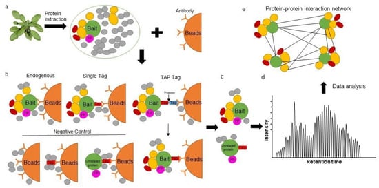
The study of protein–protein interactions (PPIs) is fundamental in understanding the unique role of proteins within cells and their contribution to complex biological systems. Affinity purification coupled to mass spectrometry (AP-MS) and proximity labeling coupled to mass spectrometry (PL-MS) are two powerful techniques that have significantly enhanced our understanding of PPIs. Relying on the specific binding properties of a protein to an immobilized ligand, AP is a fast, sensitive and targeted approach used to detect interactions between bait (protein of interest) and prey (interacting partners) under near-physiological conditions. 

affinity purification proximity labeling plant protein complex protein-protein interactions

1. Introduction

The study of biomolecular complexes is crucial in understanding the molecular mechanisms underpinning biological processes, protein function and subcellular protein localization [1][2][3][4]. Biomolecular complexes are principally formed by proteins interacting with other proteins (protein–protein interactions, PPIs), however complexes can also arise through the interaction of proteins with ligands such as nucleic acids, sugars, lipids and hormones [2][3][4]. As the biological function of a protein is defined by its interactions in the cell, an important step in investigating, disrupting or modulating biological processes lies in understanding how and why PPIs occur [1][4]. Advantages of protein complex formation are myriad, starting from greater proximity between substrate and catalyst to enhanced efficiency of whole biochemical pathways.
The field of proteomics has witnessed the development of many innovative methods for the identification and characterization of PPIs [1][3][4]. As method preferences to study protein complexes have changed over time, so too have the possibilities to obtain annotated or predicted protein complexes and composition. Over recent years, proteome-wide studies and computational approaches both point toward a scenario with an increasing number of heteromeric protein complexes being identified [5][6]. The methodology used to predict or identify protein complexes can be categorized in two ways: experimental and computational. Computational or in silico approaches are used to predict PPIs via computer simulations and are dependent on the algorithm used [7]. These predictions are based on high throughput proteomics data (binary or mass spectrometry-based methods), primary structure, 3D structure, domain, evolutionary relationship, genomic methods or a combination of these methods [7][8][9][10]. Experimental approaches are either performed in vitro or in vivo. While in vitro studies are generally performed on a low throughput scale, in vivo studies can be carried out in a high throughput manner. The most common methods used in the study of PPIs are biochemical protein purification or separation (2D gel electrophoresis, 2-DE [11]; blue native polyacrylamide gel electrophoresis, BN-PAGE; size exclusion chromatography, SEC) followed by mass spectrometry (MS), genetic engineering of cellular systems (yeast two hybrid (Y2H) assays and their variants; phage display), arrays (protein arrays or peptides microarrays), structural studies (NMR spectrometry, X-ray crystallography, cryoelectron microscopy) or fluorescence imaging (fluorescence resonance energy transfer, FRET; bimolecular fluorescence complementation BiFC) [1][3][4][12].
Recent studies highlight significant progress in the use of affinity purification and proximity labeling approaches combined with MS-based quantitative proteomics in studying PPIs [5][13][14][15]. Affinity purification mass spectrometry (AP-MS) is a fast, sensitive and targeted approach used to detect interactions between bait (protein of interest) and prey (interacting partners) under near-physiological conditions [16]. This method can be applied to large-scale studies and has been demonstrated to have high intra-and inter-laboratory reproducibility [17]. Similarly, proximity-dependent labeling methods are being increasingly used to detect transient PPIs under native conditions in living cells [14]. As the name suggests, proximity labeling (PL) relies on the principle that proteins must be physically close in order for them to interact and is predicted to be more precise in determining interacting partners [18].
Both AP-MS and PL-MS are powerful techniques that have significantly enhanced our understanding of PPIs. While these methods have become increasingly popular in animal systems, application of these techniques in plants remain underutilized. Combined, AP-MS and PL-MS have the potential to reveal an unprecedented spatial and temporal protein interaction network that better understands biological processes relevant to many fields of interest. For example, AP-MS can be theoretically used to detect transient PPIs as well as interactions involving potentially insoluble proteins such as membrane-associated proteins. Furthermore, PL-MS has the potential to detect hydrophobic interactions under native conditions and has been recently used to investigate membrane contact sites between the endoplasmic reticulum and mitochondria in plants [19].

2. Affinity Purification Mass Spectrometry in Plants

Similar to immunopurification or immunoprecipitation (IP), AP utilizes antibodies which can be targeted to the bait, or to a standardized fusion moiety often referred to as an epitope tag [6]. Using protein-specific antibodies, AP-MS has the theoretical advantage of capturing protein complexes under native conditions from plant lysates [5]. However, with limited availability of plant protein antibodies, different bait isoforms that can occlude antibody interaction sites and differing specificities of antibodies, the ability to obtain reliable protein interaction networks remains challenging [4][6]. Therefore, fusion of the bait to various affinity tags has greatly increased the efficacy of this method. Once the bait protein interacts with its respective prey, the resulting complex can be purified from the cell lysate using a matrix that specifically recognizes the affinity tag. Both stable protein complexes and weak PPIs between bait and prey have been detected by AP-MS [20][21]. A critical aspect of this technique lies in protein separation, purification and digestion to reduce the presence of contaminants. Specific protein antibodies can be used to immunoprecipitate the protein of interest under native conditions; however, this approach has only been successfully demonstrated by a few laboratories [5]. While several affinity tags have been developed to allow co-precipitation of prey and bait proteins under native conditions (Table 1), the use of such tags comes with its challenges. Introduction of an epitope tag can result in non-native folding of the tagged protein or steric hindrance of interactions. As bait fused affinity tags generally need to be overexpressed, such expression can influence the physiological properties of the bait or stoichiometry of the complex. Epitope tags can also result in incorrect localization or alternative localization of the protein of interest. It has been shown that overexpression of the bait may result in false positive interactions [6][22]. For these reasons, it is highly recommended that researchers confirm that the chosen epitope tag does not interfere with the endogenous function, localization, or properties of the bait by complementation of the mutant plant line [3][6]. However, these recommendations are not widely utilized due to the time-consuming nature of producing stable transgenic lines and cannot be followed if wild-type plants are used. The use of clustered regularly interspaced short palindromic repeats (CRISPR) technology could help to improve these limitations. Such technology provides researchers with the ability to directly insert affinity tags into endogenous loci without changing the genomic context of the gene and also maintain the native environment to which protein interactions can then be characterized [3][23].
Table 1. Affinity tags successfully used to investigate plant protein–protein interactions.

Tag

Sequence/Size

Affinity Resin

Elution Conditions

Reference

TAPi tag

45 kDa

Calmodulin binding peptide with two protein A domain

Protein A/low pH

[22][23]

Streptavidin binding peptide (SBP)

WSHPQFEK

Streptavidin

Desthiobiotin

[23][24]

GSyellow

37 kDa

Streptavidin-binding peptide tag with citrine yellow fluorescent protein

Desthiobiotin/pH

[23][25][26]

Fluorescent protein (GFP, YFP)

26.9 kDa

Anti-GFP

pH

[13][23][25][26]

GSrhino tag

21.9 kDa

two IgG-binding domains of protein G and a SBP tag

Streptavidin elution buffer [5]

[5][23][27]

Alternative TAP (TAPa)

26 kDa

2 xIgG-BD with 6 XHis and 9 Xmyc

HR3C cleavage/Imidazole/low pH

[23][28]

Given the increased sensitivity of MS and the application of novel bioinformatic approaches for accurate data analysis, affinity-based methods have improved considerably in recent years [5][29]. While single tag AP-MS is now widely used in large scale studies, selection of the epitope tag and positioning of the tag at either the N- or C-terminus of protein remains critical. In addition to being an efficient purification handle, some affinity tags also provide benefits such as information regarding subcellar localization of the PPI. For example, fluorescent tags (i.e., green fluorescence protein (GFP), yellow fluorescence protein (YFP) and the mFruits family of monomeric red fluorescent proteins (mRFPs)) allow for localization studies to be performed in parallel to AP-MS studies. The ability to simultaneously monitor both protein localization and expression is useful in investigating whether the recombinant protein occurs under native conditions and if the preyed interactions are biologically relevant. For example, differences in the metabolic roles of glycolytic and TCA cycle enzymes fused with C-terminal GFP were observed in the cytosol and mitochondria respectively [30][31]. In addition, one benefit of using epitope tags is that several proteins can be fused with the same epitope and purified with same method. As a result, background contamination should be consistent across all purifications and should enable the use of the same negative controls, including tag-only constructs or wild-type plants. As shown in Table 1, several types of epitope tags have been successfully applied to AP-MS in plants.
The main disadvantage of AP-MS however, remains in the ability to fully characterize affinity matrix/epitope tag interaction properties. The identification of non-specific bound proteins is one of the main disadvantages of a single-step purification approach and contaminant proteins associated with either the solid-phase or the epitope tag are hard to distinguish from positive interactors. Thus, the use of proper negative controls such as protein extracts from wild-type plants, mutant lines, or tag-only expressing plants is critical (Figure 1). In principle, unspecific proteins identified in these controls can be simply subtracted from the list of interactors that are identified by the bait. However, given the limitations of AP enrichment and liquid chromatography–mass spectrometry (LC–MS), false positives are still likely. Alternatively, various algorithms can be applied. For example, the SAINT algorithm [32] allows researchers to determine fold change abundance (FC-A), which can be used to filter out potential false positives. Possible interactions can also be evaluated based on the ratio of spectral counts of the bait versus overexpression of an unrelated protein or tag-only controls [33]. Moreover, a second purification step can be introduced to reduce the amount of non-specific binding proteins [5][21]. In tandem affinity purification (TAP), two types of affinity tags linked by a protease cleavage site are fused to a bait protein and expressed in plants. Two affinity purification steps are then performed to obtain reliable interacting partners (Figure 1b). Interestingly, an Arabidopsis plant cell culture system has been developed for TAP technology which allows for the high-throughput identification of protein complexes, even with very low sample volumes (25 mg total protein) [5]. GS tags and their derivatives are the most frequently and successfully used TAP tags in plant research [5][34]. A GS tag consists of two immunoglobulin domains of a streptavidin-binding peptide and protein G linked by a unique cleavage site that is recognized by the etch virus protease from tobacco (Nicotiana tabacum). Following an initial affinity purification step with immunoglobulin G agarose beads, protein complexes can be incubated with the tobacco etch virus protease to release the complex from the matrix. In a subsequent purification step, the bait protein complex associates with a streptavidin-conjugated bead trap. Following several washing steps, the protein complex is eluted and determined by LC-MS (Figure 1b; [5][21]). In addition, a multifunctional TAP tag (GSyellow) has been developed that combines the fluorescent properties of citrine YFP with a streptavidin-binding peptide tag. This double affinity tag can not only be used to determine the subcellular localization of proteins in vivo but also the potential function of the protein through AP [26].
Figure 1. Overview of affinity purification strategies. (a) Total protein extraction for affinity purification. (b) Bait specific antibodies are linked to beads for protein complex immunoprecipitation under native conditions. Such beads can be used to detect endogenous proteins within a plant, proteins fused displaying a single tag (single affinity purification) or proteins expressing a double (TAP) tag (double affinity purification). Suggested controls used to reduce background contaminants and thus the identification of false positives include using a wild-type plant extract, purification from cells expressing the tag only, or unrelated proteins fused with a tag. (c) Several washing steps are used to reduce non-specific interactions. (d) Proteins are measured by LC-MS. (e) Data analysis to determine a protein–protein interaction network. FP: false positive; UP: unrelated protein.

3. The Proximity Labeling Method

PL-MS is a high-throughput approach for the systematic analysis of PPIs in vivo. While PL-MS is already firmly established in mammalian and unicellular eukaryote systems, application of this technique in planta remains challenging. PL utilizes enzymes that produce reactive molecules that covalently interact with proteins in close proximity. Labeled proteins can be isolated using conventional affinity purification methods and identified via immunoblot analysis or by protein mass spectrometry, Proximity labeling overcomes some of the limitations of AP-MS and Y2H, as abundant soluble proteins as well as insoluble membrane proteins can be effectively enriched under stringent denaturing conditions, which in turn, facilitates their identification. PL can detect weak, transient or hydrophobic PPIs in their native state and provides an unedited spatial and temporal protein interaction network for better understanding of a specific biological process. In addition, fusion of PL enzymes to a minimal targeting motif that restricts proteins to a particular subcellular location or structure, can be used to map the protein population therein [35]. While application of PL-MS to plant systems remains in its infancy, we summarize the recent development of this technology and highlight its potential in studying plant PPIs (Figure 2).
Figure 2. Overview of proximity labeling system. (a) Transient and stable protein with proximity-labeling (PL) enzyme transformation. (b) PL assay based on the tagged PL enzyme. A biotin ligase or APEX PL enzyme is fused to the target protein and expressed in plants. Upon the addition of a substrate, such as biotin or biotin-phenol and hydrogen peroxide (H2O2), proteins or RNAs are tagged by biotin. (c) Interacting pairs are fused to the PL enzyme at either the N- and C-terminus to investigate the composition of protein complexes. As two proteins interact in cells, the two halves of a split-PL are reorganized as a full PL enzyme and initiate the labeling of proximal partners of the protein complex. After protein extraction and incubating with streptavidin beads, biotin-labeled proteins or RNAs can be enriched for subsequent LC-MS/MS or high-throughput sequencing analysis.

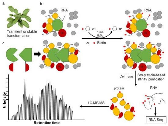
4. Combining Proximity Labeling and Affinity Purification-Mass Spectrometry

While AP-MS results in the identification of proteins that form stable complexes, PL enables the identification of proteins that are in close proximity to the bait, which results in overlapping yet distinct protein identifications. By integrating AP- and PL-MS data, one has the ability to comprehensively characterize a protein’s molecular context and so several combined AP and PL experiments have been trialed. Enzyme combinations allow for both AP-MS and BioID analysis within a single construct and with almost identical protein purification and mass spectrometry (MS) identification procedures such as FLAG-BirA* tag [36][37], Multiple Approaches Combined (MAC)-tag [16] and Strep-Tactin [27] have now been developed. However, there are limitations in combining these two approaches due to the large size of BirA* and the small affinity purification peptide of a Flag or His tag. This strategy of combining AP and PL has not been used in plants to date; however, the generation of specific antibodies for PL tags may facilitate the combination of these two methods in the future.

References

  1. Zhang, Y.; Fernie, A.R. Stable and temporary enzyme complexes and metabolons involved in energy and redox metabolism. Antioxid. Redox Signal. 2020.
  2. Chae, L.; Lee, I.; Shin, J.; Rhee, S.Y. Towards understanding how molecular networks evolve in plants. Curr. Opin. Plant Biol. 2012, 15, 177–184.
  3. Lampugnani, E.R.; Wink, R.H.; Persson, S.; Somssich, M. The toolbox to study protein–protein interactions in plants. Crit. Rev. Plant Sci. 2018, 37, 308–334.
  4. Struk, S.; Jacobs, A.; Sánchez Martín-Fontecha, E.; Gevaert, K.; Cubas, P.; Goormachtig, S. Exploring the protein–protein interaction landscape in plants. Plant Cell Environ. 2019, 42, 387–409.
  5. Van Leene, J.; Eeckhout, D.; Cannoot, B.; De Winne, N.; Persiau, G.; Van De Slijke, E.; Vercruysse, L.; Dedecker, M.; Verkest, A.; Vandepoele, K.; et al. An improved toolbox to unravel the plant cellular machinery by tandem affinity purification of arabidopsis protein complexes. Nat. Protoc. 2015, 10, 169–187.
  6. Dunham, W.H.; Mullin, M.; Gingras, A.C. Affinity-purification coupled to mass spectrometry: Basic principles and strategies. Proteomics 2012, 12, 1576–1590.
  7. Zhang, Y.; Gao, P.; Yuan, J.S. Plant protein-protein interaction network and interactome. Curr. Genom. 2010, 11, 40–46.
  8. Dong, S.; Lau, V.; Song, R.; Ierullo, M.; Esteban, E.; Wu, Y.; Sivieng, T.; Nahal, H.; Gaudinier, A.; Pasha, A. Proteome-wide, structure-based prediction of protein-protein interactions/new molecular interactions viewer. Plant Physiol. 2019, 179, 1893–1907.
  9. Jiang, M.; Niu, C.; Cao, J.; Ni, D.A.; Chu, Z. In silico-prediction of protein–protein interactions network about mapks and pp2cs reveals a novel docking site variants in brachypodium distachyon. Sci. Rep. 2018, 8, 1–11.
  10. De Bodt, S.; Proost, S.; Vandepoele, K.; Rouzé, P.; Van de Peer, Y. Predicting protein-protein interactions in arabidopsis thaliana through integration of orthology, gene ontology and co-expression. BMC Genom. 2009, 10, 1–15.
  11. Barnouin, K. Two-dimensional gel electrophoresis for analysis of protein complexes. Protein-Protein Interact. 2004, 261, 479–497.
  12. Zhang, Y.; Fernie, A.R. On the detection and functional significance of the protein–protein interactions of mitochondrial transport proteins. Biomolecules 2020, 10, 1107.
  13. Zhang, Y.; Natale, R.; Domingues, A.P.J.; Toleco, M.R.; Siemiatkowska, B.; Fàbregas, N.; Fernie, A.R. Rapid identification of protein-protein interactions in plants. Curr. Protoc. Plant Biol. 2019, 4, e20099.
  14. Yang, X.; Wen, Z.; Zhang, D.; Li, Z.; Li, D.; Nagalakshmi, U.; Dinesh-Kumar, S.P.; Zhang, Y. Proximity labeling: An emerging tool for probing in planta molecular interactions. Plant Commun. 2020, 100137.
  15. Mair, A.; Xu, S.-L.; Branon, T.C.; Ting, A.Y.; Bergmann, D.C. Proximity labeling of protein complexes and cell-type-specific organellar proteomes in arabidopsis enabled by turboid. Elife 2019, 8, e47864.
  16. Liu, X.; Salokas, K.; Weldatsadik, R.G.; Gawriyski, L.; Varjosalo, M. Combined proximity labeling and affinity purification− mass spectrometry workflow for mapping and visualizing protein interaction networks. Nat. Protoc. 2020, 15, 3182–3211.
  17. Varjosalo, M.; Sacco, R.; Stukalov, A.; Van Drogen, A.; Planyavsky, M.; Hauri, S.; Aebersold, R.; Bennett, K.L.; Colinge, J.; Gstaiger, M. Interlaboratory reproducibility of large-scale human protein-complex analysis by standardized ap-ms. Nat. Methods 2013, 10, 307.
  18. Kim, D.I.; Birendra, K.; Zhu, W.; Motamedchaboki, K.; Doye, V.; Roux, K.J. Probing nuclear pore complex architecture with proximity-dependent biotinylation. Proc. Natl. Acad. Sci. USA 2014, 111, E2453–E2461.
  19. Kwak, C.; Shin, S.; Park, J.-S.; Jung, M.; Nhung, T.T.M.; Kang, M.-G.; Lee, C.; Kwon, T.-H.; Park, S.K.; Mun, J.Y. Contact-id, a tool for profiling organelle contact sites, reveals regulatory proteins of mitochondrial-associated membrane formation. Proc. Natl. Acad. Sci. USA 2020, 117, 12109–12120.
  20. Bauer, A.; Kuster, B. Affinity purification-mass spectrometry. Eur. J. Biochem. 2003, 270, 570–578.
  21. Li, Y. The tandem affinity purification technology: An overview. Biotechnol. Lett. 2011, 33, 1487–1499.
  22. Rohila, J.S.; Chen, M.; Cerny, R.; Fromm, M.E. Improved tandem affinity purification tag and methods for isolation of protein heterocomplexes from plants. Plant J. 2004, 38, 172–181.
  23. Bontinck, M.; Van Leene, J.; Gadeyne, A.; De Rybel, B.; Eeckhout, D.; Nelissen, H.; De Jaeger, G. Recent trends in plant protein complex analysis in a developmental context. Front. Plant Sci. 2018, 9, 640.
  24. Wu, S.-C.; Wong, S.-L. Structure-guided design of an engineered streptavidin with reusability to purify streptavidin-binding peptide tagged proteins or biotinylated proteins. PLoS ONE 2013, 8, e69530.
  25. Née, G.; Tilak, P.; Finkemeier, I. A versatile workflow for the identification of protein–protein interactions using gfp-trap beads and mass spectrometry-based label-free quantification. In Plant Proteomics; Springer: Heidelberg, Germany, 2020; pp. 257–271.
  26. Besbrugge, N.; Van Leene, J.; Eeckhout, D.; Cannoot, B.; Kulkarni, S.R.; De Winne, N.; Persiau, G.; Van De Slijke, E.; Bontinck, M.; Aesaert, S. Gsyellow, a multifaceted tag for functional protein analysis in monocot and dicot plants. Plant Physiol. 2018, 177, 447–464.
  27. Schmidt, T.G.; Skerra, A. The strep-tag system for one-step purification and high-affinity detection or capturing of proteins. Nat. Protoc. 2007, 2, 1528.
  28. Rubio, V.; Shen, Y.; Saijo, Y.; Liu, Y.; Gusmaroli, G.; Dinesh-Kumar, S.P.; Deng, X.W. An alternative tandem affinity purification strategy applied to arabidopsis protein complex isolation. Plant J. 2005, 41, 767–778.
  29. Struk, S.; Braem, L.; Walton, A.; De Keyser, A.; Boyer, F.-D.; Persiau, G.; De Jaeger, G.; Gevaert, K.; Goormachtig, S. Quantitative tandem affinity purification, an effective tool to investigate protein complex composition in plant hormone signaling: Strigolactones in the spotlight. Front. Plant Sci. 2018, 9, 528.
  30. Zhang, Y.; Sampathkumar, A.; Kerber, S.M.; Swart, C.; Hille, C.; Seerangan, K.; Graf, A.; Sweetlove, L.; Fernie, A.R. A moonlighting role for enzymes of glycolysis in the co-localization of mitochondria and chloroplasts. Nat. Commun. 2020, 11, 4509.
  31. Zhang, Y.J.; Beard, K.F.M.; Swart, C.; Bergmann, S.; Krahnert, I.; Nikoloski, Z.; Graf, A.; Ratcliffe, R.G.; Sweetlove, L.J.; Fernie, A.R.; et al. Protein-protein interactions and metabolite channelling in the plant tricarboxylic acid cycle. Nat. Commun. 2017, 8, 1–11.
  32. Mellacheruvu, D.; Wright, Z.; Couzens, A.L.; Lambert, J.-P.; St-Denis, N.A.; Li, T.; Miteva, Y.V.; Hauri, S.; Sardiu, M.E.; Low, T.Y. The crapome: A contaminant repository for affinity purification–mass spectrometry data. Nat. Methods 2013, 10, 730.
  33. Nesvizhskii, A.I. Computational and informatics strategies for identification of specific protein interaction partners in affinity purification mass spectrometry experiments. Proteomics 2012, 12, 1639–1655.
  34. Bürckstümmer, T.; Bennett, K.L.; Preradovic, A.; Schütze, G.; Hantschel, O.; Superti-Furga, G.; Bauch, A. An efficient tandem affinity purification procedure for interaction proteomics in mammalian cells. Nat. Methods 2006, 3, 1013–1019.
  35. Kim, D.I.; Roux, K.J. Filling the void: Proximity-based labeling of proteins in living cells. Trends Cell Biol. 2016, 26, 804–817.
  36. Hesketh, G.G.; Youn, J.-Y.; Samavarchi-Tehrani, P.; Raught, B.; Gingras, A.-C. Parallel exploration of interaction space by bioid and affinity purification coupled to mass spectrometry. In Proteomics; Comai, L.K.J., Mallick, P., Eds.; Springer: New York, NY, USA, 2017; Volume 1550, pp. 115–136.
  37. Lambert, J.-P.; Tucholska, M.; Go, C.; Knight, J.D.; Gingras, A.-C. Proximity biotinylation and affinity purification are complementary approaches for the interactome mapping of chromatin-associated protein complexes. J. Proteom. 2015, 118, 81–94.
More
Information
Subjects: Plant Sciences
Contributor MDPI registered users' name will be linked to their SciProfiles pages. To register with us, please refer to https://encyclopedia.pub/register :
View Times: 610
Revisions: 2 times (View History)
Update Date: 06 Jul 2021
1000/1000