Submitted Successfully!
To reward your contribution, here is a gift for you: A free trial for our video production service.
Thank you for your contribution! You can also upload a video entry or images related to this topic.
Version Summary Created by Modification Content Size Created at Operation
1 + 4002 word(s) 4002 2021-06-15 08:46:21 |
2 Fix a spacing issue between two chapters. Meta information modification 4002 2021-06-20 19:34:09 | |
3 format correct Meta information modification 4002 2021-06-21 04:41:51 | |
4 format correct Meta information modification 4002 2021-10-12 05:16:18 | |
5 format correct Meta information modification 4002 2021-10-12 05:16:36 |

Video Upload Options

Do you have a full video?

Confirm

Are you sure to Delete?
Cite
If you have any further questions, please contact Encyclopedia Editorial Office.
Lavrik, O. Nucleotide Excision Repair. Encyclopedia. Available online: https://encyclopedia.pub/entry/11043 (accessed on 25 April 2024).
Lavrik O. Nucleotide Excision Repair. Encyclopedia. Available at: https://encyclopedia.pub/entry/11043. Accessed April 25, 2024.
Lavrik, Olga. "Nucleotide Excision Repair" Encyclopedia, https://encyclopedia.pub/entry/11043 (accessed April 25, 2024).
Lavrik, O. (2021, June 20). Nucleotide Excision Repair. In Encyclopedia. https://encyclopedia.pub/entry/11043
Lavrik, Olga. "Nucleotide Excision Repair." Encyclopedia. Web. 20 June, 2021.
Nucleotide Excision Repair
Edit

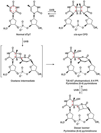
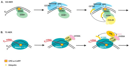
Nucleotide excision repair (NER) is the most versatile DNA repair pathway, which can remove diverse bulky DNA lesions destabilizing a DNA duplex. NER substrates are UV photoproducts, e.g., cyclobutane pyrimidine dimers (CPDs), pyrimidine-pyrimidone-(6-4)-photoproducts (6-4PPs), intrastrand crosslinks, and bulky adducts of DNA bases with reactive metabolites of some chemical carcinogens or chemotherapeutic agents. These kinds of lesions can be substrates for two NER sub-pathways—global genome NER (GG-NER) and transcription-coupled NER (TC-NER)—that overlap, except for the mode of DNA damage recognition.

NER defects cause several autosomal recessive genetic disorders. Xeroderma pigmentosum (XP) is one of the NER-associated syndromes characterized by low efficiency of the removal of bulky DNA adducts generated by ultraviolet radiation. XP patients have extremely high ultraviolet-light sensitivity of sun-exposed tissues, often resulting in multiple skin and eye cancers.

nucleotide excision repair xeroderma pigmentosum neurodegeneration

1. Introduction

In all cells, DNA is the carrier of genetic information from generation to generation; thus, its integrity must be maintained to ensure the survival of the cell, the whole organism, or even the whole species. Nonetheless, DNA is constantly jeopardized by multiple external adverse factors, such as ultraviolet (UV) light, ionizing radiation, chemotherapy drugs, or environmental pollutants. DNA damage can also be caused by endogenous factors such as replication errors or cellular oxidative metabolism products from mitochondria or inflammation [1]. The lesions can disrupt the basic processes of DNA metabolism by blocking replication and transcription. To counteract these adverse effects, eukaryotic cells are equipped with several DNA repair mechanisms acting on different types of DNA damage [2][3]. In some cases, if the lesions cannot be eliminated—either because the damage load is too high or because a requisite repair pathway is deficient—the cell cycle can be arrested until the damage is repaired, and if this does not occur rapidly, the cell may be eliminated by apoptosis or may accumulate mutations and transform into a potentially cancerous cell that might proliferate uncontrollably and give rise to a tumor. Ultimately, cells can tolerate some DNA lesions owing to translesion DNA synthesis.
Typical types of DNA lesions include a variety of oxidative DNA modifications involving base or sugar damage, DNA crosslinks, strand breaks, and adducts with chemically active molecules [2][3]. Moreover, DNA can be damaged because of internal instability due to the spontaneous hydrolysis of the glycosidic bond with the formation of an abasic site (i.e., apurinic/apyrimidinic site, hereafter: AP site). As a rule, base excision repair (BER) deals with the repair of nonbulky base damage and AP sites in both the nuclear and mitochondrial cellular compartments [1][4][5].
The nucleotide excision repair (NER) pathway is the most universal repair pathway to remove a wide range of helix distorting lesions from DNA [6][7]. NER substrates are UV photoproducts, e.g., cyclobutane pyrimidine dimers (CPDs), pyrimidine-pyrimidone-(6-4)-photoproducts (6-4PPs), intrastrand crosslinks, and bulky adducts of DNA bases with reactive metabolites of some chemical carcinogens or chemotherapeutic agents [8]. These kinds of lesions can be substrates for two NER sub-pathways—global genome NER (GG-NER) and transcription-coupled NER (TC-NER)—that overlap, except for the mode of DNA damage recognition. Specific damage sensing proteins of GG-NER scan the entire genome at any moment of the cell cycle [6][7][8]. In contrast to GG-NER, TC-NER rapidly eliminates transcription-blocking lesions from actively transcribed DNA strands only. During TC-NER DNA damage can be detected in the template DNA strand when it stalls the RNA polymerase [9][10]. After the lesion has been recognized, all subsequent steps require the same NER core factors in GG-NER and TC-NER.
Mutations in NER-related genes cause several hereditary diseases, such as xeroderma pigmentosum (XP) and Cockayne syndrome (CS) [11]. Mutations in XP-related gene products (except proteins that exclusively taking part in GG-NER damage recognition) lead to the disruption of both NER sub-pathways. At the same time, mutations in CS proteins affect only TC-NER. XP is characterized by extreme sensitivity of the skin to sunlight and a dramatically increased risk of skin cancer [12][13]. A subset of XP patients develops a profound neurodegenerative condition known as XP neurological disease [14]. XP and CS are often grouped together as related diseases owing to overlapping sun sensitivity phenotypes and progressive neurodegeneration, but the specific nature of the neurological pathologies is qualitatively different between them [15][16][17].
Progressive neurodegeneration occurs when a loss of neuronal structure or function leads to a decline in the number of neurons owing to apoptotic cell death [2]. Neurons have a high metabolic load and consume large amounts of energy, which is supplied by mitochondria in the form of ATP. Byproducts of the ATP formation give rise to reactive oxygen species (ROS), which can cause many types of oxidative DNA damage to genomic and mitochondrial DNA [18][19]. Nowadays, it is widely accepted that the accumulation of oxidative DNA lesions is the cause of the neuropathology that takes place with aging as in several neurodegenerative disorders. Moreover, the accumulation of damaged mitochondria due to a decrease in mitophagy is also a hallmark of the aging process and a clinical feature of XP and CS [18][20][21].

2. Nucleotide Excision Repair

2.1. Classic NER Substrates

One of the most astonishing features of the NER pathway is its broad ability to recognize and process many structurally and chemically diverse lesions. NER is the only repair pathway that protects our skin from DNA photodamage induced by UV light. The latter is the high-energy component of sunlight that reaches the Earth surface. According to the wavelength, UV radiation can be subdivided into several ranges. Fortunately, the atmosphere blocks ~3/4 of the sun’s UV light, and its most powerful part, UV-C (100–280 nm), is completely absorbed. The ozone layer filters most of UV-B (280–315 nm). Thus, most of the UV light that reaches the Earth surface is UV-A (315–400 nm) with a small remainder of UV-B. UV-A can penetrate more deeply into the skin than UVB can because of its longer wavelength. Photodamage formation (and sunburn) in human skin starts near the boundary between UV-A and UV-B light (~315 nm) and continues during UV-B exposure. Notably, a wavelength closer to the nucleotide light absorption maximum produces more lesions in DNA.
CPDs are the major DNA photoproducts of UV light (Figure 1) [7][8][22][23]; 6-4PPs are formed in a 25–30% lower amount than CPDs and are the second most prevalent UV lesion. CPDs only minimally distort the double helix, whereas 6-4PPs produce a pronounced DNA backbone bending and base-pairing disruption. The DNA thermodynamic destabilization ability correlates with repair efficiency of these lesions; CPDs are excised by NER with much slower kinetics as compared to 6-4PPs [1][22][24][25].
Figure 1. Chemical structures of DNA photoproducts caused by sunlight. The majority of cyclobutane pyrimidine dimers (CPDs) is formed between adjacent thymine residues (TT) but can eventually arise between adjacent T and C, C and T, or C and C, depending on the wavelength, irradiation dose, and adjacent sequences. CPD can be formed with cis-syn isomer representing large majority of CPDs within duplex DNA, and trans-syn occurring exclusively within single-stranded DNA. Pyrimidine-(6-4)-pyrimidone photoproducts (6-4PPs) are generated preferentially in nucleotide pairs TC, CC, and TT, with the ratio and yields depending on irradiation wavelength and adjacent sequences [8]. This figure is based on several studies [8][22].
Another source of DNA damage is various electrophilic compounds that directly penetrate the cell from an external medium or are produced inside the organism after metabolic activation. These compounds include environmental mutagens like polycyclic aromatic hydrocarbons (benzo[a]pyrene and various aromatic amines) and adducts of cancer chemotherapeutic drugs such as cisplatin. These electrophilic compounds can react with nucleophilic atoms of DNA, especially with N7 of the guanine base [7][8].
DNA damage recognition is the first key step, which affects overall efficiency of DNA repair [6]. The fact that NER can repair so many structurally different types of DNA damage indicated early on that the system may not recognize a lesion per se but rather some specific conformational features caused by the lesion within DNA [7]. In general, a good NER substrate should be bulky and must destabilize a DNA double helix (disrupt base pairing and bend the duplex). To detect both conditions, NER has evolved special bipartite substrate discrimination: firstly, it recognizes a local thermodynamically destabilized site, and then the latter is probed for lesion presence. The double recognition allows NER to avoid processing mismatched but damage-free sites.

2.2. The Damage Recognition Step

DNA damage can be recognized by NER in one of two modes. GG-NER can search for damage anywhere in the genome throughout the cell cycle. The second mode is TC-NER, which is responsible for the accelerated repair of lesions in the template DNA strand of actively transcribed genes only.
In the case of mammalian GG-NER, lesions are recognized by xeroderma pigmentosum factor C (XPC) complexed with proteins RAD23B and centrin 2 (CETN2) [6]. Small subunits collectively stabilize the XPC structure, possibly modulate some protein–protein interactions, and stimulate the DNA binding of the major subunit of the complex (XPC) thereby increasing NER efficiency in vitro and in vivo ([26] and references within). The XPC–RAD23B–CETN2 complex (hereinafter, XPC) can detect and bind DNA sites where the regular double-helical structure is perturbed, and as a result, one or more base pairs are disrupted and/or destabilized [6][7]. X-ray crystal structure of Rad4—the yeast ortholog of XPC—has revealed a structural basis for the unique DNA damage-searching ability [27][28]. A series of subsequent biophysical studies indicates that Rad4/XPC can bind to DNA nonspecifically via a damage-independent DNA-binding domain (TGD) and freely diffuse mainly by a one-dimensional-diffusion mechanism [7][29][30]. It is noteworthy that Rad4/XPC diffuse along DNA not by “sliding” but rather by “hopping” (diffusion through repeated microscopic dissociation and reassociation with the DNA). An advantage of the hopping mode is that it allows a protein to overcome protein obstacles on DNA [29]. At a suspicious DNA site of certain single-stranded character (where DNA is “breathing” too much because of a mismatch or an AT-rich sequence, where DNA can transiently melt), Rad4/XPC are slowed down [29][30]. The presence of a helical distortion and base pair disruption enables XPC to insert two β-hairpin modules from BHD2/BHD3 domains into the DNA duplex and to form a stable protein–DNA complex. In this complex, Rad4/XPC interacts exclusively with the nucleotides on the undamaged strand and flips out damage-containing nucleotide pairs to form an “open” conformation [30]. The reason is that the damaged DNA is already destabilized and has a lower free-energy barrier for “opening,” thus increasing the probability that Rad4 (or XPC) can use the hairpin modules to sense the lesion presence and not diffuse away [7][30]. Notably, DNA duplexes containing bulky lesions on both strands are not processed by NER [31][32].
CPDs—the most abundant photolesions—are poorly recognized by XPC because they cause only a minimal distortion in DNA. These lesions are recognized by a special protein (UV-damaged DNA-binding protein (UV-DDB), a heterodimeric protein consisting of DDB1 and DDB2/XPE), which has extraordinarily high binding affinity and specificity for CPD and 6-4PP [6][7]. In contrast to XPC, DDB2 interacts directly with UV light–induced photolesions in DNA, introduces a kink into the duplex, and creates a more suitable substrate for XPC (Figure 2A). Structural studies have revealed that DDB2 flips out the two nucleotides of CPD into a shallow binding pocket, which can accommodate such lesions as CPDs or 6-4PPs via shape complementarity [6][7]. In addition, DDB2 is thought to facilitate XPC recruitment within chromatinized DNA through the ability to promote chromatin reorganization. Moreover, DDB1 is also a connector protein for ubiquitin ligase CUL4–RBX1 [7][33]. The ubiquitin ligase is activated upon DDB2 binding and ubiquitinates DDB2 and XPC [33]. The ubiquitination of DDB2 launches its proteasomal degradation after extraction from NER complexes. By contrast, the XPC ubiquitination increases its DNA-binding activity [34]. Damage handover from DDB2 to XPC coincides with the arrival of the TFIIH complex, which further promotes DDB2 dissociation [33][35]. It should be noted that both UV-DDB and XPC proteins are also targets of poly(ADP-ribosyl)ation catalyzed by poly(ADP-ribose)polymerase 1 (PARP1) in response to UV-irradiation. Taking in account that PARP1 participates in the UV-induced chromatin decondensation and PARP1 activity promotes DDB2 interaction with XPC, this modification can facilitate lesion recognition in the chromatin context (reviewed in [36]).
Figure 2. An overview of the damage recognition step of nucleotide excision repair (NER). (A) Global genome NER (GG-NER) can search for damage anywhere in the genome throughout the cell cycle. The UV-DDB protein recognizes CPD or 6-4PP, directly binds to it through its DDB2 subunit, and facilitates efficient recognition of the lesion by the XPC–RAD23B–CETN2 complex. The DDB1 subunit is also a connector protein for ubiquitin ligase CUL4, which ubiquitinates DDB2 and XPC [33]. (B) Transcription-coupled NER (TC-NER) is responsible for accelerated repair of lesions in the template DNA strand of actively transcribed genes only. The CSB protein and then proteins CSA and UVSSA bind to DNA damage stalled RNAPII. CSB and CSA associate with CRL ubiquitin ligase and contribute to the ubiquitination of the RNAPII RPB1 subunit at K1268. This ubiquitination stimulates the association of TFIIH with the stalled RNAPII through a transfer mechanism that also involves UVSSA-K414 ubiquitination [9][35].
TC-NER is initiated by the stalling of elongating RNA polymerase II (RNAPII) at DNA lesions (Figure 2B). The CSB protein (Cockayne syndrome group B protein, a member of the SNF2 family of DNA-dependent ATPases) interacts loosely with the elongating RNAPII and stimulates transcription but becomes more tightly bound after transcription arrest [10]. It is suggested that CSB participates in RNAPII backtracking to make a DNA lesion accessible to repair proteins. Upon RNAPII stalling at a lesion, the RNAPII-bound CSB recruits the CSA protein (Cockayne syndrome group A protein), and both together contribute to the polyubiquitination of the K1268 residue of RPB1, a subunit of RNAPII [35][37][38]. The RPB1 ubiquitination acts as a master switch for the alternation of transcription, RNAPII degradation, and initiation of DNA repair [9]. At the next step, CSA facilitates the association of UVSSA (UV-sensitive syndrome protein A) with the stalled RNAPII. UVSSA is the key factor that recruits the TFIIH complex [37].
When the RPB1 K1268 residue is mutated or some of CSB/CSA/UVSSA accessory proteins are absent, TC-NER cannot start. In this situation, transcription does not shut down, leading to multiple transcription restarts (resulting primarily in the transcription of short genes) and subsequent RNAPII stalling instances; therefore, eventually, the RNAPII pool is depleted, and transcription is dysregulated ([38] and reviewed in [9]).
A recent study has discovered that RNAPII stalling could follow by nascent RNA hybridization with DNA template strand generating an RNA–DNA hybrid and displaced ssDNA. Such kind of structures called an R-loop could occur physiologically during an early step in transcription elongation (especially are abundant at promoters) and transcription termination [39][40]. Moreover, it is proposed that R-loops can regulate gene expression through multiple context-dependent mechanisms. At the same time, R-loop can be problematic for cells as it blocks efficient transcription and replication. The accumulation of R-loops is associated with cancer and several neurological diseases. It was shown that NER proteins participate in R-loops resolving process, but the mechanism of R-loop resolution is not clear.
The TFIIH (transcription factor IIH) complex is a multifunctional protein machine required for transcription initiation and NER [41]. Depending on a context, its composition changes from a core of seven subunits, including the XPB translocase and XPD helicase, to 10 subunits, through the addition of three CAK (Cdk-activating-kinase module) kinase subunits. Recent advances in breakthrough cryo-electron microscopy give investigators a unique opportunity to investigate the TFIIH structure [41][42][43]. TFIIH assumes an arch-like conformation with subunits curving from XPD on the one end to XPB located on the second end. The CAK components close the ends of this structure and stabilize the arch. It is suggested that TFIIH core structure becomes more flexible after CAK module dissociation, and this arrangement may be sufficient for subsequent functioning during NER [7]. The release of the CAK complex from core TFIIH transforms TFIIH from a transcription factor into a repair factor [37].

2.3. Damage Verification and Pre-Incision Complex Formation

The TFIIH complex is the key protein for the damage verification step. TFIIH probes the lesion itself and unwinds the DNA duplex around the lesion, thereby making room for the subsequent assembly of a repair machine; we simply could even say that NER machinery is built around TFIIH.
In the case of GG-NER (Figure 3A), TFIIH is recruited via XPB engagement to the DNA duplex and interaction with the C terminus of XPC as well as an additional interaction of the p62 subunit with XPC’s N terminus [7]. After that, the XPD helicase may get loaded on the DNA because of its location on the other end of the TFIIH arch. A striking similarity between GG-NER and TC-NER is that XPC and UVSSA share an interaction surface on the p62 subunit of TFIIH, suggesting that the two pathways at least partially share a mechanism for the engagement of TFIIH with the lesion site [41].
Figure 3. Schematic view of the damage verification step of NER and pre-incision complex formation. (A) GG-NER. TFIIH initially interacts with XPC’s N terminus by means of p62 subunits, then tumbles to XPC’s C terminus, where the interaction with XPB promotes its binding to a duplex part. XPA releases an inhibitory CAK module and together with XPG stimulates XPD activity [44]. The XPD helicase binds to the damaged strand and starts to a repair bubble formation [7]. When XPD gets to the lesion and stalls on it, XPC is displaced, and XPG binds to the 3′ edge of the repair bubble. (B) TC-NER. XPC and UVSSA share an interaction surface on the p62 subunit of TFIIH [41]. RNAPII moves in the 3′→5′ direction on the damaged strand, then, after its lesion stalling and assembly of factors CSB, CSA, and UVSSA, the latter promotes TFIIH binding downstream of RNAPII [37]. Thereafter, XPA and XPG stimulate XPD activity, and TFIIH starts to move in the 5′→3′ direction and may “push” RNAPII for a backtracking movement. (C) The NER pre-incision complex (PIC): TFIIH stalls on the lesion-bearing strand, RPA covers the undamaged strand, XPA marks the 5′ edge of the repair bubble, XPG marks the 3′ edge of the repair bubble, and XPF–ERCC1 binds behind XPA.
Notably, human TFIIH binds downstream of RNAPII (which moves in the 3′→5′ direction) in the transcription pre-initiation complex [45]. In line with these data, we propose that in TC-NER initiation, TFIIH should also bind downstream of RNAPII for subsequent movement on the same damaged strand in the opposite 5′→3′ direction (Figure 3B). In the case of R-loop formation behind the RNAPII, it cannot be easily displaced by TFIIH to make a space for repair process. The sequence of events in this situation should be a subject for future investigations.
TFIIH structure flexibility allows XPD to unwind DNA while tracking along in the 5′→3′ direction [7]. During the tracking process, XPD pulls the DNA through a narrow tunnel that is too small for bulky DNA lesions to pass through [7]. This “damage filtration” process is simple but effective.
The release of the CAK module from core TFIIH is triggered by the association of repair factors XPA and XPG (XP factors A and G) [44]. Biochemical data show that XPA can stimulate the overall helicase activity of TFIIH, and on the contrary, can inhibit the helicase activity in the presence of lesions; therefore, XPA also contributes to damage verification [46]. Moreover, XPA has some bulky-damage recognition ability as well and especially prefers to bind kinked and branched DNA structures [47][48]. It was shown recently by atomic force microscopy, scanning force microscopy, and mathematical modeling that XPA undergoes episodic one-dimensional diffusion to search DNA for damage [49]. Furthermore, biochemical research revealed that XPA is located on the 5′ side from a lesion at the damaged bubbled DNA [47]. Cryo-electron microscopy data have extended our knowledge of the modulation of TFIIH activity by XPA and XPG [44]: (1) XPA and XPG stabilize an alternative conformation of TFIIH, where the XPD helicase is opened for functioning; (2) XPA and XPG also stimulate XPB and XPD, and this event may facilitate DNA opening; consequently, they are present in a ternary complex in the lesion-scanning mode; (3) XPA interacts with an XPB subunit in the TFIIH–DNA complex and marks the DNA at the 5′ edge of the repair bubble; (4) XPA forms a bridge between XPB and XPD and thus possibly facilitates XPD positioning on the single-stranded 3′ extension.
Immediately after forming single-stranded undamaged DNA inside the repair bubble, it binds to the replication protein A (RPA) [50]. RPA interacts with the undamaged strand and protects it from a nuclease attack [47]. The size of the NER-excised fragment coincides with maximal length of the single-stranded–DNA platform for RPA binding (approximately 30 nt), to which RPA binds tightly with defined 5′→3′ polarity [51]. RPA tightly interacts with XPA inside the repair bubble, and they together regulate the correct orientation and activation of NER nucleases [52]. Moreover, an ability of RPA and XPA to form a complex in the absence of DNA as well as a ternary complex with DNA was reported, and XPA interaction with RPA is indispensable for NER [51]. Crystal structure of Ustilago maydis RPA stably bound to single-stranded DNA was resolved some time ago [53]. These data revealed that single-stranded DNA in complex with RPA is also U-shaped; for this reason, the 5′ edge and 3′ edge of the repair bubble are pulled together.
During the lesion scanning by TFIIH, XPG “rides” on the XPD subunit [44]. After XPD stalls on the lesion, XPG binds to the 3′ edge of the repair bubble (possibly by simultaneous displacement of XPC). NER pre-incision complex formation is completed by the engagement of XPF–ERCC1, which is recruited by XPA [54].
Thus, the interior of the NER pre-incision complex is as follows: TFIIH stalls at the lesion, RPA covers the undamaged opposite strand, XPA marks the 5′ edge of the repair bubble, XPG marks the 3′ edge of the repair bubble, and XPF–ERCC1 binds behind XPA. The XPA is a central component in the pre-incision complex room because it interacts with all its compartments: with the damage recognition proteins XPC and DDB2, verifies the damage and interacts with TFIIH and RPA, promotes correct positioning of both nucleases. Patients with reported mutations in the XPA gene have the severest form of XP (we discuss it in the next chapter). Today, XPA is considered as organizing or scaffold component of the pre-incision complex, which makes sure that all the NER factors are in the right place for the incision to occur [23][48].

2.4. Dual Incision, Resynthesis, and Ligation

Two endonucleases XPF–ERCC1 and XPG can now incise the lesion-containing DNA strand (Figure 4). The DNA incision is first carried out by XPF–ERCC1 from the 5′ side to the damage site with the formation of a free 3′-OH group [55]. Next, replication machinery can be loaded to start repair synthesis [56]. RPA promotes the arrival and positioning of RFC and enhances repair synthesis with possible help of XPA as it interacts with PCNA [57][58].
Figure 4. Late NER stages: dual incision, resynthesis, and ligation. The first incision is carried out by XPF–ERCC1 from the 5′ side to the lesion site. Then, replication machinery is loaded, and repair synthesis can be initiated. Possible sets of replication machines: DNA polymerase δ, PCNA, and RFC; DNA polymerase ε, PCNA, and a modified form of RFC; or DNA polymerase κ, ubiquitinated PCNA, and XRCC1. Halfway gap resynthesis is followed by a second incision by XPG. After repair synthesis is completed, nick sealing is performed by DNA ligase I or by the DNA ligase IIIα–XRCC1 complex.
Repair synthesis can proceed halfway through the gap in the absence of an XPG-made incision [23]. The XPG-made 3′ incision is possibly triggered by PCNA–XPG interaction [59]. The lesion-containing oligonucleotide (~30 nt) is released from the repair bubble in complex with TFIIH [60]. Then, after ATP binding, TFIIH slowly dissociates from the excised oligonucleotide, and the latter is bound by RPA or degraded by cellular nucleases.
Repair synthesis may be performed by different sets of replication machines: DNA polymerases δ/sliding clamp PCNA/clamp loader RFC or DNA polymerases ε/PCNA/ a modified form of RFC or DNA polymerases κ/ubiquitinated PCNA/XRCC1 [23][61]. Which set of replication factors will be loaded possibly depends on the cell cycle but in general remains unknown. The XPG-made incision leaves a 5′-phosphate that is utilized in the nick-sealing reaction by DNA ligase I or by DNA ligase IIIα (with XRCC1) [62]. Now, NER is completed.

References

  1. Kumar, N.; Moreno, N.C.; Feltes, B.C.; Menck, C.F.; Houten, B.V. Cooperation and interplay between base and nucleotide excision repair pathways: From DNA lesions to proteins. Genet Mol. Biol. 2020, 43 (Suppl. 1), e20190104.
  2. Jeppesen, D.K.; Bohr, V.A.; Stevnsner, T. DNA repair deficiency in neurodegeneration. Prog. Neurobiol. 2011, 94, 166–200.
  3. Kumar, N.; Raja, S.; Van Houten, B. The involvement of nucleotide excision repair proteins in the removal of oxidative DNA damage. Nucleic Acids Res. 2020, 48, 11227–11243.
  4. Krokan, H.E.; Bjørås, M. Base excision repair. Cold Spring Harb. Perspect. Biol. 2013, 5, a012583.
  5. Limpose, K.L.; Corbett, A.H.; Doetsch, P.W. BERing the burden of damage: Pathway crosstalk and posttranslational modification of base excision repair proteins regulate DNA damage management. DNA Repair (Amst.) 2017, 56, 51–64.
  6. Kusakabe, M.; Onishi, Y.; Tada, H.; Kurihara, F.; Kusao, K.; Furukawa, M.; Iwai, S.; Yokoi, M.; Sakai, W.; Sugasawa, K. Mechanism and regulation of DNA damage recognition in nucleotide excision repair. Genes Environ. 2019, 41, 2.
  7. Mu, H.; Geacintov, N.E.; Broyde, S.; Yeo, J.E.; Schärer, O.D. Molecular basis for damage recognition and verification by XPC-RAD23B and TFIIH in nucleotide excision repair. DNA Repair (Amst.) 2018, 71, 33–42.
  8. Gillet, L.C.; Scharer, O.D. Molecular mechanisms of mammalian global genome nucleotide excision repair. Chem. Rev. 2006, 106, 253–276.
  9. Son, K.; Schärer, O.D. Repair, Removal, and Shutdown: It All Hinges on RNA Polymerase II Ubiquitylation. Cell 2020, 180, 1039–1041.
  10. Hanawalt, P.C.; Spivak, G. Transcription-coupled DNA repair: Two decades of progress and surprises. Nat. Rev. Mol. Cell Biol. 2008, 9, 958–970.
  11. Rapin, I.; Lindenbaum, Y.; Dickson, D.W.; Kraemer, K.H.; Robbins, J.H. Cockayne syndrome and xeroderma pigmentosum. Neurology 2000, 55, 1442–1449.
  12. DiGiovanna, J.J.; Kraemer, K.H. Shining a light on xeroderma pigmentosum. J. Investig. Dermatol. 2012, 132, 785–796.
  13. Fassihi, H.; Sethi, M.; Fawcett, H.; Wing, J.; Chandler, N.; Mohammed, S.; Craythorne, E.; Morley, A.M.; Lim, R.; Turner, S.; et al. Deep phenotyping of 89 xeroderma pigmentosum patients reveals unexpected heterogeneity dependent on the precise molecular defect. Proc. Natl. Acad. Sci. USA 2016, 113, E1236–E1245.
  14. Bradford, P.T.; Goldstein, A.M.; Tamura, D.; Khan, S.G.; Ueda, T.; Boyle, J.; Oh, K.S.; Imoto, K.; Inui, H.; Moriwaki, S.; et al. Cancer and neurologic degeneration in xeroderma pigmentosum: Long term follow-up characterises the role of DNA repair. J. Med. Genet. 2011, 48, 168–176.
  15. Brooks, P.J. The Case for 8, 5′-Cyclopurine-2′-Deoxynucleosides as Endogenous DNA Lesions That Cause Neurodegeneration in Xeroderma Pigmentosum. Neuroscience 2007, 145, 1407–1417.
  16. Okur, M.N.; Fang, E.F.; Fivenson, E.M.; Tiwari, V.; Croteau, D.L.; Bohr, V.A. Cockayne syndrome proteins CSA and CSB maintain mitochondrial homeostasis through NAD+ signaling. Aging Cell 2020, 19, e13268.
  17. Tiwari, V.; Baptiste, B.A.; Okur, M.N.; Bohr, V.A. Current and emerging roles of Cockayne syndrome group B (CSB) protein. Nucleic Acids Res. 2021, 49, 2418–2434.
  18. Saki, M.; Prakash, A. DNA damage related crosstalk between the nucleus and mitochondria. Free Radic. Biol. Med. 2017, 107, 216–227.
  19. Roca-Portoles, A.; Tait, S.W.G. Mitochondrial quality control: From molecule to organelle. Cell Mol. Life Sci. 2021. Online ahead of print.
  20. Scheibye-Knudsen, M.; Fang, E.F.; Croteau, D.L.; Bohr, V.A. Contribution of defective mitophagy to the neurodegeneration in DNA repair-deficient disorders. Autophagy 2014, 10, 1468–1469.
  21. Patel, J.; Baptiste, B.A.; Kim, E.; Hussain, M.; Croteau, D.L.; Bohr, V.A. DNA damage and mitochondria in cancer and aging. Carcinogenesis 2020, 41, 1625–1634.
  22. Yokoyama, H.; Mizutani, R. Structural biology of DNA (6-4) photoproducts formed by ultraviolet radiation and interactions with their binding proteins. Int. J. Mol. Sci. 2014, 15, 20321–20338.
  23. Schärer, O.D. Nucleotide excision repair in eukaryotes. Cold Spring Harb. Perspect. Biol. 2013, 5, a012609.
  24. Kim, J.K.; Patel, D.; Choi, B.S. Contrasting structural impacts induced by cis-syn cyclobutane dimer and (6-4) adduct in DNA duplex decamers: Implication in mutagenesis and repair activity. Photochem. Photobiol. 1995, 62, 44–50.
  25. Reardon, J.T.; Sancar, A. Recognition and repair of the cyclobutane thymine dimer, a major cause of skin cancers, by the human excision nuclease. Genes Dev. 2003, 17, 2539–2551.
  26. Krasikova, Y.S.; Rechkunova, N.I.; Maltseva, E.A.; Craescu, C.T.; Petruseva, I.O.; Lavrik, O.I. Influence of centrin 2 on the interaction of nucleotide excision repair factors with damaged DNA. Biochemistry 2012, 77, 346–353.
  27. Min, J.H.; Pavletich, N.P. Recognition of DNA damage by the Rad4 nucleotide excision repair protein. Nature 2007, 449, 570–575.
  28. Paul, D.; Mu, H.; Zhao, H.; Ouerfelli, O.; Jeffrey, P.D.; Broyde, S.; Min, J.H. Structure and mechanism of pyrimidine-pyrimidone (6-4) photoproduct recognition by the Rad4/XPC nucleotide excision repair complex. Nucleic Acids Res. 2019, 47, 6015–6028.
  29. Cheon, N.Y.; Kim, H.S.; Yeo, J.E.; Scharer, O.D.; Lee, J.Y. Single-molecule visualization reveals the damage search mechanism for the human NER protein XPC-RAD23B. Nucleic Acids Res. 2019, 47, 8337–8347.
  30. Paul, D.; Mu, H.; Tavakoli, A.; Dai, Q.; Chen, X.; Chakraborty, S.; He, C.; Ansari, A.; Broyde, S.; Min, J.H. Tethering-facilitated DNA’ opening’ and complementary roles of β-hairpin motifs in the Rad4/XPC DNA damage sensor protein. Nucleic Acids Res. 2020, 48, 12348–12364.
  31. Buterin, T.; Meyer, C.; Giese, B.; Naegeli, H. DNA quality control by conformational readout on the undamaged strand of the double helix. Chem. Biol. 2005, 12, 913–922.
  32. Lukyanchikova, N.V.; Petruseva, I.O.; Evdokimov, A.N.; Koroleva, L.S.; Lavrik, O.I. DNA Bearing Bulky Fluorescent and Photoreactive Damage in Both Strands as Substrates of the Nucleotide Excision Repair System. Mol. Biol. (Mosk.) 2018, 52, 277–288.
  33. Ribeiro-Silva, C.; Sabatella, M.; Helfricht, A.; Marteijn, J.A.; Theil, A.F.; Vermeulen, W.; Lans, H. Ubiquitin and TFIIH-stimulated DDB2 dissociation drives DNA damage handover in nucleotide excision repair. Nat. Commun. 2020, 11, 4868.
  34. Sugasawa, K.; Okuda, Y.; Saijo, M.; Nishi, R.; Matsuda, N.; Chu, G.; Mori, T.; Iwai, S.; Tanaka, K.; Tanaka, K.; et al. UV-induced ubiquitylation of XPC protein mediated by UV-DDB-ubiquitin ligase complex. Cell 2005, 121, 387–400.
  35. Nakazawa, Y.; Hara, Y.; Oka, Y.; Komine, O.; van den Heuvel, D.; Guo, C.; Daigaku, Y.; Isono, M.; He, Y.; Shimada, M.; et al. Ubiquitination of DNA Damage-Stalled RNAPII Promotes Transcription-Coupled Repair. Cell 2020, 180, 1228–1244.e24.
  36. Rechkunova, N.I.; Maltseva, E.A.; Lavrik, O.I. Post-translational Modifications of Nucleotide Excision Repair Proteins and Their Role in the DNA Repair. Biochemistry 2019, 84, 1008–1020.
  37. van der Weegen, Y.; Golan-Berman, H.; Mevissen, T.E.T.; Apelt, K.; González-Prieto, R.; Goedhart, J.; Heilbrun, E.E.; Vertegaal, A.C.O.; van den Heuvel, D.; Walter, J.C.; et al. The cooperative action of CSB, CSA, and UVSSA target TFIIH to DNA damage-stalled RNA polymerase II. Nat. Commun. 2020, 11, 2104.
  38. Tufegdžić Vidaković, A.; Mitter, R.; Kelly, G.P.; Neumann, M.; Harreman, M.; Rodríguez-Martínez, M.; Herlihy, A.; Weems, J.C.; Boeing, S.; Encheva, V.; et al. Regulation of the RNAPII Pool Is Integral to the DNA Damage Response. Cell 2020, 180, 1245–1261.
  39. Crossley, M.P.; Bocek, M.; Cimprich, K.A. R-Loops as Cellular Regulators and Genomic Threats. Mol. Cell 2019, 73, 398–411.
  40. Shivji, M.K.K.; Renaudin, X.; Williams, Ç.H.; Venkitaraman, A.R. BRCA2 Regulates Transcription Elongation by RNA Polymerase II to Prevent R-Loop Accumulation. Cell Rep. 2018, 22, 1031–1039.
  41. Tsutakawa, S.E.; Tsai, C.L.; Yan, C.; Bralić, A.; Chazin, W.J.; Hamdan, S.M.; Schärer, O.D.; Ivanov, I.; Tainer, J.A. Envisioning how the prototypic molecular machine TFIIH functions in transcription initiation and DNA repair. DNA Repair (Amst.) 2020, 96, 102972.
  42. Schilbach, S.; Hantsche, M.; Tegunov, D.; Dienemann, C.; Wigge, C.; Urlaub, H.; Cramer, P. Structures of transcription pre-initiation complex with TFIIH and Mediator. Nature 2017, 551, 204–209.
  43. Greber, B.J.; Toso, D.B.; Fang, J.; Nogales, E. The complete structure of the human TFIIH core complex. eLife 2019, 8, e44771.
  44. Kokic, G.; Chernev, A.; Tegunov, D.; Dienemann, C.; Urlaub, H.; Cramer, P. Structural basis of TFIIH activation for nucleotide excision repair. Nat. Commun. 2019, 10, 2885.
  45. He, Y.; Yan, C.; Fang, J.; Inouye, C.; Tjian, R.; Ivanov, I.; Nogales, E. Near-atomic resolution visualization of human transcription promoter opening. Nature 2016, 533, 359–365.
  46. Li, C.L.; Golebiowski, F.M.; Onishi, Y.; Samara, N.L.; Sugasawa, K.; Yang, W. Tripartite DNA Lesion Recognition and Verification by XPC, TFIIH, and XPA in Nucleotide Excision Repair. Mol. Cell 2015, 59, 1025–1034.
  47. Krasikova, Y.S.; Rechkunova, N.I.; Maltseva, E.A.; Petruseva, I.O.; Lavrik, O.I. Localization of xeroderma pigmentosum group A protein and replication protein A on damaged DNA in nucleotide excision repair. Nucleic Acids Res. 2010, 38, 8083–8094.
  48. Sugitani, N.; Sivley, R.M.; Perry, K.E.; Capra, J.A.; Chazin, W.J. XPA: A key scaffold for human nucleotide excision repair. DNA Repair (Amst.) 2016, 44, 123–135.
  49. Beckwitt, E.C.; Jang, S.; Carnaval Detweiler, I.; Kuper, J.; Sauer, F.; Simon, N.; Bretzler, J.; Watkins, S.C.; Carell, T.; Kisker, C.; et al. Single molecule analysis reveals monomeric XPA bends DNA and undergoes episodic linear diffusion during damage search. Nat. Commun. 2020, 11, 1356.
  50. Chen, R.; Wold, M.S. Replication protein A: Single-stranded DNA’s first responder: Dynamic DNA-interactions allow replication protein A to direct single-strand DNA intermediates into different pathways for synthesis or repair. Bioessays 2014, 36, 1156–1161.
  51. Krasikova, Y.S.; Rechkunova, N.I.; Maltseva, E.A.; Lavrik, O.I. RPA and XPA interaction with DNA structures mimicking intermediates of the late stages in nucleotide excision repair. PLoS ONE 2018, 13, e0190782.
  52. Topolska-Woś, A.M.; Sugitani, N.; Cordoba, J.J.; Le Meur, K.V.; Le Meur, R.A.; Kim, H.S.; Yeo, J.E.; Rosenberg, D.; Hammel, M.; Schärer, O.D.; et al. A key interaction with RPA orients XPA in NER complexes. Nucleic Acids Res. 2020, 48, 2173–2188.
  53. Fan, J.; Pavletich, N.P. Structure and conformational change of a replication protein A heterotrimer bound to ssDNA. Genes Dev. 2012, 26, 2337–2347.
  54. Tsodikov, O.V.; Ivanov, D.; Orelli, B.; Staresincic, L.; Shoshani, I.; Oberman, R.; Schärer, O.D.; Wagner, G.; Ellenberger, T. Structural basis for the recruitment of ERCC1-XPF to nucleotide excision repair complexes by XPA. EMBO J. 2007, 26, 4768–4776.
  55. Fagbemi, A.F.; Orelli, B.; Schärer, O.D. Regulation of endonuclease activity in human nucleotide excision repair. DNA Repair (Amst.) 2011, 10, 722–729.
  56. Staresincic, L.; Fagbemi, A.F.; Enzlin, J.H.; Gourdin, A.M.; Wijgers, N.; Dunand-Sauthier, I.; Giglia-Mari, G.; Clarkson, S.G.; Vermeulen, W.; Schärer, O.D. Coordination of dual incision and repair synthesis in human nucleotide excision repair. EMBO J. 2009, 28, 1111–1120.
  57. Overmeer, R.M.; Moser, J.; Volker, M.; Kool, H.; Tomkinson, A.E.; van Zeeland, A.A.; Mullenders, L.H.; Fousteri, M. Replication protein A safeguards genome integrity by controlling NER incision events. J. Cell Biol. 2011, 192, 401–415.
  58. Gilljam, K.M.; Müller, R.; Liabakk, N.B.; Otterlei, M. Nucleotide excision repair is associated with the replisome and its efficiency depends on a direct interaction between XPA and PCNA. PLoS ONE 2012, 7, e49199.
  59. Yu, S.L.; Kang, M.S.; Kim, H.Y.; Gorospe, C.M.; Kim, T.S.; Lee, S.K. The PCNA binding domain of Rad2p plays a role in mutagenesis by modulating the cell cycle in response to DNA damage. DNA Repair (Amst.) 2014, 16, 1–10.
  60. Kemp, M.G.; Reardon, J.T.; Lindsey-Boltz, L.A.; Sancar, A. Mechanism of release and fate of excised oligonucleotides during nucleotide excision repair. J. Biol. Chem. 2012, 287, 22889–22899.
  61. Sertic, S.; Mollica, A.; Campus, I.; Roma, S.; Tumini, E.; Aguilera, A.; Muzi-Falconi, M. Coordinated Activity of Y Family TLS Polymerases and EXO1 Protects Non-S Phase Cells from UV-Induced Cytotoxic Lesions. Mol. Cell 2018, 70, 34–47.e4.
  62. Moser, J.; Kool, H.; Giakzidis, I.; Caldecott, K.; Mullenders, L.H.; Fousteri, M. Sealing of chromosomal DNA nicks during nucleotide excision repair requires XRCC1 and DNA ligase III alpha in a cell-cycle-specific manner. Mol. Cell 2007, 27, 311–323.
More
Information
Contributor MDPI registered users' name will be linked to their SciProfiles pages. To register with us, please refer to https://encyclopedia.pub/register :
View Times: 1.5K
Revisions: 5 times (View History)
Update Date: 12 Oct 2021
1000/1000