Submitted Successfully!
To reward your contribution, here is a gift for you: A free trial for our video production service.
Thank you for your contribution! You can also upload a video entry or images related to this topic.
Version Summary Created by Modification Content Size Created at Operation
1 + 3107 word(s) 3107 2021-05-27 04:29:43 |
2 format correct Meta information modification 3107 2021-05-28 03:45:32 | |
3 move into EC Meta information modification 3107 2021-08-04 11:18:27 | |
4 move out from EC Meta information modification 3107 2021-09-22 02:33:30 |

Video Upload Options

Do you have a full video?

Confirm

Are you sure to Delete?
Cite
If you have any further questions, please contact Encyclopedia Editorial Office.
López Malo, D. Curcumin in Retinal Diseases. Encyclopedia. Available online: https://encyclopedia.pub/entry/10186 (accessed on 24 April 2024).
López Malo D. Curcumin in Retinal Diseases. Encyclopedia. Available at: https://encyclopedia.pub/entry/10186. Accessed April 24, 2024.
López Malo, Daniel. "Curcumin in Retinal Diseases" Encyclopedia, https://encyclopedia.pub/entry/10186 (accessed April 24, 2024).
López Malo, D. (2021, May 27). Curcumin in Retinal Diseases. In Encyclopedia. https://encyclopedia.pub/entry/10186
López Malo, Daniel. "Curcumin in Retinal Diseases." Encyclopedia. Web. 27 May, 2021.
Curcumin in Retinal Diseases
Edit

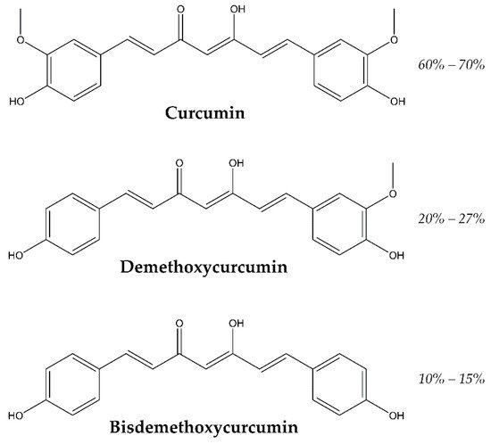
The retina is subjected to oxidative stress due to its high vascularization, long time light exposition and a high density of mitochondria. Oxidative stress can lead to pathological processes, like cell apoptosis, angiogenesis and inflammation ending in retinal pathologies. Curcumin, a major bioactive component obtained from the spice turmeric (Curcuma longa) rhizome has been used for centuries in Asian countries for cooking and for curing all kinds of diseases like dysentery, chest congestion and pain in general, due to its antioxidant effects. Curcumin prevents the formation of reactive oxygen species and so it is a good protective agent. Curcumin has shown also anti-inflammatory, and antitumor properties. Curcumin is a natural product, which can be a therapeutic option in a variety of retinal diseases due to its pleiotropic properties. Some drawbacks are its poor solubility, bioavailability and lack of stability at physiological conditions; which have been shown in curcumin skeptical publications.

curcumin oxidative stress retina retinal diseases

1. Introduction

The retina, located in the posterior segment of the ocular globe, in contact with the vitreous humor and the retinal pigment epithelium (RPE) and anteriorly with the ciliary epithelium of the pars plana. A structure present in the eye of most vertebrates and some mollusks, which embryonically outgrows, with the optic nerve, from the diencephalon. In relation to this fact, the retina is regarded as part of the central nervous system (CNS). The retina has several cell types, among them there are two types of photoreceptors: cones and rods. Cones, located in the macula lutea, detect the fine shape, colors and the motion of objects. Cones require high intensity of light and work in photopic light conditions (>10 lux). Rods, placed at the periphery, lack the color discrimination feature but are much more sensitive to dim light, hence rods work in scotopic light conditions (<0.1 lux, night vision). Half of the neurons that form the optic nerve, retinal ganglion cells (RGCs) are in this region, where bipolar cells, horizontal cells, amacrine cells and Müller cells can also be found [1].
In the retina, photoreceptors and RGCs are particularly vulnerable to oxidative stress due to the environment of high oxygen, glucose oxidation and lipid polyunsaturated fat (PUFA) content, coupled with photo-transduction, leading to increased reactive oxygen species (ROS) production [2]. It has been observed that ROS imbalance is involved in many retinal diseases. Among the primary reactive species are superoxide anion, singlet oxygen, hydroxyl radical, hydrogen peroxide (H2O2), peroxynitrite or nitric oxide. While the oxidation of PUFA can lead to the formation of malondialdehyde (MDA) and 4-hydroxynonenal (4-HNE) [3]. All these ROS can vanquish the endogenous antioxidant enzymes, like superoxide dismutase (SOD), glutathione peroxidase (GPx) and catalase (CAT) [4].
In most of the retinal pathological processes like angiogenesis, apoptosis or inflammation, ROS play an important role. Subsequently, numerous exogenous antioxidant compounds have been used in the literature to avoid the effects of ROS. Different natural compounds stand out as potential nutraceuticals, such as, docosahexaenoic acid [5], carotenoids like lutein [6] and zeaxanthin [7]; saffron [8][9], catechins [10] and ginkgo biloba extract [11]. Especially curcumin has drawn researcher’s attention during the last years [12][13][14][15][16].
Turmeric is the curcuma longa’s rhizome, which has curcuminoids as curcumin and mono-demethoxycurcumin and bis-demethoxycurcumin, shown in Figure 1, and also sesquiterpenoids as curcumenes and turmerones. All these derivatives have been identified by capillary GC–MS (gas chromatography–mass spectrometry) and HPLC (high performance liquid chromatography) analysis in other species of curcuma as well [17].
Figure 1. Structure and relative abundance of major curcuminoids.
Curcumin also known as diferuloylmethane, E100 or Natural Yellow 3 (IUPAC name is (1E, 6E)-1,7-bis(4-hydroxy-3-methoxyphenyl)-1,6-heptadiene-3,5-dione) is an orange-yellow solid at room temperature and its chemical formula C21H20O6 (MW 368.38 g/mol). Curcumin has two aromatic ring systems containing o-methoxy phenolic groups, which are linked with a seven-carbon linker consisting of an α-, β- unsaturated β-diketone moiety.
Curcumin exists in two tautomeric forms, the keto–enol and di-keto tautomers. The predominant tautomer of curcumin is the keto–enol form when it is present in polar organic solvents. This tautomer possesses intramolecular hydrogen bonding in the keto–enol moiety and π-conjugation is kept across the molecule, which results in an ultraviolet-visible (UV-Vis) absorption peak around 420 nm [18]. Curcumin is a hydrophobic molecule being insoluble in water, only approximately 30 nM, and readily soluble in polar solvents such as methanol, ethanol, acetonitrile, chloroform, dimethyl sulfoxide (DMSO) and ethyl acetate. Curcumin has high potential to scavenge reactive oxygen species, which makes it an important therapeutic and antioxidant molecule [19]. Most free radical oxidants participate in electron transfer reactions and hydrogen abstraction. Reports suggest that during free radical reactions with curcumin, the hydrogen of phenol-OH group is readily abstractable, producing phenoxyl radicals, which are resonance stabilized across the keto–enol structure [20].

2. Diabetic Retinopathy (DR)

One of the main secondary complications of type 1 and type 2 diabetes is diabetic retinopathy [21]. Diabetic retinopathy (DR) has associated retina edema, hemorrhage, ischemia, microaneurysms, augmented neovascularization and neuronal degeneration in the retina [22]. The progress of this disorder alters the photoreceptors and the blood vessels of the retina. The retina has a high content in polyunsaturated fatty acids (PUFA) and a high oxygen and glucose uptake compared with other tissues, making the retina more prone to oxidative stress. It has been demonstrated that oxidative stress not only renders DR condition, but even after the glycemic levels are back to homeostatic ones it hinders the remission of the DR. Several metabolic pathways are involved in the reactive oxygen species imbalance: polyol pathway, hexosamine pathway, advanced glycation end product (AGE) pathway and protein kinase C (PCK) pathway [23]. One of the main angiogenic factors implicated in ocular neovascularization is vascular endothelial growth factor (VEGF) [24]. Another investigation led by Chiu described the activation of nuclear enzyme poly(ADP-ribose) polymerase (PARP) after an oxidative insult in PARP−/− mice and diabetic rats; and how it is related to increase in extracellular matrix protein expression and its direct relation with the activation of transcription factors as nuclear factor kappa-light-chain-enhancer of activated B cells (NFκB) and extracellular matrix proteins by upregulating endothelin-1 (ET-1) [25].
There are studies on streptozotocin-induced diabetic rat that describe that the use curcumin decreases oxidative stress and prevented the loss of the chaperone function of alpha-crystallin [26], other authors describe the inhibition of VEGF expression in retina under hyperglycemic conditions [27]. In the same year Kowluru and Kanwar evaluated the total antioxidant capacity and the levels of glutathione (GSH), oxidatively modified DNA (8-OHdG), nitrotyrosine, interleukin 1 beta (IL-1β), NFκB and VEGF in Lewis streptozotocin-induced diabetic rats after the administration of curcumin (0.5 g/Kg) during six weeks; total antioxidant capacity was restored, GSH levels were partly restored and the levels of inflammatory markers were diminished when compared with matched weight controls [28]. In the same line, Bharara et al. claimed that glucose induced upregulation of DNA excision repair protein (ERCC1) and DNA repair endonuclease XPF (ERCC4) lead to a rise in fibronectin production via p300-dependent pathway in endothelial cells, retina and kidney of streptozotocin-induced diabetic rats [29].
Curcumin has also been investigated in vitro on human retinal pigment epithelial immortalized cell line derived from Amy Aotaki-Keen eyes (ARPE-19), human retinal endothelial cells (HRECs) and human retinal pericytes (HRPCs) and in vivo on male New Zealand white rabbits. Showing that curcumin administration protected ARPE-19 cells from H2O2 oxidative damage in a dose-dependent manner, being statistically significant at 100 µM. Regarding HERCs cells, treatment with 10 µM curcumin reverted the effect of 40 mM glucose leading to a significant decrease (p < 0.01) of tumor necrosis factor (TNF), similar to control cells, and on HRPCs cells the use of 10 µM curcumin prevented cell death caused by 40 mM glucose. On the in vivo test, different commercial available products containing curcumin were assessed, namely, curcumin formulation with a polyvinylpyrrolidone-hydrophilic carrier, curcumin-phosphatidylcholine complex and curcumin + piperine, after the intake at different times, the animals were sacrificed and the retinae were analyzed by HPLC-MS/MS showing that just the formulation with polyvinylpyrrolidone-hydrophilic carrier was able to reach the retina with a tMAX of 6 h after oral administration [30].
Bucolo et al. studied the protective effect of curcumin against high glucose insult on ARPE-19 cells. The administration of curcumin improved the cell viability in a dose dependent manner, significantly using 15 and 20 µM concentration of curcumin. The proposed mechanism involves the modulation of extracellular signal–regulated kinases (ERK1/2) pathway, upregulating nuclear respiratory factor 2 (Nrf2) expression and HO-1 activity. It was later confirmed using a mitogen-activated protein kinase (MAPK) inhibitor (PD98059) and the apoptotic cleaved caspase-3 protein [31].

3. Eye Antitumor Activity (Retinal and Choroidal Tumors)

The main malignancy located in the retina of children is retinoblastoma [32]. The trigger of this disease is the inactivation of the RB1 gene, a tumor suppressor gene. The most common early symptom is leukocoria, followed by strabismus. There are multiple treatment options, of which the most used currently is the more eye conservative ones, chemotherapy in situ, in detriment of systemic chemotherapy, radiotherapy and eye enucleation [33]. Yu et al. found than the activation of c-Jun N-terminal kinase (JNK) and p38 MAPK on the retinoblastoma Y79 cell line was induced by curcumin. This finding was corroborated by the use of SP600125 and SB203580, specific inhibitors of JNK and p38 MAPK respectively, which inhibited the apoptosis of the retinoblastoma cell line and suppressed the activation of caspase-9 and -3 [34]. Li et al. studied the effect of curcumin on SO-Rb50 and Y79 cells, retinoblastoma cell lines, and found out that curcumin inhibited cell proliferation, induced apoptosis and inhibited the migratory and invasive capacities of the retinoblastoma cell lines. The authors proposed that curcumin deactivates the Janus kinase-signal transducer and activator of transcription (JAK/STAT) signaling pathway via regulation of microRNA-99, which was supported by the lack of activity of curcumin in microRNA-99a-silenced cells [35].
Although not directly generated in the retina, some cases of intraocular lymphoma affect the eye secondarily by metastasis, which tend to migrate to the retinal ganglion cell layer or can be originated within the eye and cause visual diseases [36]. It has been stated in the literature that curcumin possesses anticancer activity, because of the combination of its antioxidant, anti-inflammatory proapoptotic, immunomodulatory and anti-angiogenic properties [37].
Lu et al. studied the growth inhibition effect of curcumin in N18 mouse-rat hybrid retina ganglion cells in vitro, they explained the effect by G2/M phase cell cycle arrest and the induction of apoptosis combined with up-regulation of Bcl-2-associated X protein (BAX) and down regulation of Bcl-2. Curcumin also up-regulated the active form of caspase-8, -9 and -3 suggesting that both mitochondrial and death receptor pathways are implicated by promoting the levels of apoptosis antigen 1 (Fas) and Fas-associated protein with death domain (FADD) [38]. These same authors studying the same cell line concluded that curcumin also inhibited DNA repair genes expression such as ATM, ATR, BRCA1, 14-3-3r, DNA-dependent protein kinase (DNA-PK) and O-6-methylguanine-DNA methyltransferase (MGMT) [39].
Lin et al. explored the effect of curcumin on the migration and invasion of the same cell line. They observed a dose- and time-dependent protection with best results with a concentration of curcumin of 15 µM administered for 48 h. The authors observed an inhibition in the levels of microRNA (miRNA) in N18 cells, with a decreased expression levels of matrix metalloproteinases (MMPs) MMP-2, MMP-7, focal adhesion kinase (FAK), ras homolog family member A (Rho A) and rho-associated, coiled-coil-containing protein kinase 1 (ROCK1); they also noticed lower levels of growth factor receptor bound protein 2 (GRB2), Ras, protein kinase C (PKC), MKK7, FAK, Rho A, ROCK1, MMP-2, MMP-9, inducible nitric oxide synthase (iNOS), NF-κB p65, Prostaglandin-endoperoxide synthase 2 (COX-2), JNK1/2 and ERK1/2 when curcumin was administered, so they attributed the action of curcumin to an inhibition of MMP-2 and 9 [40].
Burugula et al. studied a murine retinal ganglion cell line (RGC-5) treated with various doses of protein kinase inhibitor staurosporine (SS) and curcumin. Two optimal doses, which were SS (12.5 and 100 nM) or curcumin (2.5 and 100 µM), were injected in C57BL/6 mice. In the in vitro test the results indicated that curcumin diminished SS-mediated cell death at low doses, whereas high doses were toxic; while in vivo a 10 µM dose of curcumin attenuated the protease-mediated death of RGCs and amacrine cells significantly. The authors outlined that curcumin offered a protective effect by restoring NFκB [41].

4. Curcumin Delivery

Different approaches have been attempted during the last few years in order to avoid the first of the aforementioned issues and improve the potential of curcumin as oral administered drug.

4.1. Encapsulation

One of these approaches has been the encapsulation of curcumin with the aim to increase its solubility, stability and bioavailability in physiological conditions [42]. Curcumin has been integrated on different systems, such as folic acid tagged aminated starch/ZnO coated iron oxide nanoparticles [43], curcumin incorporating with Fe3O4 loaded into: polyethylene glycol–poly lactic acid-co-glycolic acid (PLGA-PEG) co-polymer [44], disulfide-linked hydrophobic backbone of a PEGylated amphiphilic diblock copolymer (biotin poly(ethylene glycol)–poly(curcumin-dithio dipropionic acid)) conjugated with chemotherapeutic agent paclitaxel [45], superparamagnetic iron oxide nanoparticles [46], graphene oxides nanocomposites [47], curcumin-loaded graphene quantum dots [48], cyclodextrin-metal organic frameworks [49], different polymeric nanoparticles like: poly(ethylene glycol)-poly(ε-caprolactone) (PEG-PCL) copolymer [50], PEG-β-cyclodextrin, curcumin solid lipid nanoparticles [51], PCL stabilized with C18-HbPG [52], cholesterol-conjugated poly(D,L-lactide)-based micelles [53] and curcumin-loaded embryonic stem cell exosomes [54]; sequential delivery of curcumin-docosahexaenoic acid loaded carriers towards promoting neuronal survival [55]. We will focus on those applied to retinal pathologies.
Granata et al. synthesized a micellar nanoaggregate of the calix[4]arene as a nanocarrier for curcumin ocular delivery, which was able to enhance curcumin solubility by 9000 fold and increase its stability 7.5 times; the calix[4]arene nanoaggregate protected curcumin from degradation and also mediated cellular uptake due to the calix[4]arene capability to cross cellular membranes. The effect of the entrapment on curcumin anti-inflammatory and antioxidant properties was assessed in vitro on J774A.1 cell line under lipopolysaccharide (LPS) induced oxidative stress; reducing IκB-α, NF-κB p65 nuclear translocation, COX2 and iNOS expression, to a higher extent compared to free curcumin. A rat model of anterior uveitis, induced by LPS was also evaluated with the same results, the application of curcumin/calix[4]arene decreased ocular inflammation and reduced inflammation proteins to a higher extent than free curcumin. It also reduced ICAM-1 by the suppression of NF-κB [56].
Davis et al. proposed a curcumin nanocarrier comprising TPGS and Pluronic F127, a non-ionic copolymer surfactant, valid as a topical formulation in the treatment of eye diseases, which increased curcumin solubility by a factor of 400,000 and the potential to be transported across ocular barriers; being neuroprotective against glutamate and cobalt chloride induced injury in immortalized retinal cultures in vitro (R28 cells) and preserving the RGCs in ocular hypertension (OHT) and partial optic nerve transection (pONT) murine models [57]. Lim et al. proposed a system using an albumin based nanoformulation in order to increase the solubility of different natural products with low solubility: curcumin, rosmarinic acid (RosA), or ursolic acid (UrsA). Although the antioxidant activity of the different nanoformulations was more efficient for RosA and UrsA when compared to curcumin on ARPE-19 cells under H2O2 oxidative insult. The authors also performed an ex vivo assay on rabbit corneas and retinas under the same insult to evaluate the protective effects of each nanoformulation; the results showed no signs of tissue damage [58]. The same group proposed the inclusion of curcumin or tetrahydrocurcumin (THC) in hydroxypropyl-γ-cyclodextrin (HP-γCD) and hydroxypropyl-β-cyclodextrin (HP-βCD), which improved water solubility of both drugs, and also studied the drug release rate, with 50% released after 1 h for HP-βCD and almost 2 h for HP-γCD, total drug release was achieved within 8 h in all cases. Regarding cytotoxicity concentrations up to 200 μM did not diminished cell viability in HCE and RPE cells. The inclusion of the drugs was higher on RPE than in HCE and THC showed more permeability than curcumin. Antioxidant activity on both epithelial cells was assessed after an H2O2 oxidative insult by monitoring SOD1, CAT1 and HMOX1; results showed that the combination of HP-γCD and THC provided higher antioxidant activity and improved bioavailability. An ex vivo assay on rabbit corneas pretreated with the drug inclusion complexes was also performed under H2O2 oxidative insult; pretreated corneas did not show significant damage. Worth to note, corneas treated with curcumin/cyclodextrin turned yellow whereas THC/cyclodextrin showed no coloration after 12 h, which indicates that the most suitable combination was THC/cyclodextrin [59]. Another proposed carrier was a thermosensitive chitosan-gelatin-based hydrogel containing curcumin-loaded nanoparticles in combination with latanoprost, a drug to increase uveoscleral outflow, in glaucoma treatment. The hydrogel was assayed in human trabecular meshwork cells as a post treatment application after an H2O2-induced oxidative insult showed reduced levels of inflammation-related gene (TNF, IL-1α, IL-6 and MMP-13), apoptosis, and ROS expression. The hydrogel showed a continuous release profile for 7 days. Biocompatibility was studied in rabbits with no negative effects after the topical use of the hydrogel [60].

4.2. Curcumin Analogues

Another approach to improve the stability and the bioavailability of curcumin is the modification of the molecule in order to avoid the problems associated with its oral administration. The poor stability and bioavailability may result from the high reactivity of the β-diketone moiety of its structure [61].
Pittalà et al. designed and studied the effects of nitric oxide-releasing curcumin (VP10/12) and caffeic acid phenethyl ester (CAPE; VP10/39) on oxidative stress caused by H2O2 oxidative insult in ARPE-19 cells. It was shown that both decreased ROS concentration in a dose dependent manner, being more efficient the CAPE derivative, but not achieving negative control values in either case. It was seen that VP10/12 presented significant cell toxicity at the highest concentration assayed (100 μM). Furthermore, VP10/39 could induce HMOX1 expression [62].
Yet, another proposal by Wang et al. includes the synthesis of the prodrug diphosphorylated curcumin (Cur-2p), and its posterior enzymatic activation, which resulted in a great improvement in terms of stability and a lower aggregation in aqueous media. When administered as intraocular injection, Cur-2p displays good biocompatibility with no morphological or functional alterations of the retina [63]. Using the same approach, Muangnoi et al. synthesized another curcumin prodrug, curcumin diethyl disuccinate (curDD), in order to improve the poor stability of curcumin in physiological pH. The compound was tested on undifferentiated and differentiated ARPE-19 cells. Cell viability was not compromised using up to 10 μM of curDD. Under H2O2 induced ROS, both curcumin and curDD, when used as pretreatment for 24 h showed protective effects on undifferentiated and differentiated ARPE-19 cells. The molecular mechanism was also evaluated, due to the variations of phosphorylated p44/42, ERK1/2 pathway was proposed. Apoptosis was also inhibited by curcumin and curDD modulating BAX/Bcl2 expression at a transcriptional level. Protein expression of HMOX1 and NAD(P)H quinone dehydrogenase 1 (NQO1) was also increased with curcumin or curDD pretreatment. In all cases curDD also showed a slightly greater protection than curcumin [64].

References

  1. Levine, B.; Klionsky, D.J. Development by Self-Digestion: Molecular Mechanisms and Biological Functions of Autophagy. Dev. Cell 2004, 6, 463–477.
  2. Wright, A.F.; Chakarova, C.F.; Abd El-Aziz, M.M.; Bhattacharya, S.S. Photoreceptor degeneration: Genetic and mechanistic dissection of a complex trait. Nat. Rev. Genet. 2010, 11, 273–284.
  3. Beatty, S.; Koh, H.-H.; Phil, M.; Henson, D.; Boulton, M. The Role of Oxidative Stress in the Pathogenesis of Age-Related Macular Degeneration. Surv. Ophthalmol. 2000, 45, 115–134.
  4. Saccà, S.C.; Roszkowska, A.M.; Izzotti, A. Environmental light and endogenous antioxidants as the main determinants of non-cancer ocular diseases. Mutat. Res. 2013, 752, 153–171.
  5. Arnal, E.; Johnsen-Soriano, S.; Lopez-Malo, D.; Perez-Pastor, G.; Vidal-Gil, L.; Morillas, N.; Sancho-Pelluz, J.; Romero, F.; Barcia, J. Docosahexaenoic Acid Protects against High Glucose-Induced Oxidative Stress in Human Retinal Pigment Epithelial Cells. React. Oxyg. Species 2016, 2, 298–307.
  6. Kamoshita, M.; Toda, E.; Osada, H.; Narimatsu, T.; Kobayashi, S.; Tsubota, K.; Ozawa, Y. Lutein acts via multiple antioxidant pathways in the photo-stressed retina. Sci. Rep. 2016, 6, 30226.
  7. Arnal, E.; Miranda, M.; Johnsen-Soriano, S.; Alvarez-Nlting, R.; Daz-Llopis, M.; Araiz, J.; Cervera, E.; Bosch-Morell, F.; Romero, F.J. Beneficial effect of docosahexanoic acid and lutein on retinal structural, metabolic, and functional abnormalities in diabetic rats. Curr. Eye Res. 2009, 34, 928–938.
  8. Di Marco, S.; Carnicelli, V.; Franceschini, N.; Di Paolo, M.; Piccardi, M.; Bisti, S.; Falsini, B. Saffron: A Multitask Neuroprotective Agent for Retinal Degenerative Diseases. Antioxidants 2019, 8, 224.
  9. Fernández-Albarral, J.A.; Ramírez, A.I.; de Hoz, R.; López-Villarín, N.; Salobrar-García, E.; López-Cuenca, I.; Licastro, E.; Inarejos-García, A.M.; Almodóvar, P.; Pinazo-Durán, M.D.; et al. Neuroprotective and Anti-Inflammatory Effects of a Hydrophilic Saffron Extract in a Model of Glaucoma. Int. J. Mol. Sci. 2019, 20, 4110.
  10. Yang, Y.; Qin, Y.J.; Yip, Y.W.Y.; Chan, K.P.; Chu, K.O.; Chu, W.K.; Ng, T.K.; Pang, C.P.; Chan, S.O. Green tea catechins are potent anti-oxidants that ameliorate sodium iodate-induced retinal degeneration in rats. Sci. Rep. 2016, 6, 29546.
  11. Martínez-Solís, I.; Acero, N.; Bosch-Morell, F.; Castillo, E.; González-Rosende, M.E.; Muñoz-Mingarro, D.; Ortega, T.; Sanahuja, M.A.; Villagrasa, V. Neuroprotective Potential of Ginkgo biloba in Retinal Diseases. Planta Med. 2019.
  12. Pescosolido, N.; Giannotti, R.; Plateroti, A.M.; Pascarella, A.; Nebbioso, M. Curcumin: Therapeutical potential in ophthalmology. Planta Med. 2014, 80, 249–254.
  13. Liu, X.F.; Hao, J.L.; Xie, T.; Mukhtar, N.J.; Zhang, W.; Malik, T.H.; Lu, C.W.; Zhou, D.D. Curcumin, a potential therapeutic candidate for anterior segment eye diseases: A review. Front. Pharmacol. 2017, 8, 66.
  14. Peddada, K.V.; Brown, A.; Verma, V.; Nebbioso, M. Therapeutic potential of curcumin in major retinal pathologies. Int. Ophthalmol. 2019, 39, 725–734.
  15. Farajipour, H.; Rahimian, S.; Taghizadeh, M. Curcumin: A new candidate for retinal disease therapy? J. Cell. Biochem. 2019, 120, 6886–6893.
  16. Saberi-Karimian, M.; Katsiki, N.; Caraglia, M.; Boccellino, M.; Majeed, M.; Sahebkar, A. Vascular endothelial growth factor: An important molecular target of curcumin. Crit. Rev. Food Sci. Nutr. 2019, 59, 299–312.
  17. Uehara, S.; Yasuda, I.; Takeya, K.; Itokawa, H. Terpenoids and curcuminoids of the rhizoma of Curcuma xanthorrhiza Roxb. Yakugaku Zasshi 1992, 112, 817–823.
  18. Chignell, C.F.; Bilskj, P.; Reszka, K.J.; Motten, A.G.; Sik, R.H.; Dahl, T.A. Spectral And photochemical properties of curcumin. Photochem. Photobiol. 1994, 59, 295–302.
  19. Ahsan, H.; Parveen, N.; Khan, N.U.; Hadi, S.M. Pro-oxidant, anti-oxidant and cleavage activities on DNA of curcumin and its derivatives demethoxycurcumin and bisdemethoxycurcumin. Chem. Biol. Interact. 1999, 121, 161–175.
  20. Agnihotri, N.; Mishra, P.C. Scavenging mechanism of curcumin toward the hydroxyl radical: A Theoretical study of reactions producing ferulic acid and vanillin. J. Phys. Chem. A 2011, 115, 14221–14232.
  21. Diabetes Control and Complications Trial Research Group; Nathan, D.M.; Genuth, S.; Lachin, J.; Cleary, P.; Crofford, O.; Davis, M.; Rand, L.; Siebert, C. The Effect of Intensive Treatment of Diabetes on the Development and Progression of Long-Term Complications in Insulin-Dependent Diabetes Mellitus. N. Engl. J. Med. 1993, 329, 977–986.
  22. Ciulla, T.A.; Amador, A.G.; Zinman, B. Diabetic retinopathy and diabetic macular edema: Pathophysiology, screening, and novel therapies. Diabetes Care 2003, 26, 2653–2664.
  23. Kowluru, R.A.; Chan, P.-S. Oxidative Stress and Diabetic Retinopathy. Exp. Diabetes Res. 2007, 2007, 43603.
  24. Premanand, C.; Rema, M.; Sameer, M.Z.; Sujatha, M.; Balasubramanyam, M. Effect of curcumin on proliferation of human retinal endothelial cells under in vitro conditions. Investig. Ophthalmol. Vis. Sci. 2006, 47, 2179–2184.
  25. Chiu, J.; Xu, B.Y.; Chen, S.; Feng, B.; Chakrabarti, S. Oxidative stress-induced, poly(ADP-ribose) polymerase-dependent upregulation of ET-1 expression in chronic diabetic complications. Can. J. Physiol. Pharmacol. 2008, 86, 365–372.
  26. Kumar, P.A.; Haseeb, A.; Suryanarayana, P.; Ehtesham, N.Z.; Reddy, G.B. Elevated expression of αA—And αB-crystallins in streptozotocin-induced diabetic rat. Arch. Biochem. Biophys. 2005, 444, 77–83.
  27. Mrudula, T.; Suryanarayana, P.; Srinivas, P.N.B.S.; Reddy, G.B. Effect of curcumin on hyperglycemia-induced vascular endothelial growth factor expression in streptozotocin-induced diabetic rat retina. Biochem. Biophys. Res. Commun. 2007, 361, 528–532.
  28. Kowluru, R.A.; Kanwar, M. Effects of curcumin on retinal oxidative stress and inflammation in diabetes. Nutr. Metab. 2007, 4, 8.
  29. Rajpathak, S.N.; Gunter, M.J.; Wylie-rosett, J.; Ho, G.Y.F.; Kaplan, R.C.; Muzumdar, R.; Rohan, T.E.; Strickler, H.D. The role of insulin-like growth factor-I and its binding proteins in glucose homeostasis and type 2 diabetes. Diabetes Metab. Res. Rev. 2009, 28, 3–12.
  30. Platania, C.B.M.; Fidilio, A.; Lazzara, F.; Piazza, C.; Geraci, F.; Giurdanella, G.; Leggio, G.M.; Salomone, S.; Drago, F.; Bucolo, C. Retinal protection and distribution of curcumin in vitro and in vivo. Front. Pharmacol. 2018, 9, 670.
  31. Bucolo, C.; Drago, F.; Maisto, R.; Romano, G.L.; D’Agata, V.; Maugeri, G.; Giunta, S. Curcumin prevents high glucose damage in retinal pigment epithelial cells through ERK1/2-mediated activation of the Nrf2/HO-1 pathway. J. Cell. Physiol. 2019, 234, 17295–17304.
  32. Dimaras, H.; Corson, T.W. Retinoblastoma, the visible CNS tumor: A review. J. Neurosci. Res. 2019, 97, 29–44.
  33. Munier, F.L.; Beck-Popovic, M.; Chantada, G.L.; Cobrinik, D.; Kivelä, T.T.; Lohmann, D.; Maeder, P.; Moll, A.C.; Carcaboso, A.M.; Moulin, A.; et al. Conservative management of retinoblastoma: Challenging orthodoxy without compromising the state of metastatic grace. “Alive, with good vision and no comorbidity. Prog. Retin. Eye Res. 2019.
  34. Yu, X.; Zhong, J.; Yan, L.; Li, J.; Wang, H.; Wen, Y.; Zhao, Y. Curcumin exerts antitumor effects in retinoblastoma cells by regulating the JNK and p38 MAPK pathways. Int. J. Mol. Med. 2016, 38, 861–868.
  35. Li, Y.; Sun, W.; Han, N.; Zou, Y.; Yin, D. Curcumin inhibits proliferation, migration, invasion and promotes apoptosis of retinoblastoma cell lines through modulation of miR-99a and JAK/STAT pathway. BMC Cancer 2018, 18, 1230.
  36. Karakawa, A.; Taoka, K.; Kaburaki, T.; Tanaka, R.; Shinozaki-Ushiku, A.; Hayashi, H.; Miyagi-Maeshima, A.; Nishimura, Y.; Uekusa, T.; Kojima, Y.; et al. Clinical features and outcomes of secondary intraocular lymphoma. Br. J. Haematol. 2018, 183, 668–671.
  37. Bar-Sela, G.; Epelbaum, R.; Schaffer, M. Curcumin as an Anti-Cancer Agent: Review of the Gap Between Basic and Clinical Applications. Curr. Med. Chem. 2009, 17, 190–197.
  38. Lu, H.-F.; Lai, K.-C.; Hsu, S.-C.; Lin, H.-J.; Yang, M.-D.; Chen, Y.-L.; Fan, M.-J.; Yang, J.-S.; Cheng, P.-Y.; Kuo, C.-L.; et al. Curcumin induces apoptosis through FAS and FADD, in caspase-3-dependent and -independent pathways in the N18 mouse-rat hybrid retina ganglion cells. Oncol. Rep. 2009, 22, 97–104.
  39. Lu, H.F.; Yang, J.S.; Lai, K.C.; Hsu, S.C.; Hsueh, S.C.; Chen, Y.L.; Chiang, J.H.; Lu, C.C.; Lo, C.; Yang, M.D.; et al. Curcumin-induced DNA damage and inhibited dna repair genes expressions in mouse-rat hybrid retina neuroblastoma cells ganglion cells (n18). Neurochem. Res. 2009, 34, 1491–1497.
  40. Lin, H.-J.; Su, C.-C.; Lu, H.-F.; Yang, J.-S.; Hsu, S.-C.; Ip, S.-W.; Wu, J.-J.; Li, Y.-C.; Ho, C.-C.; Wu, C.-C.; et al. Curcumin blocks migration and invasion of mouse-rat hybrid retina ganglion cells (N18) through the inhibition of MMP-2, -9, FAK, Rho A and Rock-1 gene expression. Oncol. Rep. 2010, 23, 665–670.
  41. Burugula, B.; Ganesh, B.S.; Chintala, S.K. Curcumin attenuates staurosporine-mediated death of retinal ganglion cells. Investig. Ophthalmol. Vis. Sci. 2011, 52, 4263–4273.
  42. Anand, P.; Kunnumakkara, A.B.; Newman, R.A.; Aggarwal, B.B. Bioavailability of curcumin: Problems and promises. Mol. Pharm. 2007, 4, 807–818.
  43. Saikia, C.; Das, M.K.; Ramteke, A.; Maji, T.K. Controlled release of curcumin from thiolated starch-coated iron oxide magnetic nanoparticles: An in vitro evaluation. Int. J. Polym. Mater. Polym. Biomater. 2017, 66, 349–358.
  44. Sadeghzadeh, H.; Pilehvar-Soltanahmadi, Y.; Akbarzadeh, A.; Dariushnejad, H.; Sanjarian, F.; Zarghami, N. The Effects of Nanoencapsulated Curcumin-Fe3O4 on Proliferation and hTERT Gene Expression in Lung Cancer Cells. Anticancer Agents Med. Chem. 2017, 17, 1363–1373.
  45. Wang, J.; Wang, F.; Li, F.; Zhang, W.; Shen, Y.; Zhou, D.; Guo, S. A multifunctional poly(curcumin) nanomedicine for dual-modal targeted delivery, intracellular responsive release, dual-drug treatment and imaging of multidrug resistant cancer cells. J. Mater. Chem. B 2016, 4, 2954–2962.
  46. Lachowicz, D.; Szpak, A.; Malek-Zietek, K.E.; Kepczynski, M.; Muller, R.N.; Laurent, S.; Nowakowska, M.; Zapotoczny, S. Biocompatible and fluorescent superparamagnetic iron oxide nanoparticles with superior magnetic properties coated with charged polysaccharide derivatives. Colloids Surf. B Biointerfaces 2017, 150, 402–407.
  47. Hou, L.; Shi, Y.; Jiang, G.; Liu, W.; Han, H.; Feng, Q.; Ren, J.; Yuan, Y.; Wang, Y.; Shi, J.; et al. Smart nanocomposite hydrogels based on azo crosslinked graphene oxide for oral colon-specific drug delivery. Nanotechnology 2016, 27, 315105.
  48. Some, S.; Gwon, A.R.; Hwang, E.; Bahn, G.H.; Yoon, Y.; Kim, Y.; Kim, S.H.; Bak, S.; Yang, J.; Jo, D.G.; et al. Cancer therapy using ultrahigh hydrophobic drug-loaded graphene derivatives. Sci. Rep. 2014, 4, 6314.
  49. Moussa, Z.; Hmadeh, M.; Abiad, M.G.; Dib, O.H.; Patra, D. Encapsulation of curcumin in cyclodextrin-metal organic frameworks: Dissociation of loaded CD-MOFs enhances stability of curcumin. Food Chem. 2016, 212, 485–494.
  50. Danafar, H.; Davaran, S.; Rostamizadeh, K.; Valizadeh, H.; Hamidi, M. Biodegradable m-PEG/PCL core-shell micelles: Preparation and characterization as a sustained release formulation for curcumin. Adv. Pharm. Bull. 2014, 4, 501–510.
  51. Jourghanian, P.; Ghaffari, S.; Ardjmand, M.; Haghighat, S.; Mohammadnejad, M. Sustained release curcumin loaded solid lipid nanoparticles. Adv. Pharm. Bull. 2016, 6, 17–21.
  52. Zs. Nagy, N.; Varga, Z.; Mihály, J.; Kasza, G.; Iván, B. Kiss Highly efficient encapsulation of curcumin into and pH-controlled drug release from poly(ε-caprolactone) nanoparticles stabilized with a novel amphiphilic hyperbranched polyglycerol. Express Polym. Lett. 2020, 14, 90–101.
  53. Kumari, P.; Muddineti, O.S.; Rompicharla, S.V.K.; Ghanta, P.; Adithya, K.B.B.N.; Ghosh, B.; Biswas, S. Cholesterol-conjugated poly(D, L-lactide)-based micelles as a nanocarrier system for effective delivery of curcumin in cancer therapy. Drug Deliv. 2017, 24, 209–223.
  54. Kalani, A.; Chaturvedi, P.; Kamat, P.K.; Maldonado, C.; Bauer, P.; Joshua, I.G.; Tyagi, S.C.; Tyagi, N. Curcumin-loaded embryonic stem cell exosomes restored neurovascular unit following ischemia-reperfusion injury. Int. J. Biochem. Cell Biol. 2016, 79, 360–369.
  55. Guerzoni, L.P.B.; Nicolas, V.; Angelova, A. In Vitro Modulation of TrkB Receptor Signaling upon Sequential Delivery of Curcumin-DHA Loaded Carriers Towards Promoting Neuronal Survival. Pharm. Res. 2017, 34, 492–505.
  56. Granata, G.; Paterniti, I.; Geraci, C.; Cunsolo, F.; Esposito, E.; Cordaro, M.; Blanco, A.R.; Cuzzocrea, S.; Consoli, G.M.L. Potential Eye Drop Based on a Calix[4]arene Nanoassembly for Curcumin Delivery: Enhanced Drug Solubility, Stability, and Anti-Inflammatory Effect. Mol. Pharm. 2017, 14, 1610–1622.
  57. Davis, B.M.; Pahlitzsch, M.; Guo, L.; Balendra, S.; Shah, P.; Ravindran, N.; Malaguarnera, G.; Sisa, C.; Shamsher, E.; Hamze, H.; et al. Topical Curcumin Nanocarriers are Neuroprotective in Eye Disease. Sci. Rep. 2018, 8, 11066.
  58. Kim, D.; Maharjan, P.; Jin, M.; Park, T.; Maharjan, A.; Amatya, R.; Yang, J.; Min, K.A.; Shin, M.C. Potential Albumin-Based Antioxidant Nanoformulations for Ocular Protection against Oxidative Stress. Pharmaceutics 2019, 11, 297.
  59. Maharjan, P.; Jin, M.; Kim, D.; Yang, J.W.; Maharjan, A.; Shin, M.C.; Cho, K.H.; Kim, M.S.; Min, K.A. Evaluation of epithelial transport and oxidative stress protection of nanoengineered curcumin derivative-cyclodextrin formulation for ocular delivery. Arch. Pharm. Res. 2019, 42, 909–925.
  60. Cheng, Y.H.; Ko, Y.C.; Chang, Y.F.; Huang, S.H.; Liu, C.J. ling Thermosensitive chitosan-gelatin-based hydrogel containing curcumin-loaded nanoparticles and latanoprost as a dual-drug delivery system for glaucoma treatment. Exp. Eye Res. 2019, 179, 179–187.
  61. Noureddin, S.A.; El-Shishtawy, R.M.; Al-Footy, K.O. Curcumin analogues and their hybrid molecules as multifunctional drugs. Eur. J. Med. Chem. 2019, 182, 111631.
  62. Pittalà, V.; Salerno, L.; Fidilio, A.; Lazzara, F.; Platania, C.B.M.; Drago, F.; Bucolo, C.; Foresti, R. Effects of Novel Nitric Oxide-Releasing Molecules against Oxidative Stress on Retinal Pigmented Epithelial Cells. Oxidative Med. Cell. Longev. 2017, 2017, 1420892.
  63. Wang, J.; Zhou, J.; He, H.; Wu, D.; Du, X.; Xu, B.; Xiong, T.; Li, X. Enzymatic formation of curcumin in vitro and in vivo. Nano Res. 2018, 11, 3453–3461.
  64. Muangnoi, C.; Sharif, U.; Ratnatilaka Na Bhuket, P.; Rojsitthisak, P.; Paraoan, L. Protective Effects of Curcumin Ester Prodrug, Curcumin Diethyl Disuccinate against H2O2-Induced Oxidative Stress in Human Retinal Pigment Epithelial Cells: Potential Therapeutic Avenues for Age-Related Macular Degeneration. Int. J. Mol. Sci. 2019, 20, 3367.
More
Information
Subjects: Biology
Contributor MDPI registered users' name will be linked to their SciProfiles pages. To register with us, please refer to https://encyclopedia.pub/register :
View Times: 536
Revisions: 4 times (View History)
Update Date: 22 Sep 2021
1000/1000