- Subjects: Neurosciences
- |
- Contributor:
- Evgenia Sitnikova
- WAG/Rij rats
- absence epilepsy
- alpha2-adrenoreceptors
- pharmacological provocation
- epidural recordings
1. Pharmacological Provocation of Continuous Spike-wave Discharges (SWDs) in Genetically Prone WAG/Rij rat with i.p. injection of 2% Xylazine
This video demonstrates the acute effect of i.p. injection of 2% Xylazine in 16 months old female WAG/Rij rat. The rat was placed in a Plexiglas cage immediately after the i.p. injection of 2% Xylazine (Xyla, 20 mg/ml Xylazine hydrochloride, Interchemie Werken De Adelaar, the Netherlands) in dose of 2 mg/kg. Three-channels EEG signals were recorded using a multi-channel amplifier (PowerLab 4/35, ADInstruments), band-pass filtered between 0.5 and 200 Hz and digitized with 400 samples/second/per channel. The rat's behavior was recorded using a Genius eFace 1325R video camera.
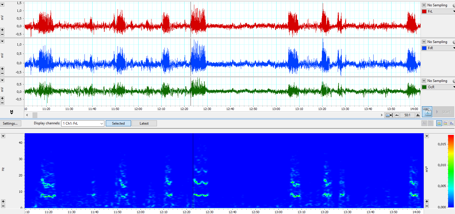
Spike-wave discharges (SWDs) in the electroencephalograms are hallmark of absence epilepsy [1][2][3]. In rats with genetic predisposition to absence epilepsy, SWDs appeared spontaneously as a sequence of high voltage spikes and waves with a frequency of 8–10 Hz [4][5]. Figure 1 displays a three-channel electrocorticogram recording from a WAG/Rij rat. Epidural electrodes were implanted over the frontal cortex (symmetrically over the left and right sides) and over the right occipital cortex.

Figure 1. Spontaneous SWDs recorded in an 8-month-old female WAG/Rij rat and visualized in LabChart 8.0. Three-channel epidural EEG was recorded and abbreviated as FrL and FrR – frontal cortical left and right leads; OcR – occipital cortical lead. Time is given in mm:ss. The bottom graph shows an FFT power spectrum computed with 1024 size and 95,5 % window overlap. From [6] Supplementary under the terms of CC BY license 4.0.
Agonists of alpha2-adrenoreceptors (such as xylazine, clonidine, dexmedetomidine) are known to aggravate SWDs in genetically prone rats [6][7][8][9][10][11][12][13]. Xylazine is widely used in veterinary medicine as a sedative with analgesic and muscle relaxant properties [14][15]. In therapeutic doses, Xylazine induces continuous SWDs in genetically prone WAG/Rij rats, revealing its potential as a substance capable of pharmacologically provoking SWDs in rats.
2. ECoG examination in rats
Epidural electrodes were made of stainless-steel screws (screw: shaft length = 2.0 mm, head diameter = 2.0 mm, shaft diameter = 0.8 mm) providing recordings of electrical brain activity (EEG). The surgery was performed under isofluran anesthesia in a Kopf stereotaxic apparatus. Tree active epidural screw electrodes was implanted over the left/right frontal cortex (AP -/+2; L 2.5) and occipital cortex (AP −6; L 3); the fours reference screw electrode was placed over the right cerebellum. Coordinates are given in mm relative to the bregma. After the surgery, animals were housed individually under 12:12 h light:dark cycle (light on 8 a.m.) with free access to food and tap water. Recovery period lasted 10–14 days.
- Koutroumanidis, M.; Tsiptsios, D.; Kokkinos, V.; Kostopoulos, G.K. Focal and Generalized EEG Paroxysms in Childhood Absence Epilepsy: Topographic Associations and Distinctive Behaviors during the First Cycle of Non-REM Sleep. Epilepsia 2012, 53, 840–849, doi:10.1111/j.1528-1167.2012.03424.x.
- Bilo, L.; Pappatà, S.; De Simone, R.; Meo, R. The Syndrome of Absence Status Epilepsy: Review of the Literature. Epilepsy Res. Treat. 2014, 2014, 1–8, doi:10.1155/2014/624309.
- Sadleir, L.G.; Scheffer, I.E.; Smith, S.; Carstensen, B.; Farrell, K.; Connolly, M.B. EEG Features of Absence Seizures in Idiopathic Generalized Epilepsy: Impact of Syndrome, Age, and State. Epilepsia 2009, 50, 1572–1578, doi:10.1111/J.1528-1167.2008.02001.X.
- Coenen, A.M.L.; van Luijtelaar, E.L.J.M. The WAG/Rij Rat Model for Absence Epilepsy: Age and Sex Factors. Epilepsy Res. 1987, 1, 297–301, doi:10.1016/0920-1211(87)90005-2.
- van Luijtelaar, G.; van Oijen, G. Establishing Drug Effects on Electrocorticographic Activity in a Genetic Absence Epilepsy Model: Advances and Pitfalls. Front. Pharmacol. 2020, 11, 395, doi:10.3389/fphar.2020.00395.
- Sitnikova, E.; Pupikina, M.; Rutskova, E. Alpha2 Adrenergic Modulation of Spike-Wave Epilepsy: Experimental Study of Pro-Epileptic and Sedative Effects of Dexmedetomidine. Int. J. Mol. Sci. 2023, 24, doi:10.3390/ijms24119445.
- Buzsáki, G.; Kennedy, B.; Solt, V.B.; Ziegler, M. Noradrenergic Control of Thalamic Oscillation: The Role of Alpha-2 Receptors. Eur. J. Neurosci. 1991, 3, 222–229, doi:10.1111/j.1460-9568.1991.tb00083.x.
- Micheletti, G.; Warter, J.-M.; Marescaux, C.; Depaulis, A.; Tranchant, C.; Rumbach, L.; Vergnes, M. Effects of Drugs Affecting Noradrenergic Neurotransmission in Rats with Spontaneous Petit Mal-like Seizures. Eur. J. Pharmacol. 1987, 135, 397–402, doi:10.1016/0014-2999(87)90690-X.
- Sitnikova, E.; van Luijtelaar, G. Reduction of Adrenergic Neurotransmission with Clonidine Aggravates Spike-Wave Seizures and Alters Activity in the Cortex and the Thalamus in WAG/Rij Rats. Brain Res. Bull. 2005, 64, 533–540, doi:10.1016/j.brainresbull.2004.11.004.
- van Luijtelaar, E.L.J.M. Spike-Wave Discharges and Sleep Spindles in Rats. Acta Neurobiol. Exp. (Wars). 1997, 57, 113–121.
- Yavuz, M.; Akkol, S.; Onat, F. Alpha-2a Adrenergic Receptor Activation in Genetic Absence Epilepsy: An Absence Status Model? Epilepsia Open 2024, 9, 534–547, doi:10.1002/EPI4.12879.
- Sitnikova, E. Adrenergic Mechanisms of Absence Status Epilepticus. Front. Neurol. 2023, 14, 1–8, doi:10.3389/fneur.2023.1298310.
- Sitnikova, E.; Rutskova, E.; Smirnov, K. Alpha2-Adrenergic Receptors as a Pharmacological Target for Spike-Wave Epilepsy. Int. J. Mol. Sci. 2023, 24, 1477, doi:10.3390/ijms24021477.
- Greene, S.A.; Thurmon, J.C. Xylazine – a Review of Its Pharmacology and Use in Veterinary Medicine. J. Vet. Pharmacol. Ther. 1988, 11, 295–313, doi:10.1111/j.1365-2885.1988.tb00189.x.
- Alexander, R.S.; Canver, B.R.; Sue, K.L.; Morford, K.L. Xylazine and Overdoses: Trends, Concerns, and Recommendations. Am. J. Public Health 2022, 112, doi:10.2105/AJPH.2022.306881.
