Submitted Successfully!
To reward your contribution, here is a gift for you: A free trial for our video production service.
Thank you for your contribution! You can also upload a video entry or images related to this topic.
Version Summary Created by Modification Content Size Created at Operation
1 + 2293 word(s) 2293 2021-09-09 08:06:31 |
2 Format change Meta information modification 2293 2021-09-24 06:19:03 |

Video Upload Options

Do you have a full video?

Confirm

Are you sure to Delete?
Cite
If you have any further questions, please contact Encyclopedia Editorial Office.
Kim, C. Peripheral Blood-Based Biomarkers. Encyclopedia. Available online: https://encyclopedia.pub/entry/14503 (accessed on 26 April 2024).
Kim C. Peripheral Blood-Based Biomarkers. Encyclopedia. Available at: https://encyclopedia.pub/entry/14503. Accessed April 26, 2024.
Kim, Chan. "Peripheral Blood-Based Biomarkers" Encyclopedia, https://encyclopedia.pub/entry/14503 (accessed April 26, 2024).
Kim, C. (2021, September 24). Peripheral Blood-Based Biomarkers. In Encyclopedia. https://encyclopedia.pub/entry/14503
Kim, Chan. "Peripheral Blood-Based Biomarkers." Encyclopedia. Web. 24 September, 2021.
Peripheral Blood-Based Biomarkers
Edit

As cancer immunotherapy using immune checkpoint inhibitors (ICIs) is rapidly evolving in clinical practice, it is necessary to identify biomarkers that will allow the selection of cancer patients who will benefit most or least from ICIs and to longitudinally monitor patients’ immune responses during treatment. Various peripheral blood-based immune biomarkers are being identified with recent advances in high-throughput multiplexed analytical technologies. The identification of these biomarkers, which can be easily detected in blood samples using non-invasive and repeatable methods, will contribute to overcoming the limitations of previously used tissue-based biomarkers. Here, we discuss the potential of circulating immune cells, soluble immune and inflammatory molecules, circulating tumor cells and DNA, exosomes, and the blood-based tumor mutational burden, as biomarkers for the prediction of immune responses and clinical benefit from ICI treatment in patients with advanced cancer. 

peripheral blood biomarker cancer immunotherapy

1. Introduction

In the past decade, cancer immunotherapy using immune checkpoint inhibitors (ICIs) has demonstrated promising clinical efficacy in the treatment of various malignancies. It enabled the complete regression of advanced tumors, resulting in long-term survival in a fraction of patients [1][2][3][4]. However, this potent immunotherapeutic efficacy is not always possible due to an immunosuppressive tumor microenvironment (TME) that lacks or excludes anti-tumor effector T cells [5][6][7][8][9]. Therefore, the identification of patients who are likely or unlikely to respond to ICI treatment before or as early as possible during treatment is crucial to elicit optimal immunotherapeutic efficacy [2][10][11].
Tumor programmed death-ligand1 (PD-L1) expression was initially suggested as a plausible biomarker for predicting the response to anti-PD-1/PD-L1 therapy. A multitude of clinical studies have demonstrated the enhanced efficacy of immune checkpoint blockade in patients with higher levels of intratumoral PD-L1 [2][4][11][12]. However, tumoral PD-L1 positivity alone is insufficient for patient stratification because some PD-L1 negative patients also respond to immunotherapy and PD-L1 levels show spatiotemporal variations during cancer treatment [10][13]. Other tumor factors associated with enhanced clinical benefit from ICIs include mismatch repair protein deficiency, microsatellite instability (MSI), high tumor mutational burden (TMB), and the effector T-cell gene signature [2][4][10][12][14].
Although tumor tissue-based biomarkers partially aid in identifying patients who will benefit more from ICIs, many challenges still exist in clinical practice [4][11][12][15][16][17]. First, tumor biopsies are generally invasive, and obtaining patient tissue samples using biopsy is severely limited by tumor accessibility and the condition of the patient. Furthermore, repeated tissue biopsy could increase the likelihood of procedure-related complications and delay cancer treatment. Moreover, because of tumor heterogeneity, the local immune response within one metastatic lesion may not represent the systemic anti-cancer immunity of the patient. Lastly, although cancer immunity may constantly change during ICI treatment, longitudinal immune monitoring using repeated tissue biopsy is usually not feasible in the clinic, especially during rapid clinical deterioration. Therefore, clinicians usually make decisions based on a singe-timepoint tumor biopsy at the time of treatment initiation, rather than performing repeated tumor biopsies to monitor the updated immunological profiles of the TME.
With the advent of high-throughput multiplexed analytical technologies, peripheral blood is now suitable for deeper immune profiling. As peripheral blood sampling is readily available, minimally invasive, and repeatable, the use of blood-based immune biomarkers can compensate for the abovementioned limitations of tissue-based immune biomarkers during cancer immunotherapy [11][15][18][19][20][21][22][23]. This review aimed to summarize pivotal findings related to blood-based immune biomarkers in patients with solid cancer treated with ICIs (Figure 1).
Figure 1. Summary of various circulating immune biomarkers during ICI treatment. Central memory T cells, TCM; effector memory T cells, TEM; effector T cells, Teff; T cell receptor, TCR; regulatory T cells, Tregs; myeloid-derived suppressor cells, MDSCs; polymorphonuclear-MDSCs, PMN-MDSCs; tumor mutation burden, TMB; circulating tumor cells, CTCs; circulating tumor DNA, ctDNA; monocytic-MDSCs, M-MDSCs; natural killer cell, NK cell.

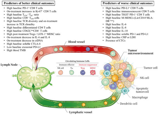
2. Circulating Immune Cells

2.1. Memory T Cells

T cells undergo a natural differentiation since antigen recognition and are phenotypically subdivided based on the following surface markers: naïve T cells (TN, CD45 RO CCR7+), central memory T cells (TCM, CD45 RO+ CCR7+), effector memory T cells (TEM, CD45 RO+ CCR7 ), and terminal effector T cells (TTE, CD45 RO CCR7) [24][25]. T cells at each stage of differentiation could have a distinct role in ICI-induced restoration of anti-tumor immunity and show varied potential as predictive biomarkers of ICI treatment outcomes. It seems that a higher frequency of memory T cells compared to that of effector T cells (Teff) in the peripheral blood at baseline were associated with better response and prognosis after ICI treatment [26][25]. However, this finding needs further validation in prospective studies due to the heterogeneous population studied and inconsistent sample processing.
Some researchers have reported the predictive potential of TCM cells in ICI treatment response. Circulating CD4+ TCM cells seem to be associated with a good response and better prognosis in NSCLC and RCC patients receiving anti-PD-1 inhibitors [27]. Furthermore, Manjarrez-Orduno et al. found that patients whose tumors exhibited increased inflamed signature and PD-L1 expression showed higher levels of CD4+ and CD8+ TCM cells compared to those of Teff cells in the peripheral blood of patients with melanoma and NSCLC [26]. Intriguingly, a high TCM/Teff ratio at baseline was associated with longer PFS (p < 0.05) in a NSCLC cohort treated with nivolumab. However, there were no major changes in the TCM/Teff ratio in the follow-up blood samples at 3 months.
Other studies have revealed the potential role of TEM cells in predicting the clinical benefits of ICIs. In patients with melanoma treated with PD-1 inhibitors, a higher frequency of circulating CD8+ TEM cells and lower frequency of CD4+ TEM cells and naïve CD8+ T cells at baseline were observed in responders [28]. Consistent results were also reported in melanoma patients treated with the anti-CTLA-4 antibody, ipilimumab. Wistuba-Hamprecht et al. demonstrated that high frequencies of CD8 effector-memory type 1 T cells defined as CD45 RA CCR7 CD27+ CD28+ in the peripheral blood within 4 weeks of treatment initiation were associated with higher clinical response and longer OS [29]. However, late stage-differentiated effector memory CD8 cells (CD45 RA+ CDR7CD27CD28) were inversely related to OS. Furthermore, other researchers have simultaneously investigated the functional status and phenotype. Kim et al. reported that a lower frequency of CD8+ TEM cells and a higher frequency of severely exhausted T cells (TIGIT+ cells among PD-1+ CD8+ T cells) at baseline were associated with HPD and shorter OS in NSCLC patients treated with PD-(L)1 inhibitors [30].
Overall, memory T cells in the peripheral blood at a certain differentiation stage could be considered when predicting responses to ICIs.

2.2. TCR Clonality and Diversity of PD-1+ CD8+ T Cells

Some researchers have focused on the role of TCR clonality in circulating T cells as a predictive determinant of ICI response. In NSCLC, dominant TCR expansion was observed not only within the tumor tissue but also in circulating T cells, and early and sustained TCR clonal expansions in the blood were present in ICI responders [31][32] Recently, Han et al. reported that pretreatment PD-1+ CD8+ TCR diversity in the peripheral blood was higher in patients with disease control than in those with disease progression, and high PD-1+ CD8+ TCR diversity (>3.14) was associated with better response, longer PFS, and OS in NSCLC patients treated with PD-(L)1 inhibitors [33]. Increasing PD-1+ CD8+ TCR clonality at 4–6 weeks after ICI treatment initiation was associated with a higher disease control rate, longer PFS, and OS. Moreover, TCR diversity was an independent prognostic factor for both PFS and OS. The higher TCR diversity of PD-1+ CD8 T cells might reflect a higher probability of neoantigen recognition. After ICI exposure, the dominant clonal expansion of tumor-specific T cells indicated a good clinical response. Furthermore, it differentiated pseudo-PD from true PD. Thus, monitoring the TCR repertoire could help predict the ICI benefit. Expansion of TCR clones in the peripheral blood after ICI treatment may not completely reflect the true diversity of the TCR repertoire within the tumor and may not be sufficient to suppress tumor growth. A recent study by El Meskini et al. elucidated this by using genetically engineered mouse melanoma models. They demonstrated that the tumor response to ICI therapy requires not only clonal expansion of the TCR repertoire but also tumor access to adequate TCRs [34]. Therefore, the post-treatment expansion of TCR clones in both the blood and tumor seems critical for the therapeutic response to ICIs. The dynamic changes in TCR clonality and diversity during ICI treatment could be validated in future prospective studies.

2.3. CD4 T Cells

CD4 T cells can promote anti-tumor immunity by supporting the priming, migration, and survival of CD8 T cells [35]. Moreover, functional CD4+ T cells are necessary to restore the cytotoxicity of CD8 T cells following anti-PD-(L)1 treatment. In NSCLC patients treated with PD(L)1 inhibitors, a high proportion (>40%) of highly differentiated CD4 T cells (CD27CD28 low/negative) in peripheral blood at baseline could predict objective RR with 100% specificity and 70% sensitivity [36]. Furthermore, this was correlated with longer PFS in multivariate analysis. These CD4 T cells mainly expressed memory features with higher Ki67+ expression, but lower co-expression of PD-1 and LAG-3. Moreover, a low percentage of CD25+ FOXP3+ CD4+ Tregs was associated with a higher RR and longer PFS and OS.
A recent report showed that CD62 Llow CD4 T cells were associated with the clinical response to nivolumab in NSCLC [37]. The circulating level of CD62 Llow CD4 T cells at baseline was higher in the nivolumab responders compared to that in non-responders (p < 0.001). These cells showed the classical characteristics of Th1 cells and were positively correlated with the percentage of effector CD8 T cells and expression of PD-1 on CD8 T cells. Conversely, the percentage of CD25+ FOXP3+ CD4+ Tregs was significantly lower in nivolumab responders compared to that in non-responders. Intriguingly, durable responders maintained high percentages of CD62 Llow CD4 T cells within the peripheral blood.
Taken together, the intrinsic functionality of CD4 T cell immunity could be a key factor for restoring anti-tumor immunity and predicting outcomes in patients treated with ICIs.

2.4. Immunosuppressive Cells: Myeloid-Derived Suppressive Cells (MDSCs) and Tregs

MDSCs are a heterogeneous group of myeloid cells that fail to differentiate into granulocytes, macrophages, or dendritic cells. They show immunosuppressive activity by inhibiting T and NK cells, stimulating Tregs, and playing an important role in various malignancies [5][38]. MDSCs are classified into two phenotypes: neutrophil-like MDSCs, called granulocytic-MDSCs (G-MDSCs), also known as polymorphonuclear-MDSCs (PMN-MDSCs), and monocyte-like MDSCs (M-MDSCs). G-MDSCs are commonly identified as Lin CD11b+ CD14 CD15+ HLA-DR or Lin CD11b+ CD14 CD66b+, and M-MDSCs are defined as Lin CD11b+ CD14+ CD15 HLA-DR−/low. Along with MDSCs, Tregs (CD4+ CD25+ FoxP3+) play another key immunosuppressive role in various underlying mechanisms [5][39][40]. They are a subset of CD4 T cells that maintain immune homeostasis by hindering the activities of CD4 and CD8 effector cells, NK cells, and antigen-presenting cells [5][40]. Therefore, these immunosuppressive cells have been investigated as potential prognostic or predictive biomarkers in cancer patients receiving ICIs.
A study examined circulating M-MDSCs (Lin CD14+ CD11b+ HLA-DRlow/−) in melanoma patients receiving ipilimumab [41]. M-MDSCs at baseline were more frequent in melanoma patients than in healthy volunteers. With a cut-off of 14.9% of M-MDSCs, patients with fewer M-MDSCs at baseline or at week 6 survived longer than patients with more abundant M-MDSCs. There was a statistically significant inverse correlation between percentage changes in the CD8+ T cell number and M-MDSC frequency at week 6.
Martens et al. analyzed M-MDSCs and Tregs in a large cohort of patients with advanced melanoma treated with ipilimumab [42]. This study revealed that M-MDSCs (LinCD14+ HLA-DR−/low) at baseline were negatively correlated with OS, while Tregs were positively correlated with OS. When these two parameters, M-MDSCs ≥ 5.1% and Tregs < 1.5%, were integrated with other clinical parameters into a prognostic model, OS and ipilimumab responses could be better predicted.
Furthermore, another study in patients with advanced melanoma treated with ipilimumab as a neoadjuvant, investigated the early dynamic changes in circulating MDSCs and Tregs at week 6. It was found that early on-treatment decrease in M-MDSCs (Lin1HLA-DR CD33+ CD11b+) and an increase in Tregs at week 6 were associated with longer PFS [43].
As MDSCs share common features with neutrophils, it is challenging to clearly distinguish MDSCs from neutrophils. Recently, lectin-type oxidized LDL receptor-1 (LOX-1) has been suggested as an MDSC-specific marker in patients with cancer [44]. A recent study examined LOX-1-expressing PMN-MDSCs and Tregs in NSCLC patients treated with nivolumab [45]. In the exploratory cohort, the baseline level of LOX-1+ PMN-MDSCs in the peripheral blood did not differ between responders and non-responders, while the level of Tregs was higher in responders compared to that in non-responders. Interestingly, LOX-1+ PMN-MDSCs were significantly decreased in responders after nivolumab treatment, but the levels of Tregs were unaltered. There was an inverse correlation between LOX-1+ PMN-MDSCs and Tregs, and a ratio of Tregs to LOX-1+ PMN-MDSCs (TMR) ≥ 0.39 could distinguish the nivolumab responders from the non-responders. A high post-treatment TMR was also significantly correlated with longer PFS. Therefore, relative changes in Tregs and PMN-MDSC during PD-1 inhibition could help predict a good response to and better prognosis from ICI treatment.
Taken together, MDSCs were consistently associated with poor ICI responses and clinical outcomes. However, regarding Tregs, the results are controversial; while some studies revealed a positive correlation, another study reported a negative correlation, and other studies reported no such relationship between Tregs and ICI outcomes [46][47][28][37][42][45]. Therefore, the clinical implication of Tregs in cancer patients treated with ICIs requires further investigation.

2.5. Natural Killer (NK) Cells

NK cells induce adaptive immune responses without prior antigen sensitization during ICI treatment [48]. Mazzaschi et al. showed that the NSCLC patient group that benefited from nivolumab treatment showed higher CD56+ NK cells with a more cytotoxic phenotype (perforin, granzyme B, CD3ζ) than the non-responding group [46]. High baseline levels of circulating NK cells (>202/µL) were associated with prolonged OS in this study. A recent study reported similar results in NSCLC [49], which require further validation due to the small sample size.


References

  1. Ribas, A.; Wolchok, J.D. Cancer immunotherapy using checkpoint blockade. Science 2018, 359, 1350–1355.
  2. Murciano-Goroff, Y.R.; Warner, A.B.; Wolchok, J.D. The future of cancer immunotherapy: Microenvironment-targeting combinations. Cell Res. 2020, 30, 507–519.
  3. Waldman, A.D.; Fritz, J.M.; Lenardo, M.J. A guide to cancer immunotherapy: From T cell basic science to clinical practice. Nat. Rev. Immunol. 2020, 20, 651–668.
  4. Hegde, P.S.; Chen, D.S. Top 10 challenges in cancer immunotherapy. Immunity 2020, 52, 17–35.
  5. Lee, W.S.; Yang, H.; Chon, H.J.; Kim, C. Combination of anti-angiogenic therapy and immune checkpoint blockade normalizes vascular-immune crosstalk to potentiate cancer immunity. Exp. Mol. Med. 2020, 52, 1475–1485.
  6. Gajewski, T.F.; Schreiber, H.; Fu, Y.-X. Innate and adaptive immune cells in the tumor microenvironment. Nat. Immunol. 2013, 14, 1014–1022.
  7. Chon, H.J.; Lee, W.S.; Yang, H.; Kong, S.J.; Lee, N.K.; Moon, E.S.; Choi, J.; Han, E.C.; Kim, J.H.; Ahn, J.B.; et al. Tumor microenvironment remodeling by intratumoral oncolytic vaccinia virus enhances the efficacy of immune checkpoint blockade. Clin. Cancer Res. 2018, 25, 1612–1623.
  8. Park, J.-S.; Kim, I.-K.; Han, S.; Park, I.; Kim, C.; Bae, J.; Oh, S.J.; Lee, S.; Kim, J.H.; Woo, D.-C.; et al. Normalization of tumor vessels by Tie2 activation and Ang2 inhibition enhances drug delivery and produces a favorable tumor microenvironment. Cancer Cell 2016, 30, 953–967.
  9. Kim, C.G.; Kim, C.; Yoon, S.E.; Kim, K.H.; Choi, S.J.; Kang, B.; Kim, H.R.; Park, S.-H.; Shin, E.-C.; Kim, Y.-Y.; et al. Hyperprogressive disease during PD-1 blockade in patients with advanced hepatocellular carcinoma. J. Hepatol. 2021, 74, 350–359.
  10. Ren, D.; Hua, Y.; Yu, B.; Ye, X.; He, Z.; Li, C.; Wang, J.; Mo, Y.; Wei, X.; Chen, Y.; et al. Predictive biomarkers and mechanisms underlying resistance to PD1/PD-L1 blockade cancer immunotherapy. Mol. Cancer 2020, 19, 1–19.
  11. Nixon, A.B.; Schalper, K.A.; Jacobs, I.; Potluri, S.; Wang, I.-M.; Fleener, C. Peripheral immune-based biomarkers in cancer immunotherapy: Can we realize their predictive potential? J. Immunother. Cancer 2019, 7, 325.
  12. Tray, N.; Weber, J.; Adams, S. Predictive biomarkers for checkpoint immunotherapy: Current status and challenges for clinical application. Cancer Immunol. Res. 2018, 6, 1122–1128.
  13. Davis, A.A.; Patel, V.G. The role of PD-L1 expression as a predictive biomarker: An analysis of all US Food and Drug Administration (FDA) approvals of immune checkpoint inhibitors. J. Immunother. Cancer 2019, 7, 1–8.
  14. Bai, R.; Lv, Z.; Xu, D.; Cui, J. Predictive biomarkers for cancer immunotherapy with immune checkpoint inhibitors. Biomark. Res. 2020, 8, 1–17.
  15. Duchemann, B.; Remon, J.; Naigeon, M.; Mezquita, L.; Ferrara, R.; Cassard, L.; Jouniaux, J.M.; Boselli, L.; Grivel, J.; Auclin, E.; et al. Integrating circulating biomarkers in the immune checkpoint inhibitor treatment in lung cancer. Cancers 2020, 12, 3625.
  16. Gerlinger, M.; Rowan, A.J.; Horswell, S.; Larkin, J.; Endesfelder, D.; Gronroos, E.; Martinez, P.; Matthews, N.; Stewart, A.; Tarpey, P.; et al. Intratumor heterogeneity and branched evolution revealed by multiregion sequencing. N. Engl. J. Med. 2012, 366, 883–892.
  17. Kim, C.; Kim, E.K.; Jung, H.; Chon, H.J.; Han, J.W.; Shin, K.-H.; Hu, H.; Kim, K.S.; Choi, Y.D.; Kim, S.; et al. Prognostic implications of PD-L1 expression in patients with soft tissue sarcoma. BMC Cancer 2016, 16, 1–7.
  18. Nabet, B.Y.; Esfahani, M.S.; Moding, E.J.; Hamilton, E.G.; Chabon, J.J.; Rizvi, H.; Steen, C.B.; Chaudhuri, A.A.; Liu, C.L.; Hui, A.B.; et al. Noninvasive early identification of therapeutic benefit from immune checkpoint inhibition. Cell 2020, 183, 363–376.e13.
  19. Simonds, E.F.; Lu, E.D.; Badillo, O.; Karimi, S.; Liu, E.V.; Tamaki, W.; Rancan, C.; Downey, K.M.; Stultz, J.; Sinha, M.; et al. Deep immune profiling reveals targetable mechanisms of immune evasion in immune checkpoint inhibitor-refractory glioblastoma. J. Immunother. Cancer 2021, 9, e002181.
  20. Reyes, M.; Vickers, D.; Billman, K.; Eisenhaure, T.; Hoover, P.; Browne, E.P.; Rao, D.A.; Hacohen, N.; Blainey, P.C. Multiplexed enrichment and genomic profiling of peripheral blood cells reveal subset-specific immune signatures. Sci. Adv. 2019, 5, eaau9223.
  21. Liu, D.; Paczkowski, P.; Mackay, S.; Ng, C.; Zhou, J. Single-cell multiplexed proteomics on the IsoLight resolves cellular functional heterogeneity to reveal clinical responses of cancer patients to immunotherapies. In Methods in Molecular Biology; Springer: New York, NY, USA, 2020; Volume 2055, pp. 413–431.
  22. Spitzer, M.H.; Nolan, G.P. Mass cytometry: Single cells, many features. Cell 2016, 165, 780–791.
  23. Kim, K.H.; Kim, C.G.; Shin, E.-C. Peripheral blood immune cell-based biomarkers in anti-PD-1/PD-L1 therapy. Immune Netw. 2020, 20, e8.
  24. Mahnke, Y.D.; Brodie, T.M.; Sallusto, F.; Roederer, M.; Lugli, E. The who’s who of T-cell differentiation: Human memory T-cell subsets. Eur. J. Immunol. 2013, 43, 2797–2809.
  25. Liu, Q.; Sun, Z.; Chen, L. Memory T cells: Strategies for optimizing tumor immunotherapy. Protein Cell 2020, 11, 549–564.
  26. Manjarrez-Orduño, N.; Menard, L.C.; Kansal, S.; Fischer, P.; Kakrecha, B.; Jiang, C.; Cunningham, M.; Greenawalt, D.; Patel, V.; Yang, M.; et al. Circulating T cell subpopulations correlate with immune responses at the tumor site and clinical response to PD1 inhibition in non-small cell lung cancer. Front. Immunol. 2018, 9, 1613.
  27. Juliá, E.P.; Mandó, P.; Rizzo, M.M.; Cueto, G.R.; Tsou, F.; Luca, R.; Pupareli, C.; Bravo, A.I.; Astorino, W.; Mordoh, J.; et al. Peripheral changes in immune cell populations and soluble mediators after anti-PD-1 therapy in non-small cell lung cancer and renal cell carcinoma patients. Cancer Immunol. Immunother. 2019, 68, 1585–1596.
  28. Krieg, C.; Nowicka, M.; Guglietta, S.; Schindler, S.; Hartmann, F.J.; Weber, L.M.; Dummer, R.; Robinson, M.D.; Levesque, M.P.; Becher, B. High-dimensional single-cell analysis predicts response to anti-PD-1 immunotherapy. Nat. Med. 2018, 24, 144–153.
  29. Wistuba-Hamprecht, K.; Martens, A.; Heubach, F.; Romano, E.; Foppen, M.G.; Yuan, J.; Postow, M.; Wong, P.; Mallardo, D.; Schilling, B.; et al. Peripheral CD8 effector-memory type 1 T-cells correlate with outcome in ipilimumab-treated stage IV melanoma patients. Eur. J. Cancer 2017, 73, 61–70.
  30. Kim, C.G.; Kim, K.H.; Lee, C.Y.; Park, S.-H.; Cho, B.C.; Shim, H.S.; Shin, E.-C.; Kim, H.R.; Pyo, K.-H.; Xin, C.-F.; et al. Hyperprogressive disease during PD-1/PD-L1 blockade in patients with non-small-cell lung cancer. Ann. Oncol. 2019, 30, 1104–1113.
  31. Anagnostou, V.; Forde, P.M.; White, J.R.; Niknafs, N.; Hruban, C.; Naidoo, J.; Marrone, K.; Sivakumar, I.A.; Bruhm, D.C.; Rosner, S.; et al. Dynamics of tumor and immune responses during immune checkpoint blockade in non-small cell lung cancer. Cancer Res. 2018, 79, 1214–1225.
  32. Olugbile, S.O.; Kiyotani, K.; Park, J.H.; Hoffman, P.C.; Szeto, L.; Patel, J.D.; Vokes, E.E.; Nakamura, Y. Sustained oligoclonal T cell expansion correlates with durable response to anti-PD1 therapy. J. Clin. Oncol. 2017, 35, 3061.
  33. Han, J.; Duan, J.; Bai, H.; Wang, Y.; Wan, R.; Wang, X.; Chen, S.; Tian, Y.; Wang, D.; Fei, K. TCR repertoire diversity of peripheral PD-1+ CD8+ T cells predicts clinical out-comes after immunotherapy in patients with non-small cell lung cancer. Cancer Immunol. Res. 2020, 8, 146–154.
  34. Walz, A.; Peveri, P.; Aschauer, H.; Baggiolini, M. Purification and amino acid sequencing of NAF, a novel neutrophil-activating factor produced by monocytes. Biochem. Biophys. Res. Commun. 1987, 149, 755–761.
  35. Borst, J.; Ahrends, T.; Bąbała, N.; Melief, C.J.; Kastenmüller, W. CD4+ T cell help in cancer immunology and immunotherapy. Nat. Rev. Immunol. 2018, 18, 635–647.
  36. Zuazo, M.; Arasanz, H.; Fernández-Hinojal, G.; García-Granda, M.J.; Gato, M.; Bocanegra, A.; Martínez, M.; Hernández, B.; Teijeira, L.; Morilla, I.; et al. Functional systemic CD 4 immunity is required for clinical responses to PD-L1/PD-1 blockade therapy. EMBO Mol. Med. 2019, 11, e10293.
  37. Kagamu, H.; Kitano, S.; Yamaguchi, O.; Yoshimura, K.; Horimoto, K.; Kitazawa, M.; Fukui, K.; Shiono, A.; Mouri, A.; Nishihara, F.; et al. CD4+ T-cell immunity in the peripheral blood correlates with response to anti-PD-1 therapy. Cancer Immunol. Res. 2020, 8, 334–344.
  38. Weber, R.; Fleming, V.; Hu, X.; Nagibin, V.; Groth, C.; Altevogt, P.; Utikal, J.; Umansky, V. Myeloid-derived suppressor cells hinder the anti-cancer activity of immune checkpoint inhibitors. Front. Immunol. 2018, 9, 1310.
  39. Ohue, Y.; Nishikawa, H. Regulatory T (Treg) cells in cancer: Can Treg cells be a new therapeutic target? Cancer Sci. 2019, 110, 2080–2089.
  40. Togashi, Y.; Shitara, K.; Nishikawa, H. Regulatory T cells in cancer immunosuppression—Implications for anticancer therapy. Nat. Rev. Clin. Oncol. 2019, 16, 356–371.
  41. Kitano, S.; Postow, M.A.; Ziegler, C.; Kuk, D.; Panageas, K.S.; Cortez, C.; Rasalan, T.; Adamow, M.; Yuan, J.; Wong, P.; et al. Computational algorithm-driven evaluation of monocytic myeloid-derived suppressor cell frequency for prediction of clinical outcomes. Cancer Immunol. Res. 2014, 2, 812–821.
  42. Martens, A.; Wistuba-Hamprecht, K.; Foppen, M.G.; Yuan, J.; Postow, M.A.; Wong, P.; Romano, E.; Khammari, A.; Dreno, B.; Capone, M.; et al. Baseline peripheral blood biomarkers associated with clinical outcome of advanced melanoma patients treated with ipilimumab. Clin. Cancer Res. 2016, 22, 2908–2918.
  43. Tarhini, A.A.; Edington, H.; Butterfield, L.; Lin, Y.; Shuai, Y.; Tawbi, H.; Sander, C.; Yin, Y.; Holtzman, M.; Johnson, J.; et al. Immune monitoring of the circulation and the tumor microenvironment in patients with regionally advanced melanoma receiving neoadjuvant ipilimumab. PLoS ONE 2014, 9, e87705.
  44. Condamine, T.; Dominguez, G.A.; Youn, J.-I.; Kossenkov, A.V.; Mony, S.; Alicea-Torres, K.; Tcyganov, E.; Hashimoto, A.; Nefedova, Y.; Lin, C.; et al. Lectin-type oxidized LDL receptor-1 distinguishes population of human polymorphonuclear myeloid-derived suppressor cells in cancer patients. Sci. Immunol. 2016, 1, aaf8943.
  45. Kim, H.R.; Park, S.-M.; Seo, S.-U.; Jung, I.; Yoon, H.I.; Gabrilovich, D.I.; Cho, B.C.; Seong, S.-Y.; Ha, S.-J.; Youn, J.-I. The ratio of peripheral regulatory T cells to Lox-1+ polymorphonuclear myeloid-derived suppressor cells predicts the early response to anti-PD-1 therapy in patients with non-small cell lung cancer. Am. J. Respir. Crit. Care Med. 2019, 199, 243–246.
  46. Mazzaschi, G.; Facchinetti, F.; Missale, G.; Canetti, D.; Madeddu, D.; Zecca, A.; Veneziani, M.; Gelsomino, F.; Goldoni, M.; Buti, S.; et al. The circulating pool of functionally competent NK and CD8+ cells predicts the outcome of anti-PD1 treatment in advanced NSCLC. Lung Cancer 2019, 127, 153–163.
  47. Huang, A.C.; Postow, M.A.; Orlowski, R.J.; Mick, R.; Bengsch, B.; Manne, S.; Xu, W.; Harmon, S.; Giles, J.R.; Wenz, B.; et al. T-cell invigoration to tumour burden ratio associated with anti-PD-1 response. Nature 2017, 545, 60–65.
  48. Shimasaki, N.; Jain, A.; Campana, D. NK cells for cancer immunotherapy. Nat. Rev. Drug Discov. 2020, 19, 200–218.
  49. Cho, Y.-H.; Choi, M.G.; Kim, D.H.; Choi, Y.J.; Kim, S.Y.; Sung, K.J.; Lee, J.C.; Kim, S.-Y.; Rho, J.K.; Choi, C.-M. Natural killer cells as a potential biomarker for predicting immunotherapy efficacy in patients with non-small cell lung cancer. Target. Oncol. 2020, 15, 241–247.
More
Information
Subjects: Immunology
Contributor MDPI registered users' name will be linked to their SciProfiles pages. To register with us, please refer to https://encyclopedia.pub/register :
View Times: 416
Revisions: 2 times (View History)
Update Date: 29 Mar 2022
1000/1000