Horses are among nature's finest examples of power and grace. From galloping at breathtaking speeds to standing motionless for hours, their musculoskeletal system combines strength, endurance, and stability in ways few other mammals can match. What makes this possible is a remarkable anatomical adaptation known as the passive stay apparatus — a natural “locking system” that allows horses to support their massive bodies with minimal muscular effort. A recent study published in the International Journal of Morphology (2025) by Chilean researchers explores how this system develops throughout a horse's life.
1. The Passive Stay Apparatus: Nature's Energy-Saving Design
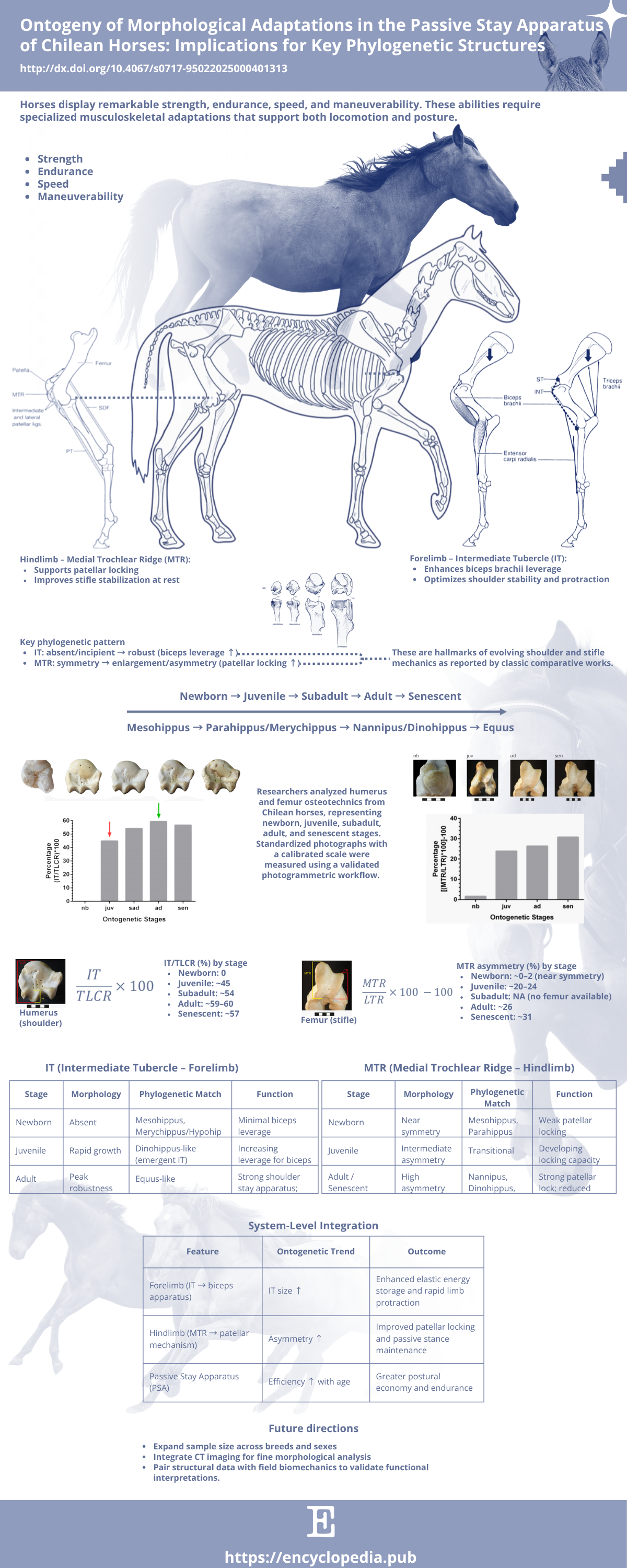
When horses stand, their joints remain stable without requiring continuous muscular contraction. This energy-saving mechanism is achieved through a combination of tendons, ligaments, and bone structures working together to “lock” the limbs in position. Two of the most critical components are:
-
The Intermediate Tubercle (IT) — a bony prominence on the humerus that provides leverage for the biceps brachii, a muscle that stabilizes the shoulder and assists in forelimb protraction.
-
The Medial Trochlear Ridge (MTR) — part of the femur that interacts with the patella to allow locking of the stifle joint (the horse’s equivalent of the human knee), preventing the hind limb from collapsing under the animal’s weight.
Together, these features act like an integrated system of biological springs and braces, enabling the horse to conserve energy whether resting or remaining vigilant against predators. Understanding how these structures develop helps explain not only individual biomechanics but also the evolutionary success of the modern horse.
2. A Life-Stage Approach to Equine Anatomy
The Chilean study analyzed humeri and femora from horses representing five distinct life stages: newborn, juvenile, subadult, adult, and senescent. Each specimen was carefully photographed using a calibrated photogrammetric workflow, a precise, non-destructive imaging technique that enables accurate morphometric analysis.
Two quantitative metrics were applied:
-
IT relative size: (IT / Trochlear Line Reference) × 100
-
MTR asymmetry: [(MTR / LTR) × 100] − 100
These ratios provided standardized measures of how the IT and MTR evolve through growth. Although each life stage was represented by a single individual — making the study exploratory — the consistent trends observed align well with broader evolutionary data.
Source: Encyclopedia Scientific Infographic (https://encyclopedia.pub/image/3837)
3. Findings: Strength Built Over Time
3.1. Shoulder Stability: The Growing Intermediate Tubercle
At birth, the IT is nearly absent, meaning the shoulder lacks the full mechanical leverage needed for efficient stabilization. During the juvenile phase, however, the IT grows rapidly, reflecting the animal’s increasing need for coordinated forelimb movement and weight-bearing. By adulthood, it reaches about 60% of the reference length, representing maximum functional efficiency.
In senescent horses, the IT shows a slight reduction, possibly due to bone remodeling or decreased muscular use — a subtle reminder that even the strongest anatomical systems are subject to aging.
This progressive development allows for greater biceps–lacertus synergy, enhancing the limb’s ability to both stabilize the shoulder and assist in forelimb protraction, a key component of stride efficiency.
3.2. Hindlimb Endurance: The Evolving Medial Trochlear Ridge
MTR–LTR asymmetry is near zero in newborns, rises to ~20–24% in juveniles, ~26% in adults, and ~31% in senescent horses.
This increasing asymmetry reinforces the patellar locking mechanism, a key feature of the stifle joint that allows horses to stand for long periods without fatigue. Essentially, the hind limb becomes a self-supporting column, transferring body weight efficiently through the skeleton rather than the muscles.
4. From Growth to Evolution: Echoes of Equine History
What makes this study particularly fascinating is how ontogeny (development) reflects phylogeny (evolution). When the researchers compared their findings with known patterns in equid evolution, a striking parallel emerged.
-
Newborn horses, lacking a defined IT and with symmetrical trochlear ridges, resemble early equids such as Mesohippus and Parahippus, species with limited stay-apparatus function.
-
Juveniles, developing these features rapidly, align with transitional forms like Dinohippus, showing intermediate shoulder and stifle mechanics.
-
Adults, with well-developed ITs and pronounced MTR asymmetry, match the anatomy of modern Equus — the fully specialized horse we know today.
This relationship supports the concept of peramorphosis, where developmental timing extends or accelerates traits that were advantageous in evolution. In this case, horses literally “grow through” their evolutionary past as they mature.
5. Why This Matters: Bridging Anatomy, Evolution, and Welfare
The implications of these findings extend beyond academic curiosity. Understanding the development of the stay apparatus offers practical insights for veterinary science, equine biomechanics, and animal welfare.
-
Training and management: Knowing when key joint structures reach maturity helps optimize the onset of physical training and minimize injury risk in young horses.
-
Veterinary diagnostics: Early detection of asymmetry or underdevelopment in the IT or MTR could indicate musculoskeletal imbalances that predispose horses to lameness or fatigue.
-
Evolutionary biology: The ontogenetic data reinforce long-standing hypotheses that evolutionary innovations often emerge from developmental modifications rather than new structures.
This connection between growth and evolution underscores a simple truth: the efficiency of modern horses was not an abrupt innovation, but a gradual refinement repeated in every individual life cycle.
6. Looking Ahead: A Blueprint for Future Research
While this study provides valuable insight, its authors emphasize the need for larger datasets encompassing multiple breeds, sexes, and environmental conditions. The integration of CT imaging and 3D morphometric modeling could further refine our understanding of how joint morphology translates into motion efficiency. Combining such anatomical studies with field biomechanics — for example, measuring joint angles during locomotion — would bridge form and function even more closely.
For more information about topic, you can view the online video entitled "Ontogeny of the Stay Apparatus in Chilean Horses".