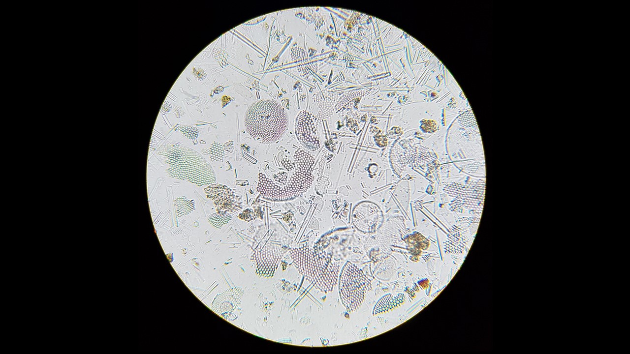


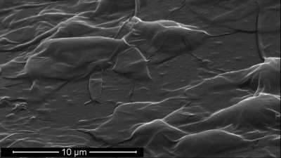
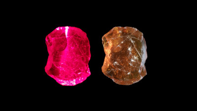
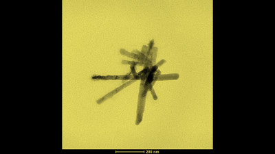
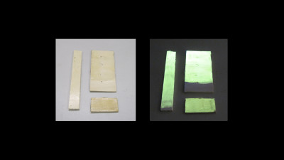
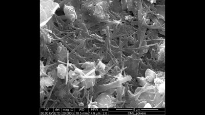
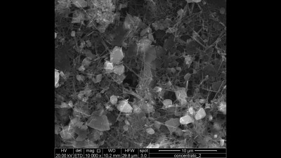

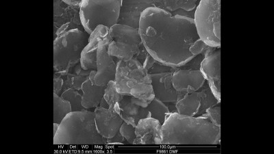
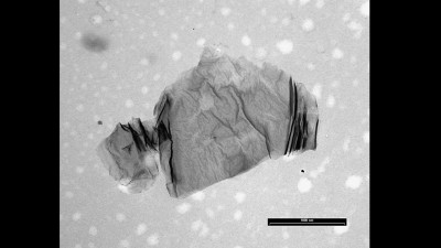
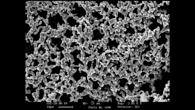
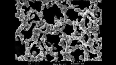
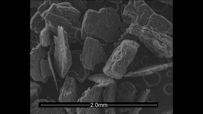
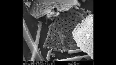
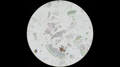
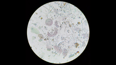
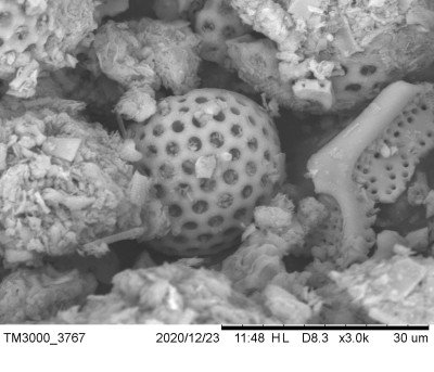
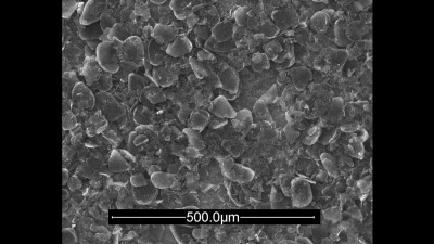
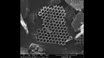
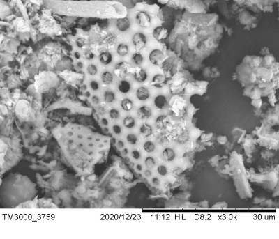
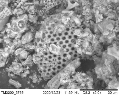
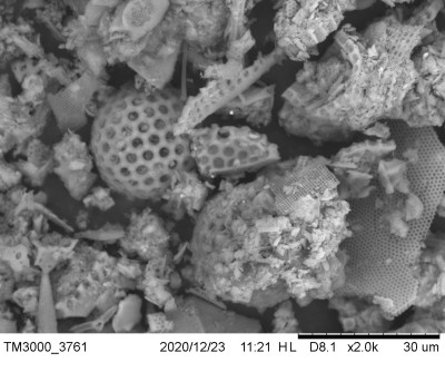
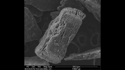
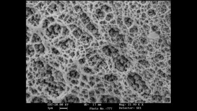

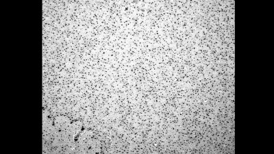
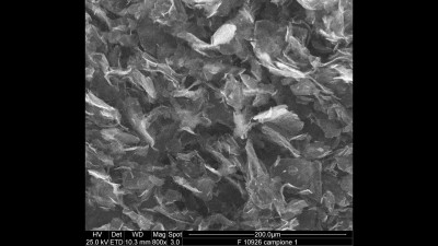
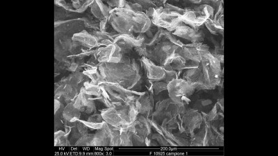
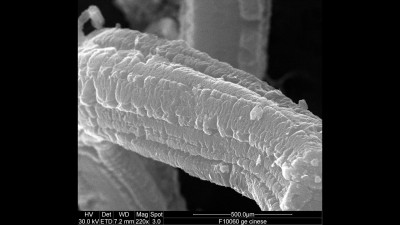
The diatomite microstructure can be conveniently visualized by using the simple transmitted light optical microscopy (OM) technique. Diatomite is a soft sedimentary rock made of siliceous shells of diatoms, a type of hard shelled unicellular microalgae, and its structure can be conveniently visualized by transmitted light microscope (namely biological microscope) because this device is typically used for investigating biological structures like cells, bacteria, fungi, etc. Actually diatomite consists mostly of tinny fragments of fossilized remains of diatoms and their ordered porosity can be clearly identified, however the inner structure of pores can be seen only at very high magnification by using a scanning electron microscope (SEM). Diatomite is a low density and high porous solid made of hydrated silica (SiO2.xH2O), it is slightly colored since it contains iron oxide and therefore it appears in transmission-light observation as colored or transparent glass pieces. Here, micrographs have been obtained simply by using the camera of a smartphone (Samsung, Galaxy S8) without adapter or additional lens.