Your browser does not fully support modern features. Please upgrade for a smoother experience.
Please note this is an old version of this entry, which may differ significantly from the current revision.
Subjects:
Oncology
|
Cell & Tissue Engineering
As the complexity of TME has called for more sophisticated human-based tumour models, organoids have allowed the dynamic study of spatiotemporal interactions between tumour cells and individual TME cell types. Here, we discuss how organoids can study the TME across cancers and how these features may improve precision I/O.
- organoid
- cancer
- immunotherapy
- tumour microenvironment
1. Investigating T Cell and ICI Responses
Although unprecedented strides have been made in the role of ICIs across multiple cancers since Allison [78] first uncovered the immunoregulatory role of CTLA-4 and Honjo [79] of PD-1 in the 1990s, a significant proportion of cancer patients still do not benefit. Amongst the responders, intrinsic [80], adaptive, and acquired [81] resistance remains an ongoing challenge. Tumour organoids are excellent platforms for studying T cells and their response to ICIs. The co-culture of dissociated NSCLC/CRC PDOs with autologous T cells from peripheral blood mononuclear cells (PBMCs) enriches the tumour-specific population that, in turn, kills the tumouroids [82,83]. A recent study shows alloreactivity-depleted engineered T cells engage with breast cancer, HNSCC, and glioma PDOs in a manner associated with type I interferon signalling [84]. MOSs [43], PDOTS [44], ALI [45], and TSCs [46,59] have all recapitulated the anti-PD-1 response ex vivo, with the potential to test novel therapeutic combinations such as ICIs + CDK4/6 inhibitors [47]. In another study, PDOs were enriched with matched immune components and treated with pembrolizumab, ipilimumab, or nivolumab for seven days before viability assays [85], which may identify responders. Interestingly, CRC organoids show that the gut microbiota may affect ICI response [86], suggesting its role in immune regulation in addition to tumourigenesis, as examined in other studies [87,88] where organoids are infected with specific bacteria with no or low-dose antibiotics in the media. The pioneering Nobel discoveries of MHC and its restriction (1980, 1996) have laid the foundation for later findings on defective antigen presentation in cancer, which facilitate the evasion of T-cell-mediated killing. Indeed, in CRC [89] and breast cancer [90] organoids, drugs that stimulate MHC-I antigen presentation on the tumour cells can increase T cell cytotoxicity and ICI effectiveness.
Future translational research on tumour organoids and T cell/ICI response may focus on two key questions. First, can ex vivo organoids predict the response of neoadjuvant, adjuvant or upfront systemic ICI, based on tumour biopsies or resected tumours [91]? It is encouraging that there are multiple clinical trials on organoid-guided therapy—such as NCT04777604 for the neoadjuvant space, NCT04736043 for the adjuvant space, and even NCT04931381 in advanced inoperable cancer (Supplementary Table S1 has a complete list)—all of which have offered great promise in validating organoid-guided I/O. Matrigel-based organoids may not serve this purpose well since the cultures may take weeks to stabilise, while PDOTS, ALI, and TSC may establish viable, TME-preserving cultures rapidly. Second, can organoids capture the spatiotemporal dynamics of checkpoint expression [9] and the changes ICI causes in the tumour cells, the TME, and the adjacent normal? This will offer insight into the characteristics of responders versus non-responders and the tumour evolution towards ICI resistance.
2. Unravelling the Functions of TME Cells
Metchnikoff might not have foreseen a century ago [92] that different macrophage populations possess distinct influences on cancers beyond phagocytosis, either in reducing the efficacy of therapies or limiting tumour growth in general. In 2004, Alberto Mantovani and colleagues proposed an M1–M2 spectrum of macrophage polarisation [93], and they subsequently defined the concept of tumour-associated macrophages (TAMs) [94]. Ovarian cancer organoids have shown that UBR5 from tumour cells drives the recruitment and immunosuppressive reprogramming of TAMs [95]. In intestinal adenoma, co-cultured macrophages promote the growth of organoids in a prostaglandin E2-dependent manner, while macrophages themselves adopt a TAM-like phenotype [96]. This reciprocal interaction is abrogated by celecoxib, a selective COX-2 inhibitor. Another co-culture of pancreatic cancer PDOs, U937 monocytes, and primary pancreatic stellate cells leads to increased PDO invasiveness and an M2-like phenotype of U937 [97], whose depletion significantly increases PDO sensitivity to chemotherapy. Finally, autologous co-culture of CRC PDOs with PBMC-derived CD8+ cytotoxic T lymphocytes (CTLs) and macrophages shows that a high sirtuin 1 (SIRT1) level in CRC cells increases macrophage infiltration and M2 polarisation, contributing to CTL dysfunction [98]. While tumour organoids have offered insight into how TAMs may act as friend or foe, caution must be taken when optimising the culture conditions (especially the external growth factor/cytokine supply), as they might shift the phenotypes of many cell types and mask the true effect of tumour–immune interaction. Future organoid-based TAM research may detail how TAMs cause T cell dysfunction [99], tumour cell inflammation, metabolic derangements [100], and increased angiogenesis [101,102] (discussed below). In fact, an M1–M2 dichotomy may be an oversimplification for TAMs in vivo, which can possess markers for both M1 and M2 at the same time [103]. We know that macrophages are pro-inflammatory during pre-tumour necroinflammation, which are subsequently polarised towards immunosuppressive cells in the tumour. However, what leads to this cascade of TAM reprogramming is not understood; thus, organoids may offer insight into this profound question.
Many years after Burnet and Medawar’s discovery of the concept and principles of immune tolerance, myeloid-derived suppressor cells (MDSCs) were described as a vital contributor to cancer immunosuppression. Several studies have shown viable MDSCs in PDOs, whose depletion sensitises ICI response [104,105]. However, detailed, longitudinal examination of MDSCs in organoids remains rare. The same holds true for dendritic cells (DCs) [106], neutrophils, and other granulocytes [107]—important TME cell types yet to be further studied in organoids.
While lymphoid organoids have been successfully generated in vitro [108,109], the recapitulation or study of B cells in tumour organoids remains challenging and hence very limited [45]. As more research has uncovered the multifarious roles of B cells in the TME [110], further work on B-cell-containing tumour organoids may be necessary.
Endothelial cells (ECs) are another critical cell type in the TME. While it is well known that angiogenesis, a hallmark of cancer [111], can be driven by tumour cells rather aberrantly [101,112,113], we now understand that the endothelium does much more than the sheer lining of blood vessels. For instance, ECs can mediate metastasis [112,114], present antigens via MHC-II [115], reprogram the myeloid phenotypes [116,117], and regulate tissue fibrosis [118]. Tumour organoids have uncovered new roles of ECs other than vascularisation. ECs utilise the tumour cell secretome and hijack the M2-like TAM polarisation for angiogenesis in HCC [101] and glioblastoma [102] organoids. Through their angiocrine functions, ECs in tumour organoids have been shown to drive a pro-inflammatory environment and M1-like TAM polarisation, antagonising the effect of M2-like TAMs [101]. Organoids have also revealed the importance of angiocrine signalling in the survival of pancreatic cancer cells [119,120] and in promoting ovarian cancer metastasis [121]. Strikingly, ovarian cancer organoids with ECs bear a close resemblance to metastatic tumour spheres in the peritoneum or ascites of patients [121,122]. Careful consideration is needed if HUVECs are used in organoids, as they may behave differently from tumour ECs and tissue-specific ECs, such as liver sinusoidal endothelial cells (LSECs) [123]. Further organoid studies are warranted on the spatiotemporal dynamics between ECs, TAMs and T cells, the vascular normalisation by anti-angiogenic drugs [124,125], and the effect of ECs in pre-cancer [126].
Tumour organoids provide the most physiologically relevant platform ex vivo and in vitro for studying CAFs. Defined as the tumour mesenchymal cells that do not bear epithelial, immune, or endothelial markers [127], CAFs in pancreatic cancer maintain their phenotypes only in organoids but not in mono-culture [120,128]. The distinction between α-SMA-positive myofibroblastic CAFs (myCAFs) and IL-6-positive inflammatory CAFs (iCAFs) [128] is driven by TGF-β and IL-1 signalling, respectively [129]. CAF plasticity in pancreatic cancer leads to distinct TME of a reactive, immune-hot subtype versus a deserted, immune-cold counterpart [130]. Intriguingly, while the immune-reactive TME CAFs promote tumour organoid growth and are associated with lower disease-free survival, the immune-deserted TME CAFs confer chemoprotection to the organoids and are associated with lower overall survival [130]. Additionally, tumour organoids for CRC [131,132], HCC [133], and prostate cancer [134] have all shown a similar trophic effect of CAFs and the conferral of therapeutic resistance, seemingly relying on paracrine signalling. It is noted that patient-derived CAFs rapidly differentiate towards myCAFs in vitro, and thus protocol optimisation is key in preserving the CAF phenotype in organoids [132]. Investigation into the recruitment and reprogramming of CAFs, and their paracrine effect on tumour cells and immune cells, will yield exciting insight into the immunoregulatory functions of CAFs and offer new therapeutic opportunities, which are discussed next.
3. Testing of Novel Precision Immuno-Oncology Strategies
The past decade has witnessed the therapeutic revolution ICI has brought to the lives of many cancer patients simply by unleashing the brakes on effector T cells. Now, the reality of tumour heterogeneity, the advance of our knowledge in the TME, and the breakthroughs of our ability to interrogate tumour biology have all called for the advent of personalised I/O. Organoids are, and will continue to be, a key driver of precision I/O with their superior accessibility, rapid throughout, and good robustness [135,136]. We begin by summarising in Figure 3 what we have discovered about different cell types from tumour organoids and whether therapies targeting each cell type have been tested on them, discussed in detail next.
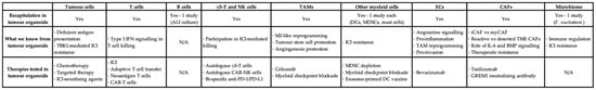
Figure 3. Tabulated summary of different cell types in tumour organoids, the knowledge added, and the therapeutics tested. TAM: tumour-associated macrophage; EC: endothelial cell; CAF: cancer-associated fibroblast; ALI: air–liquid interface; DC: dendritic cell; MDSC: myeloid-derived suppressor cell; ICI: immune checkpoint inhibitor; IFN: interferon; iCAF: inflammatory CAF; myCAF: myofibroblastic CAF; TME: tumour microenvironment; CAR: chimeric antigen receptor.
With combination immunotherapies proving superior survival outcomes across more and more cancers and even in the adjuvant setting [137], and with rising numbers of ongoing combination trials awaiting definitive results, combining ICIs and other therapeutic modalities will define a significant space in cancer immunotherapy strategies in the coming years [138]. New targets on the tumour cells have emerged that directly influence ICI-mediated cytotoxicity. PPARγ-agonists, such as pioglitazone, reprogramme the metabolism and inflammatory response of microsatellite-stable (MSS) CRC cells and increase their PD-L1 expression, sensitising these seemingly resistant organoids to ICI [139]. Targeting TBK1, a kinase with multifaceted functions in innate immunity and cell proliferation, also enhances ICI response, as seen in PDOTS of multiple cancers [140].
Combining ICIs with TME-targeting therapies may also lead to improved clinical response [141], and some of these strategies have been tested in tumour organoids. As discussed earlier, celecoxib in tumour organoids can abrogate the TAM-tumour stem cell interaction [96]. Celecoxib or other NSAIDs (e.g., aspirin in NCT00565708) may be attractive in targeting TAMs, yet their overall clinical effect remains uncertain. Depletion of MDSCs in gastric cancer PDOs increases anti-PD-1/PD-L1-induced killing [104], suggesting the immunosuppressive role of myeloid checkpoints besides PD-L1, such as arginase-1 [142] and VISTA [143,144]. Clinical trials are underway to test anti-VISTA monotherapy or combination with anti-PD-1 in advanced solid tumours (e.g., NCT05082610) [145]. Surprisingly, histamine, via H1 receptors on TAMs, confers ICI resistance by polarising TAMs towards M2 with increased VISTA expression [146]. Organoid testing for H1-antihistamines, anti-VISTA, and other myeloid checkpoint inhibitors may offer a rapid prediction, in-depth molecular insight, and longitudinal monitoring. In addition, ICI combined with bevacizumab has already shown benefits in phase 3 trials [137,147], which warrant organoid studies of EC-targeting therapies [124]. Furthermore, small molecule inhibitors or blocking antibodies targeting FGFR or TGF-β receptors may interfere with CAF activation and action [127], as seen in organoids [148]. However, their clinical benefit remains uncertain [149], suggesting the robustness of CAFs and the difficulty in targeting them. Intercepting the IL-6 axis may also abrogate tumour–CAF interaction [150], yet the effect of drugs (e.g., tocilizumab) both on the TME and systemically is concerning. Crucially, culture conditions, such as growth factor supply and hypoxia, may directly influence the readouts of organoid testing, as discussed in earlier sections.
Adoptive cell transfer of tumour infiltrating lymphocytes (TILs) or tumour-reactive T cells, especially neoantigen-specific T cells [151,152], has emerged as a promising strategy of precision I/O [153]. Lymphodepletion with fludarabine/cyclophosphamide or similar non-ablative conditioning regimens creates a favourable environment for the engraftment, survival, and optimal in vivo expansion of the infused therapeutic T cell product [153,154]. ICIs may be synergistic or additive to adoptive cell therapy [155]. Organoids can boost T cell therapy translation by rapidly enriching tumour-reactive T cells [82,83], unravelling the regulation [156,157] and heterogeneity [158] of MHC-I neoantigen presentation and, crucially, providing a platform for neoantigen validation [155] and autologous killing assays [43,159].
Chimeric antigen receptor (CAR)-T cells have shown unequivocal clinical efficacy against B cell malignancies and multiple myeloma. Testing against solid tumours has shown some encouraging clinical efficacy, although significant challenges from the immunosuppressive TME still exist [160]. The delicate design of the CAR-T cell construct and its activation mechanisms leads to their potent killing capacity and sustained presence and anti-tumour activity [161,162,163]. Organoid-killing assays have shown success in CAR-EGFRvIII T cells for glioblastoma [29], CAR-HBsAg for HBV-HCC [164], as well as CAR-CD70 for renal cell carcinoma [165]. Further investigation on organoid-CAR-T co-culture may offer solutions to CAR-T cell exhaustion in vivo and discover novel targets tackling tumour heterogeneity. For instance, the efficacy of CAR-T cells might be limited by the physical barrier of a fibrotic tumour stroma [166]. Organoid modelling of tissue fibrosis has shown the deposition of collagen and other extracellular matrix proteins [75,167]. By modifying the mechanical and biochemical properties of the matrix or hydrogels in organoid culture, we may unravel the spatiotemporal dynamics of CAR-T infiltration into and their killing of tumouroids.
Finally, NK and γδ-T cell therapies, both autologous and allogenic, have received greater attention now due to their potent anti-tumour capabilities. NK cells typically react to stress ligands via interactions including NKG2A–HLA-E and NKG2D–MIC-A/B [168], while γδ-T cells recognise phosphoantigens, such as exogenous zoledronic acid (ZOL), to exert cytotoxicity [169]. Strikingly, a recent study with ovarian cancer organoids shows that bi-specific PD-1/PD-L1 antibody activates NK cells in addition to CTLs [170], while another study with CRC organoids demonstrates the anti-tumour effect of ICIs from γδ-T cells (rather than αβ-T) when there is MHC-I defect on tumour cells [171], underlining their importance in I/O effectiveness. Organoid testing has also shown tumoricidal activity of γδ-T cells [172] and (CAR-)NK cells [173] for melanoma and mesothelioma, respectively. Importantly, while 3D killing assays have been performed and published extensively for NK and γδ-T cells, most “tumours” are only spheroid aggregates of cancer cell lines, which do not recapitulate the TME. Killing assays on organoids, on the other hand, will unveil distinct mechanisms that facilitate or inhibit cell therapies, many of which arise from interactions with the TME rather than with tumour cells alone. For instance, our group is studying the distinct phenotypes between NKG2A(+) and NKG2A(-) γδ-T cells, where tumour organoid co-culture may offer a better understanding.
This entry is adapted from the peer-reviewed paper 10.3390/cells12081165
This entry is offline, you can click here to edit this entry!